 |
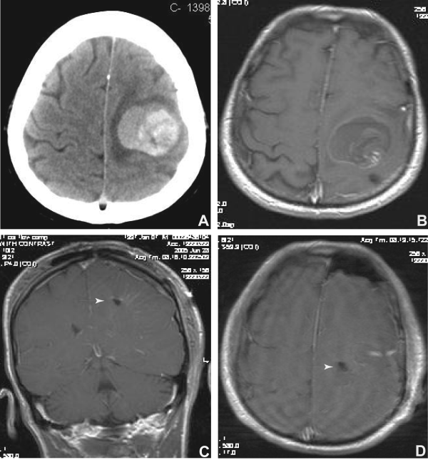
| Figure 1: Neuroimaging of the tumor before and after resection. A, Non-contrast, axial CT scan of the brain showing a hemorrhagic lesion with surrounding hypointense edema and mass effect with sulcal effacement in the posterior left frontal and anterior parietal regions. B, Gadolinium enhanced, T1-weighted axial image obtained several hours after sudden neurological deterioration showing a wisp of enhancement (white, hyperintense) and surrounding hemorrhage (dark, consistent with recent bleeding). C, D, coronal and axial T1-weighted, gadolinium enhanced images after resection, which demonstrate complete resection of tumor and clot; an arrow marks the small resection cavity remaining after resection. |