|
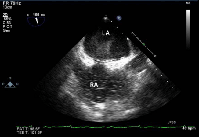
| Figure 1: A transesophageal echocardiogram, mid-esophageal view, 106 degrees; showing significant spontaneous echo contrast (SEC) in the left atrium (above), compared to a bubble study using agitated saline seen in the right atrium (below). LA: left atrium, RA: right atrium. |