|
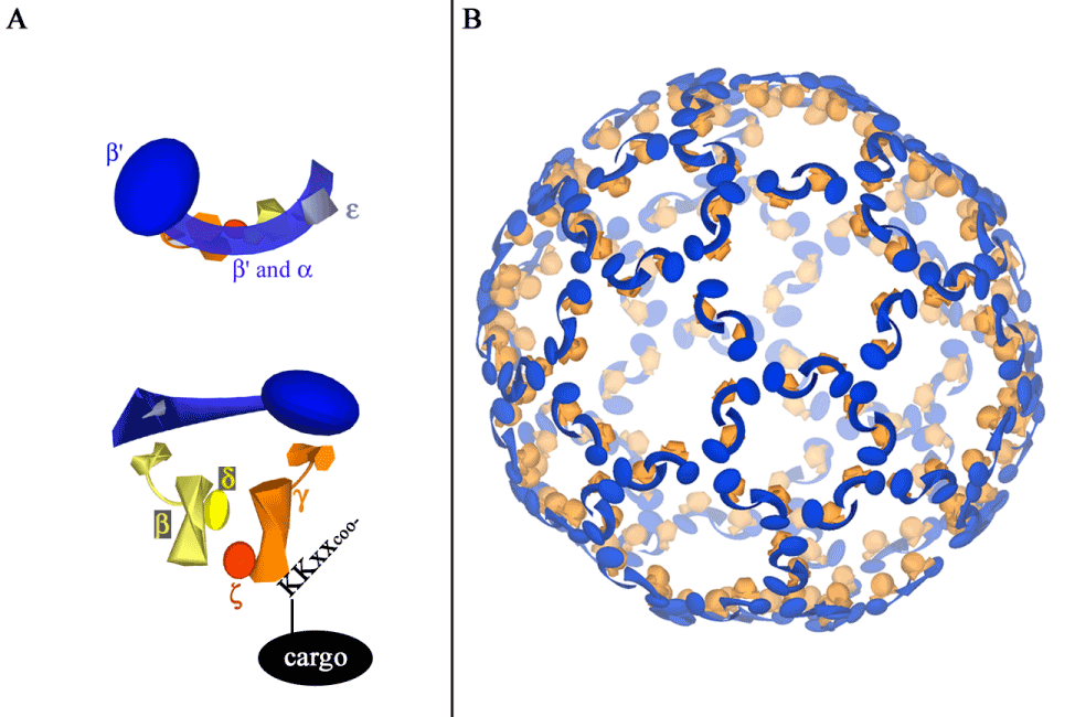
| Figure 2: Illustration of COPI subcomplexes (A) and a potential structure of the COPI cage around a spherical lipid vesicle (B). (A) As revealed by X-ray crystallography and electron microscopy, the backbone of the COPI cage is formed by the αβ’ε subcomplex (top) [11,16], which is on the cytoplasmic face of the vesicle. The βδγζ subcomplex structure (bottom) is similar to that of the adaptor protein complex, which is important for formation of clathrin-coated vesicles [12,14,15,17]. The γ subunit interacts with the KKXX domain on ER resident cargo proteins [13]. (B) In this model of the COPI lattice, the αβ’ε and βδγζ subcomplexes assemble in a manner similar to clathrin and adaptor protein complexes, respectively. The βδγζ subcomplex is located between the αβ’ε backbone and the lipid vesicle; it interacts with cargo in the vesicle membrane. Note that this is only one possible structure of a COPI cage; COPI coats can form tubular structures and vesicles of varying sizes. This structure is based on work by Lee and Goldberg [11] and Yip and Walz [16] and was created using Google SketchUp8. |