|
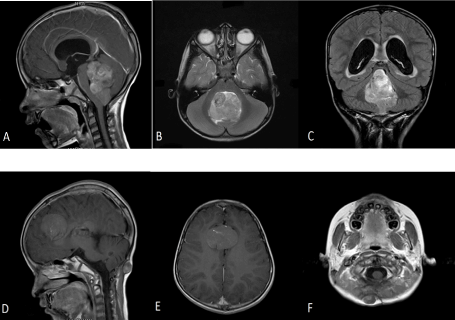
| Figure 1: Magnetic resonance imaging scan of the brain showing a 4.0x3.9 cm mass in the fourth ventricle causing non-communicating hydrocephalus in (A) sagittal, (B) axial and (C) coronal planes. Magnetic resonance imaging scan of the brain 10 months after completing radiotherapy, showing a midline 4.5 x 4.2 x 4.0 cm lesion between both frontal lobes1.7 x 2.1 x 1.2 cm soft tissue lesion in the suboccipital region in (D) sagittal and (E) (F) axial planes, respectively. |