|
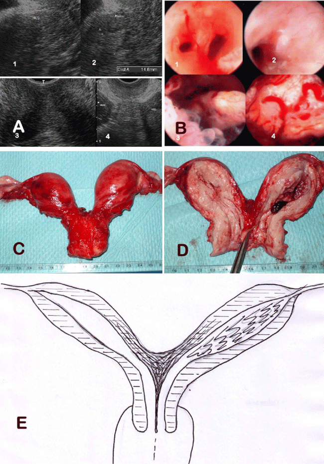
| Figure 1: A) Transvaginal ultrasonography images. 1 and 2, longitudinal cross sections of right and left hemiuterus. 3 and 4, transversal cross sections, at cervical portion in 3, at corpus of both hemiuteri in 4. B) Hysteroscopical images. 1, endocervix; 2, right hemiuterus; 3 and 4, left hemiuterus showing adenocarcinoma images. C) Surgical specimen from hysterectomy. D) Longitudinal section of the anterior wall of both hemiuteri. E) A graphical representation of the uterine malformation. |