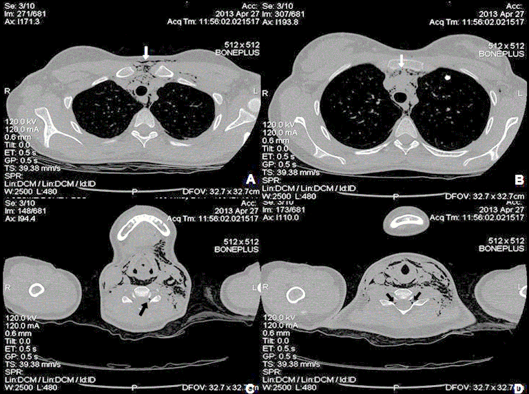
Figure 2:
CT of the thorax. A and B: confirmatory images of subcutaneous emphysema and pneumomediastinum (white arrows); C and D: images of free air in the spinal canal, consistent with a discrete pneumorrhachis (black arrows)