Case Report, Int J Cardiovasc Res Vol: 9 Issue: 7
A Rare Anatomical Anomaly: Entire Coronary Circulation Arising from the Right Sinus of Valsalva
Krishna Vedala1*, Shoaib Khan1, Ankitha Antony1 and Bennett Rudorfer2
1Department of Internal Medicine, White River Health System, Batesville AR, United States of America
2Department of Cardiology, White River Health System, Batesville AR, United States of America
*Corresponding Author: Dr. Krishna Vedala MD, Department of Internal Medicine, White River Health System, Batesville AR, United States of America Tel: 4055354723, E-mail: Kvedala@wrmc.com
Received: October 09, 2020 Accepted: November 10, 2020 Published: November 17, 2020
Citation: Vedala K, Khan S, Antony A, Rudorfer B (2020) A Rare Anatomical Anomaly: Entire Coronary Circulation Arising from the Right Sinus of Valsalva. Int J Cardiovasc Res 9:7.
Abstract
Congenital coronary anatomical anomalies pose dire negative consequences and are a significant financial burden. We report of an 82-year-old male who presented to us with dyspnea and lower extremity edema. He underwent further imaging workup and subsequent coronary angiography which revealed his entire left coronary circulation arising from the Right Sinus of Valsalva, a very rare anatomical variant. Surgical intervention was not recommended due to patient being relatively stable and asymptomatic. Anatomical variants are diverse thereby causing greater difficulty, specifically in terms of deciding between surgical treatment versus medical management. This case highlights the importance of always keeping anatomical variants as differentials for cardiac complications and utilizing advanced imaging in diagnosing such variants.
Keywords: Coronary anatomical variant; Right sinus of Valsalva; Lippincot classification; Coronary angiography.
Keywords
Coronary anatomical variant; Right sinus of Valsalva; Lippincot classification; Coronary angiography.
Introduction
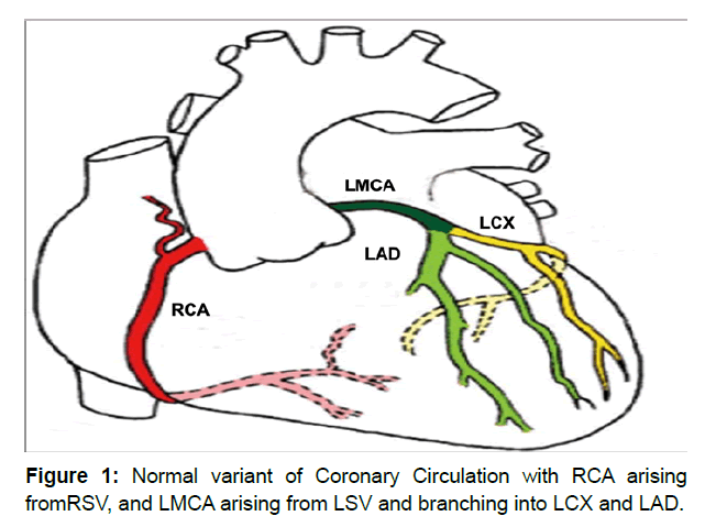
Congenital coronary anomalies have been extensively studied and documented in part due to risk of causing physiologic ischemia. Clinical symptoms can include chest pain, dyspnea, syncope, cardiomyopathy, and myocardial infarction. In addition, coronary anatomical anomalies are the second most common cause of sudden cardiac death in young athletes [1]. Despite having an incidence of only 1-2%, coronary anomalies can be a financial burden by increasing the need for greater surveillance, risk of surgical procedures, and also overall-mortality [1]. However, the wide variation in their presentation makes it difficult to assess and predict how and when certain variants can cause symptomatic complications. Anatomical variations can be classified based on three parameters: the sinusoids, the in situ vascular endothelial network and the coronary buds on aortic sinuses [1,2]. As shown in Figure 1 the conventional coronary anatomy includes the Left Main Coronary (LMCA) and the Right Coronary (RCA) arteries arising from their respective LeftSinuses of Valsalvas (LSV) and Right Sinuses of Valsalvas(RSV) [2]. Here, we describe a case where the entire coronary circulation arises from the RSV.
Case Discussion
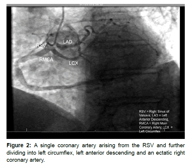
Our patient is an 82-year-old male with a past medical history of type 2 diabetes mellitus and hypertension with no history of cardiovascular events. In addition, he was a former smoker who had quit in 1980 but did have passive smoke exposure from working as a fireman. After undergoing a shoulder replacement procedure, he started to experience dyspnea and increased lower extremity edema. Transthoracic echocardiogram revealed normal systolic function with mild left ventricular hypertrophy and aortic stenosis. Following the initial visit, he then underwent a myocardial perfusion scan, which indicated decreased perfusion on inferior wall concerning for possible ischemia and an ejection fraction of 74%. Patient was then scheduled for combined left and right heart catherization. Angiographic imaging (Figure 2) revealed anomalous anatomy where the entire coronary system was derived from the RCA originating from the RSV. The RCA appeared to be a very ectatic vessel leading to disease-free posterolateral and posterior descending systems and a larger system of posterolateral LV extension branches. The first branch of the RCA was a Left Anterior Descending (LAD) that was patent proximally but showed high-grade stenosis of 95% in the mid-portion and further significant stenosis in the distal ends. The LAD also seems to have terminated prematurely before reaching the apex. The second branch of the RCA was a Left Circumflex (LCX) that seemed to have both a longer course and greater dilation than normal variant. The LCX also seems to have severe mid-stenosis of 80% but does give rise to three disease-free marginal branches. In addition, patient was noted to have elevated RA filing pressures with evidence for mild-to-moderately elevated pulmonary artery systolic hypertension. The patient tolerated the procedure well without complications. Subsequently, he followed up with a cardiothoracic surgeon for further evaluation, who recommended no need for further imaging including Coronary CT or surgical intervention, and continued surveillance monitoring. On further follow-up, lisinopril and furosemide were added to medication regimen, and strict glycemic control was recommended. Patient continues to be asymptomatic.
Discussion
In our patient, his entire coronary circulation is arising from a single coronary artery originating in the RSV. Based on Lipton’s classification of single coronary artery, our case seems to fit the subset R III-C [3]. Perhaps a more surprising notion is that despite this anomaly, our patient had a benign cardiovascular history and more importantly continues to do well since this discovery. This anomaly was discovered utilizing coronary angiography and the patient did not undergo further imaging. A 2017 retrospective study from Brigham and Women’s Hospital showed that coronary computed tomography is a very useful tool in detecting anomalous origin of coronary arteries from the opposite sinus [4]. Prevalence of such anomalies was noted to be around 1.7% [4]. It is difficult to predict whether his symptomatic presentation is a function of his age or the coronary anomaly itself. The prevalence of a single coronary artery arising from the RSV is 0.02-0.05% with prognosis being better with the anterior variant rather than the inter-arterial variant [1]. This anomaly has been associated with largely asymptomatic sudden cardiac disease until age of 20 years [1].
Venturini et al. [5] reports a 74-year-old female with LMCA and RCA coming from RSV. The case exemplifies the roles played by advanced age and coronary anomaly as the patient suffered an anterior myocardial infarction despite having disease-free vessels and no atherosclerotic plaques. Another particular variant with similar presentation could be a LMCA arising from the RSV.Ruhela et al. [6] discusses such a case about of a 45-year-old presenting with dyspnea and refractory atypical chest pain. Despite the anomaly, imaging revealed disease-free vessels. Gleeson et al. [7] reports of a 56-year-old male who was noted to have a single coronary artery arising from the RSV with a pre-pulmonic course of the left coronary artery. However, there was also noted to be a reconstitution of normal distribution at the junction of mild and distal LAD and subsequent anastomosis [7].
While the majority of patients may be asymptomatic, the risks regarding coronary anatomical anomalies are simply too significant to ignore. Sudden cardiac death, often at young ages, is perhaps the biggest concern [8]. In addition, other symptoms can include anginal chest pain, syncope, or dyspnea [8]. Such anomalies can also drastically affect quality of life specifically by limiting individual functionality, especially in athletes and military personnel [8]. Due to the range of diversity among coronary anomalies and large financial costs associated with coronary image, screening for anatomical anomalies is not recommended [9]. In the case of our patient, a major concern is that resting electrocardiograms and stress tests are usually normal in anomalies where the LMCA derives off of the RSV [10]. However, the patient’s quality of life in the short term will have minimal change as his current lifestyle lacks any physically demanding tasks. Yet, in the long-term his functionality will be dependent on aggressive risk modification and personal decision-making regarding any further surgical intervention due to his age.
Conclusion
Our case further contributes to greater understanding of coronary anomalies and their complications, but once again exemplifies the difficulty posed by such anomalies. Overall, coronary anomalies are difficult to predict regarding their complications, a task that requires us to understanding the symptoms and paying more attention to detecting them through angiography and advanced imaging.
Competing Interests
Nothing to declare.
References
- Villa AD, Sammut E, Nair A, Rajani R, Bonamini R, et al. (2016) Coronary artery anomalies overview: The normal and the abnormal. World Journal of Radiology. 8: 537-555.
- Pasaoglu L, Toprak U, Nalbant E, Yagiz G (2015) A rare coronary artery anomaly: Origin of all three coronary arteries from the right sinus of valsalva. J clin imaging sci. 5: 25.
- Christos G, Dimokritos D, Antonios N, Vasileios K, Vasileios P, et al. (2013) Percutaneous coronary intervention and stenting in a single coronary artery originating from the right sinus of valsalva. Hellenic journal of cardiology. 54: 401-407.
- Cheezum MK, Ghoshhajra B, Bittencourt MS, Hulten EA, Bhatt A, et al. (2017) Anomalous origin of the coronary artery arising from the opposite sinus: Prevalence and outcomes in patients undergoing coronary CTA. Eur Heart J Cardiovasc Imaging. 18: 224-235.
- Venturini E, Magni L (2011) Single coronary artery from the right sinus of valsalva. Heart int. 6: e5.
- Ruhela M, Chaturvedi N, Shekhawat DS (2015) Left coronary artery originating from right coronary Sinus A rare coronary artery anomaly. American Journal of Medical Case Reports. 3: 1-3.
- Gleeson T, Thiessen R, Wood D, Mayo JR, (2009) Single coronary artery from the right aortic sinus of valsalva with anomalous prepulmonic course of the left coronary artery. Canadian Journal of Cardiology. 25: e136-e138.
- Angelini P (2007) Coronary artery anomalies: An entity in search of an identity. Circulation. 115: 1296-1305.
- Angelini P, Velasco JA, Flamm S (2002) Coronary anomalies: Incidence, pathophysiology, and clinical relevance. Circulation. 105: 2449-2454.
- Bolcal C, Sargin M, Iyem H, Akay HT, Bingol H, et al. (2006) Coronary artery anomalies: Anomalous origin of the left coronary artery and circumflex branch in two patients. Exp Clin Cardiol. 11: 314-316.
 Spanish
  Chinese
  Russian
  German
  French
  Japanese
  Portuguese
  Hindi
Hindi 


