Research Article, Int J Cardiovasc Res Vol: 7 Issue: 5
Early Effect of Right Ventricular Apical Pacing On Left Ventricular Twist
Yaseen R1*, Soliman M1, Shaban G2, Elmallah S1 and Fazwy M1
1Cardiology Department, Faculty of Medicine, Menoufia University, Egypt
2National Heart Institute, Egypt
*Corresponding Author : Rehab Ibrahim Yaseen
Department of Cardiology, Menofia University, Abdel rahman Elsharkawy Street, Shebin El-Kom, Menoufia, Egypt
Tel: +01006235708
Fax: 20 (48) 222 6454
E-mail: dr_r.yaseen@hotmail.com
Received: August 01, 2018 Accepted: October 11, 2018 Published: October 17, 2018
Citation: Soliman M, Yaseen RI, Fawzy M (2018) Early Effect of Right Ventricular Apical Pacing on Left Ventricular Twist. Int J Cardiovasc Res 7:5. doi: 10.4172/2324-8602.1000364
Abstract
Abstract
Objective: The study aimed to assess the short-term effects of apical RV pacing on LV function and twist mechanics using speckle tracking echocardiography.
Background: The myofibers of the heart has geometric orientation that induces its characteristic wringing motion around its long axis during contraction leading to the systolic LV twist. This motion is created by the apical and basal rotations that are oppositely directed and has an important role in regulation of LV function. Several studies have demonstrated that LV systolic function and mechanics had delayed deterioration after RV apical pacing with limited data regarding the acute effects.
Methods: This study was conducted on Twenty-Four patients who were indicated for pacemaker insertion due to complete heart block. Echocardiographic examination was done to measure the LV dimensions and ejection fraction (EF) before and 24 hours after RV apical pacing. LV twist changes were assessed by using speckle tracking imaging modality.
Results: Our study showed that the LV end diastolic dimension has a significant reduction acutely after RV apical pacing (p value=0.001) while the end systolic dimension significantly increased (p value=0.003). EF significantly decreased after pacing (p value=0.001).Also there was a significant reduction in LV apical rotation (p value=0.001), basal rotation (p value=0.012) and torsion (p value=0.034).
Conclusion: Pacing of RV apex impairs the LV rotational mechanics acutely which subsequently have a detrimental effect on LV function.
Keywords: Apical; Pacing; Right ventricular, Left ventricular; Function; Twist
Introduction
Cardiac pacing is considered the most effective treatment in the management of bradyarrhythmia and some cases of tachyarrhythmia. It helps in improvement of symptomatic SA nodal disease and atrioventricular (AV) block [1].
Furthermore, it is considered to be the last resort in controlling symptoms of permanent, drug-refractory atrial fibrillation [2]. Finally, in selected patients with left ventricular (LV) systolic dysfunction, cardiac resynchronization therapy (CRT) can improve the cardiac function and enhance the patient’s functional capacity [3]. Large number of randomized clinical trials has studied the optimal pacing mode [4-6].
But what is more important is that these trials have suggested a strong relationship between right ventricular (RV) apical pacing and cardiac morbidity and mortality. The direct electric stimulation of the RV apex induced an abnormal activation sequence and hence asynchronous ventricular contraction [7-9]. Subsequently, LV performance is impaired leading to a decrease in the stroke volume and abnormal relaxation [10].
The apex of the heart rotates in opposite direction to the base resulting in the twisting of the left ventricle around its long axis which is determined by the contracting myofibers in the LV wall [11,12] as they are arranged in opposite directions between the subendocardial and subepicardial layers [13-15]. This twisting motion plays an essential role regulating both the LV systolic and diastolic functions [16].
The LV rotational mechanics are sensitive indicators of the cardiac performance [13,17]. Many studies demonstrated that LV systolic function and mechanics had a delayed deterioration after RV apical pacing [8-10]. This study aimed to assess the short-term effects of apical RV pacing on LV function and twist mechanics using speckle tracking echocardiography.
Patients and Methods
Patients
After approval of Menoufia Ethics Committee for the study proposal, Twenty four patients indicated for pacemaker insertion were included in the present study. In twenty three cases the indication of pacemaker insertion was to treat complete heart block and in the remaining case the indication was to treat sick sinus syndrome. We excluded patients with LV systolic dysfunction ((EF < 40%)), acute coronary syndrome, significant valvular heart diseases, myocardial diseases and bad echogenic window.
Methods
A transthoracic echocardiographic examination was performed for every patient before and 24 hours after pacemaker insertion and then off line analysis was done to measure LV twist. We used the available machine (Vivid 9, GE Vingmed Ultrasound AS, Horten, Norway) equipped with a 1.7-4 MHz phased-array transducer with simultaneous ECG tracing. All the patients were lying in the left lateral position during the examinations to allow for a better image quality [18]. 2-dimensional parasternal long axis view was obtained by placing the probe at the left sternal border in the third intercostal space with the probe marker directed to the right shoulder.
We measured the end diastolic diameter of the left ventricle, the end systolic diameter and the ejection fraction (EF) using M-mode modality with the curser perpendicular to interventricular septum and posterior wall passing through mitral valve tips [19]. 2-dimensional parasternal short axis views were obtained by rotating the probe 90 degrees clockwise while in the parasternal long axis position directing the marker towards left shoulder.
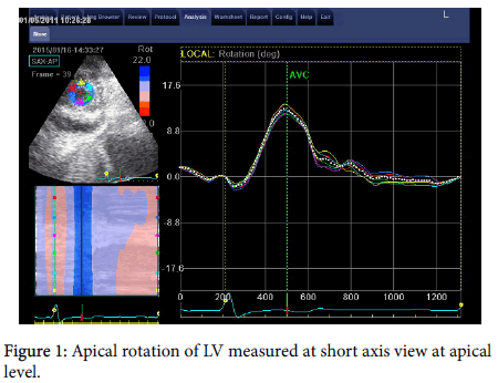
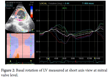
Short axis view at the level of mitral valve was obtained by tilting the probe slightly upward until we got the characteristic fish mouth appearance of the mitral valve. Apical short axis view was obtained by tilting the probe more upward until we got a cross section of the LV apex.
Speckle-tracking imaging analysis was performed using the available software (Echo PAC BT 12, GE-Vingmed; Norway). We manually traced the endocardial border of the LV and then the software automatically generated a second, larger, concentric tracing at the epicardium so that all the LV myocardium became included. Then, the software automatically transformed each LV view into 6 equal segments and performed the speckle-tracking on a frame-to-frame basis [18].
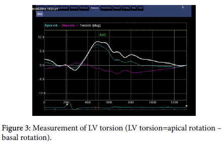
Using the average of LV rotations from the 6 segments, we measured the basal and apical rotations taking into consideration to measure the mean rotation at aortic valve closure. The apical rotation was expressed in positive values, while the basal rotation in negative values. LV twist equals the apical rotation minus the basal rotation. Rotation and twist are expressed in degrees (Figures 1-3) [18].
Statistical procedure
Data was statistically analyzed by using the well-known program statistical package for social science (SPSS) version 13 for windows (SPSS Inc., Chicago, IL, USA) and during all the analysis process a p value below 0.05 was considered statistically significant. Data are shown as mean, range or value and 95% confidence interval (95% CI) and frequency and percent. Paired t-test was done for comparing two normally distributed variables related samples to measure mean and standard deviation. The Wilcoxon signed-rank test is was used for comparing two non-normally distributed related samples.
Results
Our study included 6 males (25%) and 18 females (75%) with mean age 53.4 ± 11.7 (Table 1). Heart rate was significantly lower in pre RV apical pacing than post pacing (P value=0.001) with the mean heart rate 39.9±4.8 pre pacing (Table 2).
| Patients (n=24) | ||
|---|---|---|
| Male | Female | |
| Sex | 6 (-25%) | 18 (-75%) |
| Age (years) | Mean ± SD 53.4±11.7 | |
| Median 54.0 | ||
| Range 35.0 – 75.0 | ||
Table 1: Demographic data of the studied patients.
| Pre RV apical pacing | Post RV apical pacing | Paired t test | P value | |
|---|---|---|---|---|
| Heart rate | ||||
| Mean ± SD | 39.9 ± 4.8 | 63.3 ± 4.8 | 17.6 | 0.001 |
| Median | 40 | 60 | ||
| Range | 33-50 | 60-70 |
Table 2: Heart rate of the studied patients pre and post RV apical pacing.
Left ventricular end diastolic diameter (LVEDD) was significantly higher in pre RV apical pacing than post pacing (P value=0.001). On the contrary left ventricular end systolic diameter (LVESD) was significantly lower in pre RV apical pacing than post pacing (P value=0.003).
Lastly EF was significantly lower in post RV apical pacing than pre pacing (P value=0.001) (Table 3). Basal rotation was significantly higher in pre RV apical pacing than post pacing (P value=0.012). Moreover apical rotation was significantly higher in pre RV apical pacing than post pacing (P value=0.001) additionally LV twist was significantly higher in pre RV apical pacing than post pacing (P value=0.034) (Table 4).
| ECHO | ||||
|---|---|---|---|---|
| Pre RV apical pacing (n=24) | Post RV apical pacing (n=24) | Wilcoxon test | P value | |
| LVEDD Mean ± SD |
48.83 ± 3.3 | 46.3 ± 4.7 | 4.01 | 0.001 |
| LVESD Mean ± SD |
28.2 ± 3.3 | 29.7 ± 2.8 | 2.937 | 0.003 |
| EF Mean ± SD |
68.5 ± 4.2 |
62.5 ± 2.4 | 6.08 | 0.001 |
Table 3: Comparison between conventional echocardiographic parameters (LVEDD, LVESD, EF) pre and post RV apical pacing.
| Pre RV apical pacing | Post RV apical pacing | Wilcoxon test | P value | |
|---|---|---|---|---|
| Basal rotation Mean ± SD | -6.58 ± 2.3 | -6.03 ± 2.8 | 2.517 | 0.012 |
| Apical rotation Mean ± SD | 10.4 ± 4.1 | 8.3 ± 2.8 | 4.13* | 0.001 |
| Twist Mean ± SD | 16.4 ± 4.5 | 14.9 ± 3.6 | 2.116 | 0.034 |
Table 4: comparison of basal rotation, apical rotation and twist pre and post apical RV pacing in the studied patients.
Discussion
The long-term effects of RV apical pacing on LV function and mechanics have been detected in many previous studies, [7,8,10,20] but unfortunately no much data is available about the acute effects. Our study showed that the LVEDD decreased acutely after RV apical pacing which may be simply explained by the increase in heart rate and consequently a shorter time for LV diastole.
On the other hand the LVESD increased acutely after RV apical pacing and we contributed this to the expected decrease in the stroke volume according to Franc Starling law due to decreased myocardial stretch after pacing. The EF significantly decreased in our study after pacing in agreement with Maher Nahlawi et al. [21] study which showed a significant decrease in EF after short term (2 hours) RV apical pacing in patients with sinus rhythm, and intact AV conduction..
Our study showed that RV apical pacing also acutely affects the wringing motion of the heart leading to a significant decrease in LV twist and this is in agreement with Buchalter et al [22] study that showed a significant reduction in the LV twist during RV apical pacing in dogs by using magnetic resonance tagging imaging. Buchalter et al. [22] referred this significant changes in LV twist to the abnormal sequence of LV depolarization after RV apical pacing.
Sorger et al. [23] studied the impact of different pacing sites on left ventricular (LV) torsion in a canine model by using tagged magnetic resonance. The following pacing sites were used, right atrial pacing, right ventricular pacing, and simultaneous pacing from the RV apex and LV base. Their study showed significant reduction in LV twist in case of RV apical pacing and biventricular pacing in comparison with atrial pacing so, it was clear that activation of the heart outside the normal sequence provided by the His-Purkinje system radically alters the normal pattern of LV torsion and this also comes in agreement with our study.
Victoria Delgado et al. [24] was the first study to detect the effect of RV apical pacing on LV twist in humans using speckle tracking echocardiography and they found a significant reduction in LV twist acutely after pacing in agreement with our results. Victoria Delgado et al. added a new explanation for the reduction of LV twist which is the impairment of LV longitudinal shortening. The LV has a complex architecture that shows a characteristic pattern of deformation consisting of shortening in the longitudinal and circumferential directions during systole [25]. This pattern of deformation explains the typical wringing motion of the heart with a net counterclockwise rotation of the apex relative to the base [26]. Any abnormality in this complex pattern of deformation and twist can lead to a significant affection on cardiac performance.
There is only one difference between the results of Victoria Delgado et al, study and ours regarding the changes in apical rotation and basal rotation. Our results showed more reduction in the apical rotation than basal rotation in comparison with the results in Victoria Delgado et al study that showed more reduction in the basal rotation.
LV twist is a sensitive indicator for LV function but further studies are needed to assess the acute and chronic effects of different pacing sites on LV twist and function.
Study Limitations
Small number of patients in the study. Many patients were included in the study before pacemaker implantation but they were excluded as the pacemaker was inserted in the interventricular septum. Simpson’s role is the recommended method for assessment of EF as mentioned by the latest guidelines of the American College of Cardiology. Most of our patients were elderly and it is well known that aging itself affect LV Torsion. Lastly our patients have different heart rate before and after pacing and it is not known if this difference would affect LV torsion.
Conclusion
Pacing of RV apex impairs the LV rotational mechanics acutely which subsequently have a detrimental effect on LV function. Further studies are still needed to evaluate the early and long term effect of different pacing sites other than the RV apex on LV twist and untwist as sensitive indicators of LV function
References
- Barold SS (1996) Indications for permanent cardiac pacing in first-degree AV block: Class I, II, or III? Pacing Clin Electrophysiol 19: 747-751.
- Brignole M, Gianfranchi L, Menozzi C, Alboni P, Musso G, et al. (1997) Assessment of atrioventricular junction ablation and DDDR mode-switching pacemaker versus pharmacological treatment in patients with severely symptomatic paroxysmal atrial fibrillation. Circulation 96: 2617-2624.
- Brecker SJ, Xiao HB, Sparrow J, Gibson DG (1992) Effects of dual-chamber pacing with short atrioventricular delay in dilated cardiomyopathy. The Lancet 340: 1308-1312.
- Tops LF, Schalij MJ, Bax JJ (2009) Effect of right ventricular apical pacing and cardiac resynchronization therapy on left ventricular dyssynchrony and function. J Am Coll Cardiol 54: 764-776.
- Sweeney MO, Hellkamp AS, Ellenbogen KA, Greenspon AJ, Freedman RA, et al. (2003) Adverse effect of ventricular pacing on heart failure and atrial fibrillation among patients with normal baseline QRS duration in a clinical trial of pacemaker therapy for sinus node dysfunction. Circulation 107: 2932-2937.
- Wilkoff B (2002) Dual chamber, VVI implantable defibrillator trial investigators. Dual-chamber pacing or ventricular backup pacing in patients with an implantable defibrillator: The Dual Chamber and VVI Implantable Defibrillator (DAVID) Trial. JAMA 288: 3115-3123.
- Prinzen FW, Augustijn CH, Arts T, Allessie MA, Reneman RS (1990) Redistribution of myocardial fiber strain and blood flow by asynchronous activation. Am J Physiol 259: 300-308.
- Victor F, Mabo P, Mansour H, Pavin D, Kabalu G, et al. (2006) A randomized comparison of permanent septal versus apical right ventricular pacing: Short-term results. J Cardiovascular Electrophysiol 17: 238-242.
- Tops LF, Suffoletto MS, Bleeker GB, Boersma E, van der Wall EE, et al. (2007) Speckle-tracking radial strain reveals left ventricular dyssynchrony in patients with permanent right ventricular pacing. J Am Coll Cardiol 50: 1180-1188.
- Lieberman R, Padeletti L, Schreuder J, Jackson K, Michelucci A, et al. (2006) Ventricular pacing lead location alters systemic hemodynamics and left ventricular function in patients with and without reduced ejection fraction. J Am Coll Cardiol 48: 1634-1641.
- Sengupta PP, Tajik AJ, Chandrasekaran K, Khandheria BK (2008) Twist mechanics of the left ventricle. JACC: Cardiovasc Imaging 1: 366-376.
- Phan TT, Shivu GN, Abozguia K, Gnanadevan M, Ahmed I, et al. (2009) Left ventricular torsion and strain patterns in heart failure with normal ejection fraction are similar to age-related changes. Eur J Echocardiogr 10: 793-800.
- Ennis DB, Nguyen TC, Itoh A, Bothe W, Liang DH, et al. (2009) Reduced systolic torsion in chronic “pure” mitral regurgitation. Circulation 2: 85-92.
- Ojaghi-Haghighi Z, Mostafavi A, Moladoust H, Noohi F, Maleki M, et al. (2014) Assessment of subclinical left ventricular dysfunction in patients with chronic mitral regurgitation using torsional parameters described by tissue doppler imaging. J Tehran Heart Cent 9: 76-81.
- Shaw SM, Fox DJ, Williams SG (2008) The development of left ventricular torsion and its clinical relevance. Int J Cardiol 130: 319-325.
- Henson RE, Song SK, Pastorek JS, Ackerman JJ, Lorenz CH (2000) Left ventricular torsion is equal in mice and humans. Am J Physiol Heart Circ Physiol 278: 1117-1123.
- Moustafa SE, Kansal M, Alharthi M, Deng Y, Chandrasekaran K, et al. (2011) Prediction of incipient left ventricular dysfunction in patients with chronic primary mitral regurgitation: A velocity vector imaging study. Euro Heart J Cardiovasc Imaging 12: 291-298.
- Kocabay G, Muraru D, Peluso D, Cucchini U, Mihaila S, et al. (2014) Normal left ventricular mechanics by two-dimensional speckle-tracking echocardiography. Reference values in healthy adults. Rev Esp Cardiol (Engl Ed) 67: 651-658.
- Ommen S, Nishimura R (2003) A clinical approach to the assessment of left ventricular diastolic function by Doppler echocardiography: Update 2003. Heart 89: 18-23.
- Boerth RC, Covell JW (1971) Mechanical performance and efficiency of the left ventricle during ventricular stimulation. Am J Physiol 221: 1686-1691.
- Nahlawi M, Waligora M, Spies SM, Bonow RO, Kadish AH, et al. (2004) Left ventricular function during and after right ventricular pacing. J Am Coll Cardiol 44: 1883-1888.
- Buchalter MB, Rademakers FE, Weiss JL, Rogers WJ, Weisfeldt ML, et al. (1994) Rotational deformation of the canine left ventricle measured by magnetic resonance tagging: Effects of catecholamines, ischaemia, and pacing. Cardiovascu Res 28: 629-635.
- Sorger JM, Wyman BT, Faris OP, Hunter WC, McVeigh ER (2003) Torsion of the left ventricle during pacing with MRI tagging. J Cardiovasc Magnet Res 5: 521-530.
- Delgado V, Tops LF, Trines SA, Zeppenfeld K, Marsan NA, et al. (2009) Acute effects of right ventricular apical pacing on left ventricular synchrony and mechanics. Circ Arrhythm Electrophysiol 2: 135-145.
- Liakopoulos OJ, Tomioka H, Buckberg GD, Tan Z, Hristov N, et al. (2006) Sequential deformation and physiological considerations in unipolar right or left ventricular pacing. Euro J Cardiothorac Surg 29: S188-S197.
- Kim HK, Sohn DW, Lee SE, Choi SY, Park JS, et al. (2007) Assessment of left ventricular rotation and torsion with two-dimensional speckle tracking echocardiography. J Am Soc Echocardiogr 20: 45-53.
 Spanish
  Chinese
  Russian
  German
  French
  Japanese
  Portuguese
  Hindi
Hindi 


