Clinical Image, J Infect Dis Immune Ther Vol: 1 Issue: 1
Giant Condylomata Acuminata of Buschke and Lowenstein in a Young Cameroonian
Jan René Nkeck1*, Cleia Lidvyn Etoa1, Aude Laetitia Ndoadoumgue2 and Jeriel Pascal Nkeck1
1Faculty of Medicine and Biomedical Sciences, University of Yaoundé I, Cameroon
2Medical School of Health and Related Research, The University of Sheffield, United Kingdom
*Corresponding Author : Jan René Nkeck
Faculty of Medicine and Biomedical Sciences, University of Yaoundé I, Cameroon
E-mail: jrnkeck@gmail.com
Received: November 08, 2017 Accepted: November 16, 2017 Published: November 21, 2017
Citation: Nkeck JR, Etoa CL, Ndoadoumgue AL, Nkeck JP (2017) Giant Condylomata Acuminata of Buschke and Lowenstein in a Young Cameroonian. J Infect Dis Immune Ther 1:1.
Abstract
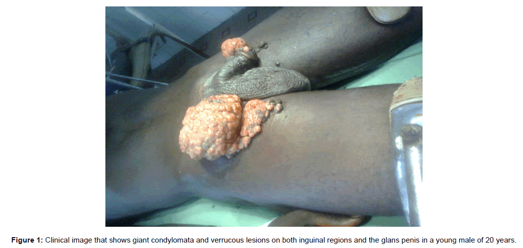
A 20 years old man presented with a budding swelling which started on his glans penis as a wart 4 years ago and then gradually spread over the inguinal regions, initially appearing as condylomata acuminata and then progressing to moderately painful cauliflower lesion with no notion of fever (Figure 1). He regularly applied traditional decoctions without effect on the tumor. He has been sexually active from the age of 15 and practices unprotected sex. His HIV, viral hepatitis B and C and syphilis serologies were negative. No history of chronic inflammation of that region, immunosuppression factors in him or his family or history of STIs were found. The management was surgical with complete removal of the budding lesions whose pathological analysis concluded on a “Giant condyloma acuminatum”. Isolated and recurrent warts were treated with podophyllin following the surgery. The evolution was favourable after 1 year. He is however regular followed-up every 6 months to assess relapse.
Keywords: Condylomata Acuminata
Case Presentation
A 20 years old man presented with a budding swelling which started on his glans penis as a wart 4 years ago and then gradually spread over the inguinal regions, initially appearing as condylomata acuminata and then progressing to moderately painful cauliflower lesion with no notion of fever (Figure 1). He regularly applied traditional decoctions without effect on the tumor. He has been sexually active from the age of 15 and practices unprotected sex. His HIV, viral hepatitis B and C and syphilis serologies were negative. No history of chronic inflammation of that region, immunosuppression factors in him or his family or history of STIs were found. The management was surgical with complete removal of the budding lesions whose pathological analysis concluded on a “Giant condyloma acuminatum”. Isolated and recurrent warts were treated with podophyllin following the surgery. The evolution was favourable after 1 year. He is however regular followed-up every 6 months to assess relapse.
Verrucous carcinomas are pseudoepitheliomatous hyperplasia that appear on the skin and mucous membranes [1]. The most common location of these tumors is oral. When verrucous carcinomas extend to the ano-genital area, is it referred to as Buscke-Lowenstein tumor (BLT) or giant condyloma acuminatum. This is a relatively rare disease which is sexually transmitted and viral in origin, caused by the papilloma virus human (HPV). Serotypes 6 and 11 are incriminated [2]. It usually occurs in young male adults who are sexually active and it is always preceded by condylomata acuminata. The main known risk factors are poor genital hygiene, uncircumcision, glans phymosis, immunosuppression, chronic inflammation, HIV infection, and low socioeconomic level [3]. The condition is characterized by a greater proliferation than that of verrucous carcinomas, deep extension and a degenerative and recurrent character. On physical examination the lesion is dyschromic with a vegetative and budding cauliflower appearance [1,4]. The low oncogenic potential of the HPV serotype 6 and 11 makes the Buscke- Lowenstein tumour benign. Its evolution is local, slow and rarely metastatic. Histologically, it is a perfectly limited squamous tumour, with epithelial hyperplasia, hyperacanthosis, hyperpapillomatosis and koilocytes. Koilocytes are pathognomonic markers of HPV infection, but their presence is not always constant. Despite being a benign tumor, the treatment of BLT is essentially surgical with extensive resection to reduce the risk of recurrence. Focus is placed on patient education and follow-up with respect to relapse. BLT has a slow evolution and may be complicated by dermatitis, infection, fistulization with neighboring organs bleeding or evolve to a malignancy in about 56% of cases [5].
References
- Martin JM, Molina I, Monteagudo C, Marti N, Lopez V, et al. (2008) Buschke-Lowenstein tumor. J Dermatol Case Rep 2: 60-62.
- Spinu D, Rădulescu A, Bratu O, Checheriţă IA, Ranetti AE,et al. (2014) Giant condyloma acuminatum - Buschke-Lowenstein disease - a literature review. Chirurgia 109: 445-450.
- Njoumi N, Tarchouli M, Ratbi MB, Elochi MR, Yamoul R, et al. (2013) The Buschke-Lowenstein tumor anorectal: to about 16 cases and review of the literature. Pan Afr Med J 16: 131.
- Elmahi H, Mernissi FZ (2016) La tumeur de Buschke-Löwenstein Buschke-Löwenstein tumor. Pan Afr Med J 25: 176.
- Chu QD, Vezeridis MP, Libbey NP, Wanebo HJ (1994) Giant condyloma acuminatum (Buschke-Lowenstein tumor) of the anorectal and perianal regions. Analysis of 42 cases. Dis Colon Rectum 37: 950-957.
 Spanish
  Chinese
  Russian
  German
  French
  Japanese
  Portuguese
  Hindi
Hindi 
