Case Report, Res J Zool Vol: 2 Issue: 1
Hypertrophic Cardiomyopathy in a Cat
Umit Ozcan, Zeynep Nurselin Kot and Handan Hilal Arslan*
Department of Internal Medicine, Faculty of Veterinary Medicine, Ondokuz Mayis University, Samsun, Turkey
*Corresponding Author : Handan Hilal Arslan
Department of Internal Medicine, Faculty of Veterinary Medicine, Ondokuz Mayis University, Samsun, Turkey
Tel: +903623121919-1231
E-mail: hharslan@omu.edu.tr
Citation: Ozcan U, Kot ZN, Arslan HH (2020) Hypertrophic Cardiomyopathy in a Cat. Res J Zool 2:1
Abstract
In this case, hypertrophic cardiomyopathy (HCM) was evaluated in a Turkis Van Cat. The cat was presented to the clinic with lethargy and anorexia. In the echocardiographic examination HCM signs were detected. Traditional pharmacological treatment and pimobendan were used. Significant clinical improve was evaluated in a short time with this therapeutic approach.
Keywords: Hypertrophic cardiomyopathy; Cat; Treatment
Introduction
Hypertrophic cardiomyopathy (HCM) is a primary myocardial disease affecting cats, people, and a variety of other species [1]. The use of the term cardiomyopathy in reference to feline heart disease seems to have first appeared in 1973. Characteristic pathologic findings of advanced feline HCM, including left ventricular hypertrophy (LVH); left ventricular fibrosis; and left atrial dilation, were described but the term cardiomyopathy was not used [2]. Spontaneously occurring models of HCM in cats and dogs with substantial structural similarities to the well-recognized disease entity in humans [3].
Hypertrophic cardiomyopathy is recognized by veterinary cardiologists as the most common cardiac disease in cats and it can lead to increased morbidity and mortality [1,4]. It is characterized by a hypertrophied left ventricular wall and a normal-size to small left ventricular chamber in the absence of other cardiac or systemic disease associated with hypertrophy [5].
The clinical manifestations of HCM in domestic cats result from impaired diastolic function, which causes an increase in left ventricular filling pressure and the development of congestive heart failure (CHF) (ie, pulmonary venous enlargement, pulmonary oedema, pleural effusion, dyspnea, lethargy, tachypnea, and tachycardia) [5].
In cats, in the absence of systemic hypertension or hyperthyroidism, diagnosis is usually based on measurement of maximal end-diastolic wall thickness on 2-D or M-mode echocardiography. Therefore, echocardiography is the gold standard for ante mortem diagnosis for HCM. It is now recognized that HCM is characterized by a broad range of phenotypic patterns of left ventricular (LV) hypertrophy ranging from localized and relatively mild wall segment thickening to extreme generalized hypertrophy [1].
Myocardial hypertrophy and reduced ventricular distensibility result in increased diastolic pressure and left ventricular filling accompanying increased left arterial (LA) and pulmonary venous pressure. Secondary right sided congenital heart failure (CHF) may occur in response to prolonged pulmonary vasoconstriction and increased pulmonary arterial pressure. LV out flow obstruction accompanying ejection murmur results from LV papillary muscle hypertrophy. Several factors contribute to myocardial ischemia, which leads to fibrosis, arrhytmia, and other so many complications. The CHF, arterial thromboembolism (ATE), and sudden cardiac death are common clinical manifestation in end-stage of feline HCM [6]. The subject of this case report is a two years old, male, Turkish Van cat with hypertrophic cardiomyopathy.
Description of the Clinical Case
The cat was presented the faculty of veterinary medicine department of internal medicine clinic with lethargy and loss of appetite. The owner reported that the cat likes walk around the house and two days ago when he came to home, they realized these sings before brought to veterinarian. At the internal medicine clinic of the faculty, a general examination was performed. Tachypnea and abnormal lung sound in the left lung lobes were detected with auscultation. Oedema was detected in back legs with the palpation examination. The cat had 3 kg BW.
The body temperature was 40°C. Haematological analyses were performed and serum enzyme levels were checked in the patient. The haematological analyses showed that white blood cell count (WBC) (31.83 109/L) and neutrophil count (29.05 109/L) were elevated, the lymphocyte count (0.93 109/L) was slightly lower than the normal value. According to biochemical analyses serum ALT, AST, ALP, GGT, creatinin, glucose, albumin, total bilirubin levels were in the references. Only serum urea level was higher than normal range (40.8 mg/dl) in the cat.
Echocardiographic Examination
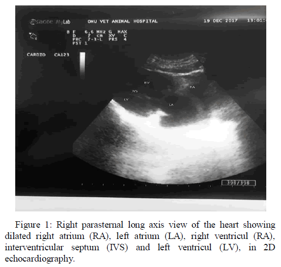
Echocardiographic data was obtained from the right parasternal long axis view via 2D and M-Mode echocardiography techniques. Echocardiography revealed severe left and right atrial dilatation. The interventricular septum (IVSd) and left ventricular posterior wall (LVPW) measured in diastole from a long-axis view at the level of mitral valve were 6.2 and 5.4 mm, respectively. The left ventricular internal diameter (LVID) was 4.9 mm in systole and 10.4 at diastole and fractional shorteining was in normal value (53%).
Treatment
First dose of furosemide at 1 mg/kg dose (Lasix 20 mg/2 ml Sanofi Aventis) was given intravenous with saline solution and then it was continuous by oral route for treatment of the oedema in the back side of the cat. Enalapril (0.5 mg/kg) and propranolol (2.5 mg/kg) were prescribed for use orally [7]. Pimobendan was started at (0.42 mg/kg) (range, 0.08-0.42) [8] orally every 24 hours and no side effect was observed (Figure 1). Because of high WBC count, antibiotics (Amoxicillin clavulanic acid 8.75 mg/kg, according to producer company, Synulox, Zoetis) were used in the cat. In addition, Hill’s h/d diet was recommended to cat’s owner.

Figure 1: Right parasternal long axis view of the heart showing dilated right atrium (RA), left atrium (LA), right ventricul (RA), interventricular septum (IVS) and left ventricul (LV), in 2D echocardiography.
Discussion
In this report HCM was detected in Turkish Van Cat. HCM has high prevalent in certain populations. The disease is known to be inherited in some breeds, most notably in The Domestic Shorthair, Turkish Van, Maine Coon, Persian, Ragdoll, Sphynx, Scottish Fold Cats, Chartreux, British Shorthair, Norwegian Forest Cat, Persian, and other breeds [6].
First HCM signs can be detected between 8 and 24 months of age young cats which is especially hereditary disease occurs in certain breed like Maine Coon cats [9]. Similarly, Turkish Van Cat was just 2 years old and his disease probably in hereditary group. On the other hand, Atkins et al. [10] reported that the mean (+/- SD) age of cats with HCM was 6.5 (4.0) years.
Echocardiography is the gold standard for ante mortem diagnosis for HCM [1]. Diagnostic criteria for HCM included a thickened (≥6 mm; measured during diastole) interventricular septum or left ventricular posterior wall in the absence of hyperthyroidism, systemic hypertension, or aortic stenosis [11]. As compatible previous indications, the echocardiographic examination was performed and interventricular septal thickness (IVSd) and left ventricular internal diameter in diastole (LVIDd) were recorded 6.2 mm and 10.4 mm respectively in this Turkish Van cat.
After that, the most appropriate treatment alternatives were scanned for the cat. Enalapril is well tolerated in cats with HCM [12]. As a classical treatment, furosemid, enalapril and propranolol were prescribed for the patient. Furosemide is a high-ceiling diuretic (higher dosages elicit a greater response) that is consistently well tolerated by clinically ill cats. ACE inhibitors are considered to be safe in cats and are pharmacologically active. No study has shown that cats receiving ACE inhibitors during CHF treatment live longer and/or better as a result of ACE inhibition. In cats with CHF, revealed a trend towards improved outcomes compared to treatment with a diuretic and either atenolol or sustained-release diltiazem, but no convincing benefit over furosemide alone. As a class, they are safe drugs and could have beneficial side effects. They can be formulated into a suspension and
the dosing interval is once a day. The researchers use enalapril or benazepril, 0.5 mg/kg PO q 24 h. In a landmark study comparing diltiazem with propranolol, which is a beta-blocker, this calcium channel blocker was found to delay recurrence of signs of CHF in cats with HCM [13]. Meanwhile, Bright et al. [14], compared between the three treatments, diltiazem, verapamil, and propranolol in cats with HCM and reported that diltiazem appears to provide a safe and effective approach for the medical management of feline HCM. According to previous reports, enalapril, furosemid and propranolol were chosen for optimal treatment alternative in this cat.
In addition to traditional pharmacological treatment, pimobendan was used for treatment to HCM in this case. Pimobendan is a positive inotropic and vasodilatory drug and it has become part of the management of CHF in dogs. Pimobendan is approved by the US FDA Center for Veterinary Medicine for use in dogs with CHF to dilated cardiomyopathy and chronic mitral valve disease [11]. But there are positive and negative approaches for pimobendan uses in the HCM treatment for cats. Such as, MacGregor et al. [8] and Gordon et al. [15] reported that pimobendan appeared to be well tolerated in cats with heart failure characterized of various causes and single-dose in the different dosage have been similar effect [16]. Addition of pimobendan to standard treatment regimens for cats with CHF secondary to HCM or HOCM appeared to confer a clear benefit in survival time in the retrospective case-control study [11]. Pimobendan has been shown to have beneficial effects on the immune system in multiple ways and in various species. Even it has a therapeutic effect in the acute stage of viral myocarditis in murine models [17].
The cat well tolerated the used medicines. After the first week of treatment, his appetite increased and he could show active habits again. He is still living as clinically healthy for two months.
Conclusion
In this case detected that classical HCM treatment regime which combined with pimobendancould be clinically effective in short period especially in young cat with hereditary HCM.
References
- Häggström J, Luis Fuentes V, Wess G (2015) Screening for hypertrophic cardiomyopathy in cats. J Vet Cardiol 17: 134-149.
- Abbott JA (2010) Feline hypertrophic cardiomyopathy: an update Vet Clin North Am Small Anim Pract 40: 685-700.
- Liu Si-Kwang, William CR, Barry JM (1993) Comparison of morphologic findings in spontaneously occurring hypertrophic cardiomyopathy in humans, cats and dogs. Am J Cardiol 72: 944-951.
- Rush JE, Freeman LM, Fenollosa NK, Brown DJ (2002) Population and survival characteristics of cats with hypertrophic cardiomyopathy: 260 cases (1990–1999). J Am Vet Med Assoc, 220: 202-207.
- Hemdon WE, Kittleson MD, Sanderson K, Drobatz KJ, Clifford CA, et al. (2002) Cardiac troponin I in feline hypertrophic cardiomyopathy. J Vet Intern Med 16: 558-564.
- Sasaki K, Mutoh T, Shirota K, Kawashima R (2017) Cardiomyopathies in Animals: Cardiomyopathies-Types and Treatments. InTec.
- Papich M (1992) Table of common drugs: approximate dosages: Kirk RV, ed. Kirk’s Current Veterinary Therapy XIII: Small AnimalPractice (13th edtn), WB Saunders Company, Philadelphia.
- MacGregor JM, Rush JE, Laste NJ, Malakoff RL, Cunningham SM (2011) Use of pimobendan in 170 cats (2006–2010). J Vet Cardiol 13: 251-260.
- Kittleson MD, Meurs KM, Munro MJ, Kittleson JA, Liu SK, et al. (1999) Familial hypertrophic cardiomyopathy in maine coon cats: an animal model of human disease. Circulation 99: 3172-3180.
- Atkins CE, Gallo AM, Kurzman ID, Cowen P (1992) Risk factors, clinical signs, and survival in cats with a clinical diagnosis of idiopathic hypertrophic cardiomyopathy: 74 cases (1985-1989). J Am Vet Med Assoc 201: 613-618.
- Reina-Doreste Y, Stern JA, Keene BW, Tou SP, Atkins CE, et al. (2014) Case-control study of the effects of pimobendan on survival time in cats with hypertrophic cardiomyopathy and congestive heart failure. J Am Vet Med Assoc 245: 534-539.
- Rush JE (1998) Therapy of feline hypertrophic cardiomyopathy. Vet Clin North Am Small Anim Pract 28: 1459-1479.
- Gordon SG and Etienne Côté (2015) Pharmacotherapy of feline cardiomyopathy: chronic management of heart failure. J Vet Cardiol 17:159-172.
- Bright JM, Golden AL, Gompf RE, Walker MA, Toal RL (1991) Evaluation of the Calcium Channelâ€ÂÂÂBlocking Agents Diltiazem and Verapamil for Treatment of Feline Hypertrophic Cardiomyopathy. J Vet Intern Med 5: 272-282.
- Gordon SG, Saunders AB, Roland RM, Winter RL, Drourr L, et al. (2012) Effect of oral administration of pimobendan in cats with heart failure. J Am Vet Med Assoc 241: 89-94.
- Yata M, McLachlan AJ, Foster DJ, Hanzlicek AS, Beijerink NJ (2016) Single-dose pharmacokinetics and cardiovascular effects of oral pimobendan in healthy cats. J Vet Cardiol 18: 310-325.
- Boyle KL and Leech E (2012) A review of the pharmacology and clinical uses of pimobendan. J Vet Emerg Crit Care 22: 398-408.
 Spanish
  Chinese
  Russian
  German
  French
  Japanese
  Portuguese
  Hindi
Hindi 
