Research Article, Res Rep Gastroenterol Vol: 3 Issue: 1
Malignant Appendeceal Carcinoid Tumour with Intestinal Obstruction and Caecal Perforation: A Case Report in Northern Ghana
1Department of Pathology, School of Medicine and Health Sciences, University for Development Studies and the Tamale Teaching Hospital, Tamale, Ghana
2Department of Surgery, School of Medicine and Health Sciences of the University for Development Studies and the Tamale Teaching Hospital, Tamale, Ghana
*Corresponding Author : Der EM
Department of Pathology, School of Medicine and Health Sciences of the University, Development Studies and the Tamale Teaching Hospital, Tamale, Ghana
E-mail: maadelle@yahoo.com
Received: September 25, 2019 Accepted: October 08, 2019 Published: October 15, 2019
Citation: Der EM, Abantanga FA (2019) Malignant Appendeceal Carcinoid Tumour with Intestinal Obstruction and Caecal Perforation: A Case Report in Northern Ghana. Res Rep Gastroenterol 3:1.
Abstract
Carcinoid tumours are the most common neoplasms of the appendix. These tumours are generally asymptomatic; however, symptomatic patients may clinically mimic acute appendicitis and or intestinal obstruction. There are a few publications in Ghana which mention carcinoid tumours. However, these publications do not give much attention to the clinico-pathological features of this tumour as an entity, or highlight the possible complications of the disease. Presented in this case report is a 43-year-old man who was seen with signs and symptoms of acute intestinal obstruction at the Medical/Surgical Emergency Department of the Tamale Teaching Hospital (TTH). He had an emergency exploratory laparotomy and right hemicolectomy. Subsequent histopathological examination of the specimen revealed a metastatic carcinoid tumour to the intestines with caecal obstruction and perforation. The origin of the carcinoid tumour was the appendix. Clinicians should exhibit a high index of suspicion concerning right-sided large bowel obstruction and thoroughly investigate the cause of the obstruction and attempt to exclude neoplasm of the appendix, which though rare, can be the cause of the obstruction as demonstrated in this case. The final diagnosis is made after an extensive histopathological examination.
Keywords: Carcinoid tumours; Hemicolectomy; Gastrointestinal tract (GIT)
Introduction
Carcinoid tumours arise from neuroendocrine cells found in the mucosa layer of the gastrointestinal tract (GIT) [1-4]. However, the common locations in descending order of frequency are: appendix, ileum, rectum, colon and the stomach. The normal neuroendocrine cells produce polypeptide hormones and biogenic amines; the tumours may do likewise [5,6]. Benign appendiceal carcinoid tumours of less than 2.0 cm are common incidental findings during appendectomy and at autopsy, with the tip as the part commonly involved in the tumour [7,8]. Malignant transformation of the benign carcinoid tumour is rare, but does occur in tumours with diameter greater than 2.0 cm; however, these are much less common than carcinomas [9].
The average age at presentation of carcinoid tumours of the GIT is 55 years; they are rare in the teens and increase in frequency thereafter [10]. However, those of the appendix have been reported to occur in relatively younger age groups [11]. Most cases of appendiceal carcinoid tumours (benign or malignant) are asymptomatic [12-14]. Symptomatic cases commonly present with clinical features of a complicated disease, such as intestinal obstruction and the carcinoid syndrome.
There are few publications in Ghana which mention carcinoid tumours. However, these publications did not give much attention to the clinico-pathological features of this tumour as an entity, or highlight the possible complications of the disease. A malignant appendiceal carcinoid tumour in a 43-year male with caecal obstruction and perforation is presented in this case report. The aim of this presentation is to remind clinicians that some rare lesions of the appendix may be masked by the exhibition of signs and symptoms of acute appendicitis; and also to lay emphasis on the need for adequate clinical assessment, including imaging and histopathological examination of appendiceal specimen removed at surgery.
Case Report
A 43-year-old man presented to the Medical/Surgical Emergency Unit of the Tamale Teaching Hospital (TTH) with a day’s history of vomiting associated with severe abdominal pain and distension. The abdominal distension and pain started about a week ago, but became severe on the day of presentation to TTH. He had noticed a change in bowel habit (constipation), but denied seeing any blood in his stools. He also said there was a right inguinal swelling that also kept increasing over a period of months. He was unspecific about the inguinal mass.
Physical examination revealed a young male adult in severe pain, dehydrated, but not pale. The abdomen was grossly distended, with generalized tenderness, guarding and rebound tenderness. There was a soft right inguinal swelling.
Investigations
Imaging
He had plain erect and supine abdominal X-rays done that showed air under the diaphragm, and distended loops of large bowel with air fluid levels.
Blood
The full blood count results were essentially normal, except a moderate rise in haemoglobin level (17.9 g/dl), due to possible dehydration. The renal and liver functions were all within normal ranges.
Initial working diagnosis
Acute abdomen? Bowel Obstruction 2˚ strangulated left inguinoscrotal hernia.
He was rehydrated with intravenous fluid and produced adequate urine. He also had an initial antibiotic cover.
Type of surgery
Exploratory laparotomy with right hemicolectomy and end-to-end ileo-colic anastomosis, and a repair of the right inguinal hernia were carried out.
Findings at surgery
Complex solid appendiceal mass
Distended caecum with perforation at the base.
Solid nodules palpable within the walls of the caecum, ileum and jejunum.
Empty hernia sac.
Pus in the pelvic cavity.
Histopathology Examination
Gross examination
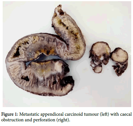
A resected segment of bowel, consisting of the terminal ileum, caecum and the ascending colon was received in the Department of Pathology of the Tamale Teaching hospital. There were multiple perforations involving the caecum and the terminal ileum. The bowel serosa was covered with greenish-cream coloured exudate.
When the bowel was opened up, it revealed fairly-defined tan nodular masses that have infiltrated the serosa and part of the muscular layers of the terminal ileum, caecum and the ascending colon. There was obstruction of the lumen of the caecum with perforation at the base. The bowel wall particularly, that of the caecum was oedematous and also showed large areas of myxoid changes.
Also received in the same container was a detached appendix with a tan nodular mass at the tip that measured 7.0 cm in its widest dimension (Figure 1).
Microscopic examination
Sections of representative portions of the appendiceal mass showed infiltration by sheets of monomorphic neoplastic cells with round hyperchromatic nuclei. Mitotic figures were present (Figures 2a and 2b).
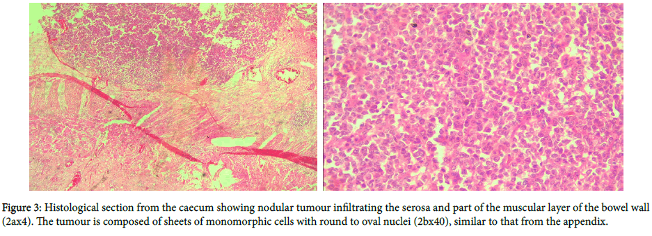
Sections of the bowel wall showed tumors morphologically and cytologically similar to that in the appendix. The tumour infiltrated the serosa, the muscular wall of the caecum and the sub mucosal layer as solid nodules. The mucosa was free of tumour. There were tumour deposits in the lymph nodes (Figures 3a and 3b).
Histopathological diagnosis
Bowel and appendix (Right hemicolectomy with bowel anastomosis): Metastatic appendiceal carcinoid tumor with large bowel obstruction.
Discussion
Carcinoid tumour of the appendix globally represents the commonest neuroendocrine malignancy of the gastrointestinal tract. Diagnosis is difficult as it is often asymptomatic and the presence of symptoms generally implies advanced disease state with some complications [15].
The present case presented with a day’s history of vomiting associated with severe abdominal pains and distension. Surgery was performed and histopathological examination of the specimen confirmed malignant carcinoid tumour involving the tip of the appendix. Carcinoid tumours of the gastrointestinal tract are rarely suspected before surgery; and more than 50.0% are diagnosed during surgery or after histopathological examination or at autopsy. This may possibly explain why the clinicians in this current case report made an initial diagnosis of bowel obstruction, without carcinoid tumour as a possible differential diagnosis. Anatomically, the primary tumour in this man involved the tip of the appendix. The organ and site of involvement of the carcinoid tumour in this case report supports reports of previous studies that found the appendix and more so its tip, to be the commonest gastrointestinal organ to be involved by carcinoid tumors [16].
The age of the man in this current case report was 43 years, relatively young. Carcinoid tumours of the gastrointestinal tract in general are diagnosed in older individuals; however, those of the appendix have been reported to occur in relatively younger age groups as in this case report. Furthermore, while there have been no gender differences regarding gastrointestinal carcinoids, others found it to be female predominant.
The patient presented with abdominal distension, abdominal pains and vomiting suggestive of bowel obstruction. These symptoms support previous publications that patients with symptomatic carcinoid tumour of the appendix may present with symptoms of bowel obstruction, early satiety, weight loss and a palpable abdominal mass [17]. From the history, the patient accepted that the illness started for an unspecified duration, but he was only brought to the hospital because of the severity of his condition. The average time of onset of symptoms to diagnosis is reported to exceed 9 years, by which time complications have set in, similar to the current case report [18,19].
Studies have shown that there is a close relation between the size of the appendiceal carcinoid tumor and the risk of malignancy and metastasis. For instance, close to 90.0% of carcinoid tumours with size less than 2.0 cm have benign characteristics, while the risk of malignant transformation increases thereof with increase in the size of the tumour. The primary tumour involving the tip of the appendix was found during gross histopathological examination to measure 7.0 cm in its widest dimension (very large). Microscopic examination revealed the cytological and histological growth patterns of the primary tumour to be similar to those taken from the caecum (point of perforation), the lymph nodes and small bowel wall. It was therefore concluded from the histopathological examination of the resected bowel that the man had malignant carcinoid tumour of the appendix that has spread to the mesenteric lymph nodes and bowels with caecal obstruction and perforation. The histopathological features of the current case are therefore in line with the findings of previous studies.
The risk of metastasis is found to be 2 fold for tumours smaller than 1 cm, but reaches up to 80 fold for lesions larger than 2 cm. The primary tumour in this current case report was 7.0 cm, a high risk factor for distant spread and this explained why he had metastases at presentation. Among all the malignant neuroendocrine tumours of the GIT, published data have indicated that, malignant carcinoid tumour of the appendix has a very good prognosis and overall high survival rate [20,21]. For instance, Thompson et al., and Kulke et al., reported an overall survival decades ago of 85% for those with regional metastases as in this current case report and 34% for those with distant metastases. This may be the reason why the patient in this current case report is said to be doing well after discharge from the hospital, despite the very large size of the tumour with the associated complications.
The patient had a confirmed case of malignant appendiceal carcinoid with metastasis to the mesoappendix, ileum and caecum with obstruction and perforation. He had surgery with bowel anastomosis as a form of surgical management, with no significant complications. A right hemicolectomy is the recommended surgical management procedure for malignant appendiceal carcinoid tumor limited to the mesoappendix, regional lymph nodes and the terminal ileum and caecum [16,17,22]. Hence the surgical management of this case report was appropriate.
What has this case report added to the existing literature?
There is the need for adequate and detailed pre-operative assessment of patients with symptoms of acute abdomen, including imaging procedures.
The first clinical presentation of appendiceal carcinoid tumour may be a complication as in this case report; not all malignant carcinoid tumours present with the syndrome.
All appendix removed at surgery must be submitted for histopathological examination to confirm the diagnosis (infectious or neoplastic) and hence administer the appropriate medical care [23,24].
Malignant carcinoid tumour of the appendices even with added complications as in this case report, has good prognosis if the right surgical and medical treatment options are initiated promptly.
Conclusion
Preoperative diagnosis of carcinoid of the appendix, a very rare condition, is very difficult to make. The condition can lead to complications such as perforations of the bowel and intestinal obstruction. Clinicians should have a high index of suspicion when dealing with conditions of the appendix to be able to investigate the patients appropriately, including imaging, and to have a differential diagnosis of tumours of the appendix in mind, even though they are rare. All resected appendices should be examined histopathologically even when they appear normal.
References
- Thompson GB, Van Heerden JA, Martin JK, Schutt AJ, Ilstrup DM, et al. (1985) Carcinoid tumors of the gastrointestinal tract: presentation, management, and prognosis. Surgery 98: 1054-1063.
- Kulke MH, Mayer RJ (1999) Carcinoid tumours. N Engl J Med 340: 858-868
- Norton JA (1994) Surgical management of carcinoid tumors: role of debulking and surgery for patients with advanced disease. Digestion 55: 98-103.
- Horton KM, Kamel I, Hofmann L, Fishman EK (2004) Carcinoid tumors of the small bowel: a multitechnique imaging approach. AJR Am J Roentgenol 182: 559-567
- Shebani KO, Souba WW, Finkelstein DM, Stark PC, Elgadi KM, et al. (1992) Prognosis and survival in patients with gastrointestinal tract carcinoid tumors. Ann Surg 229: 815-823.
- Moran CA, Suster S (2007) Neuroendocrine carcinomas (carcinoid, atypical carcinoid, small cell carcinoma, and large cell neuroendocrine carcinoma): Current concepts. Hematol Oncol Clin North Am 21: 394-407.
- Pastore C, D'Annibale A, Piazza A, Pignata G, Conte C, et al. (1997) Diagnosis and treatment of gastrointestinal carcinoid. Report of a clinical case with duodenal site. Minerva Chir 52: 283-287.
- Deans GT, Spence RA (1995) Neoplastic lesions of the appendix. Br J Surg 82: 299-306.
- Connor SJ, Hanna GB, Frizelle FA (1998) Appendiceal tumors: retrospective clinicopathologic analysis of appendiceal tumors from 7,970 appendectomies. Dis Colon Rectum 41: 75-80.
- Hermans JJ, Hermans AL, Risseeuw GA, Verhaar JC, Meradji M (1993) Appendicitis caused by carcinoid tumor. Radiology 188: 71-72.
- Martin RG (1970) Management of carcinoid tumors. Cancer 26: 547-551.
- Zarrilli L, Marzano LA, Porcelli A, D’Avanzo A, Misso C (1991) The surgical management of carcinoid tumors. J Nucl Biol Med 35: 341-342.
- Mrevlje Z, Stabuc B (2006) Pitfalls in diagnosing small bowel carcinoid tumors. J Buon 11: 83-86
- Ha J, Tan WA (2012) Gastrointestinal Carcinoid Tumours: A Review. J Gastroint Dig Syst 2: 107.
- McCusker ME, Coté TR, Clegg LX, Sobin LH (2002) Primary malignant neoplasms of the appendix: a population-based study from the surveillance, epidemiology and end-results program. Cancer 94: 3307-3312.
- Kulke MH (2007) Clinical presentation and management of carcinoid tumors. Hematol Oncol Clin North Am 21: 433-455.
- Aranha GV, Greenlee HB (1980) Surgical management of carcinoid tumors of the Gastrointestinal tract. Am Surg 46: 429-435.
- Moertel CG (1983) Treatment of the carcinoid tumor and the malignant carcinoid syndrome. J Clin Oncolo 1: 727-740.
- Makridis C, Oberg K, Juhlin C, Rastad J, Johansson H, et al. (1990) Surgical treatment of mid-gut carcinoid tumors. World J Surg 14: 377-380.
- Modlin IM, Sandor A (1997) An analysis of 8305 cases of carcinoid tumors. Cancer 79: 813-829.
- Sandor A, Modlin IM (1998) A retrospective analysis of 1570 appendiceal carcinoids. Am J Gastroenterol 93: 422-428.
- Onaitis MW, Kirshbom PM, Hayward TZ, Quayle FJ, Feldman JM, et al. (2000) Gastrointestinal carcinoids: characterization by site of origin and hormone production. Ann Surg 232: 549-556
- Der EM, Naaeder SB, Clegg-Lamptey JNA, Dakubo JN, Edusei L, et al. (2015) Anatomic categorization of gastrointestinal malignancies: a 10 year retrospective histopathologic study at the KBTH. Journal of Cancer Research and Therapy 3: 8-14.
- Der EM, Gyasi RK (2016) The trend of colorectal cancers at Korle-Bu Teaching Hospital: A retrospective histopathological study. Journal of Cancer and Tumor International 4: 1-8.
 Spanish
  Chinese
  Russian
  German
  French
  Japanese
  Portuguese
  Hindi
Hindi 


