Case Report, Res Rep Gastroenterol Vol: 1 Issue: 1
Rectal Melanoma with Duodenal Metastasis: Very Unusual Endoscopic Findings
Feldman D, Mari A*, Abu-Backer F and Kopelman Y
Gastroenterology and Hepatology Department, Hillel Yaffe Medical Center, Israel
*Corresponding Author : Amir Mari, MD
Hillel Yaffe MC, Hadera, Israel
Tel: 00972542142070, 00972 9 8781587
E-mail: amir.mari@hotmail.com
Received: October 11, 2017 Accepted: October 20, 2017 Published: October 25, 2017
Citation: Feldman D (2017) Rectal Melanoma with Duodenal Metastasis: Very Unusual Endoscopic Findings. Res Rep Gastroenterol 1:1.
Abstract
Melanoma of the GI tract is a rare and carries a poor prognosis. Most GI melanomas are metastatic from an oculo-cutaneous primary. Many hypothesize that melanoma of the GI tract is a product of spontaneous regression of an unknown primary. The etiology of primary GI melanomas is unclear. Hypothesis suggests it arises from the neural crest cells known to exist in the esophagus, stomach, small bowel, and ano-rectum.
Keywords: Rectal Melanoma; Duodenal Metastasis; Pembrolizumab therapy
Case Study
We present a case of primary rectal melanoma, aggressive and resistant to treatment, with duodenal metastases.
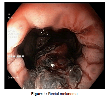
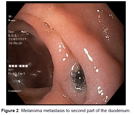
A 57-years old man was referred to our hospital at March 2015, presented with acute rectal bleeding. Colonoscopy revealed at 1 cm from the anal verge a 60 mm ulcerated lesion suspected to be rectal carcinoma (Figure 1). Histology demonstrated rectal mucosa infiltrated by malignant melanoma, stained positive for Melan A and negative for cytokeratin and CDX-2. Positive for C-KIT mutation and negative for BRAF mutation [1]. Oculo-cuatneous origin was ruled out. PET-CT demonstrated inguinal, adrenals and pulmonary metastases. Gastroscopy revealed three ulcerated pigmented lesions in the 2nd part of the duodenum [2]. Biopsy confirmed the clinical suspicion demonstrating duodenal mucosa infiltrated by metastatic malignant melanoma (Figure 2). Pembrolizumab therapy was initiated and although Improvement was observed 3 month later on PET-CT, 1 year later PET-CT demonstrated worsening of the adrenals involvement [3]. Ipilimumab therapy combined with adrenals irradiation (SBRT) was given without an objective response. Later the patient died from addisonian crises, sepsis and multi-organ failure.
References
- Blecker D, Abraham S, Furth EE, Kochman ML (1999) Melanoma in the gastrointestinal tract. Am J Gastroenterol 94: 3427–3433
- Savoia P, Fava P, Osella-Abate S (2010) Melanoma of unknown primary site: A 33-year experience at the Turin Melanoma Centre. Melanoma Res 20: 227–232.
- Kottschade LA, Grotz TE, Dronca RS (2014) Rare presentations of primary melanoma and special populations: A systematic review. Am J Clin Oncol 37: 635–641.
 Spanish
  Chinese
  Russian
  German
  French
  Japanese
  Portuguese
  Hindi
Hindi 

