Research Article, Int J Cardiovasc Res Vol: 9 Issue: 3
The Relationship of HSP-70 with Calcineurin, Sod and Catalase Post-Acute Myocardial Infarction in Wistar Rats Model
Johanes Nugroho1,2*, Christo Darius2, Maria Yolanda Probohoesodo3, Suhartono Taat Putra4 and Cornelia Ghea5
1Department of Cardiology and Vascular Medicine, Faculty of Medicine, Universitas Airlangga, Surabaya, Indonesia
2Dr. Soetomo General Hospital, Surabaya, Indonesia
3Department of Clinical Pathology, Faculty of Medicine, Airlangga University-Dr. Soetomo Hosiptal, Surabaya, Indonesia
4Department of Anatomic Pathology, Faculty of Medicine, Airlangga University-Dr. Soetomo Hosiptal, Surabaya, Indonesia
5PILAR Research and Education, Cambridge, UK
*Corresponding Author: Dr. Johannes Nugroho
Department of Cardiology and Vascular Medicine, Faculty of Medicine, Universitas Airlangga, Surabaya, Indonesia
Tel: +6282139090953
E-mail: j.nugroho.eko@fk.unair.ac.id
Received: April 07, 2020 Accepted: May 26, 2020 Published: June 02, 2020
Citation: Nugroho J, Darius C, Probohoesodo MY, Putra ST, Ghea C (2020) The Relationship of HSP-70 with Calcineurin, Sod and Catalase Post-Acute Myocardial Infarction in Wistar Rats Model. Int J Cardiovasc Res 9:3. doi: 10.37532/icrj.2020.9(3).402
Abstract
Background: HSP-70s help to reduce infarct area through unclear mechanism. It appears that HSP-70 activates calcineurin and induce antioxidant enzymes such as Superoxide Dismutase (SOD) and catalase. Therefore, we sought to investigate the relationship of HSP-70 with calcineurin, SOD and catalase post-Acute Myocardial Infarction (AMI). Methods: An experimental study involved 24 Wistar rats as models of chronic coronary occlusion. The rats were randomly divided into 4 groups: no-intervention after AMI (N), sedentary intervention after AMI (S), exercise intervention after AMI (E), and sham (C). Intervention consisted of 2 weeks of recovery then 4 weeks of sedentary for group S or exercise for group E. HSP-70, calcineurin, SOD, and catalase expression in heart were evaluated the difference among groups. Correlation between HSP-70 to other proteins was analysed also. Results: HSP-70 and calcineurin was higher in group S and E compared to group N and C. HSP-70 (MD=0.97, 95% CI 0.60 to 1.34), calcineurin (MD=1.25, 95% CI 0.68 to 1.82, p<0.05), catalase (MD=0.57, 95% CI 0.25 to 0.88, p<0.05), and SOD (0.42, 95% CI 0.14 to 0.69, p<0.05) significantly higher in Group E compared to S. Sham group had higher SOD and catalase activity than group E. HSP-70 was correlated with calcineurin (r=0.856, p<0.05). HSP-70 was correlated with catalase and SOD when the analysis included only groups who had intervention. Conclusion: HSP-70 correlated with calcineurin and stimulated an increase in calcineurin activity.
Keywords: Exercise; Myocardial infarct; HSP-70; Calcineurin; SOD; Catalase
Introduction
70-kDa Heat Shock proteins (HSP-70) acts as a molecular chaperones that assist in the correct folding of nascent and stressaccumulated mis-folded proteins and prevents their aggregation [1]. It is well established that ischemia result in an increase in HSP- 70 production in the heart and the amount of HSP-70 synthesis correlates with the extent of myocardial protection after ischemia/reperfusion injury [2]. Inducing super expression of HSP-70 reduced significantly infarcted area [3,4].
In ischemic myocardial cells, HSP-70 helps prevent cell death through binding with proteins that play a role in inhibiting cell necrosis [5]. HSP-70 activates calcineurin via a calmodulin-dependent and independent pathway [6]. HSP-70 may induce antioxidant SOD through Reactive Oxygen Species (ROS) which was increased in early phase of ischemia [7]. Myocardial catalase activity was induced as well as HSP-70 after heat shock [8,9]. No work has been done, however, to show correlation between HSP-70 and calcineurin, COD and catalase after ischemia in vivo. Therefore, we sought to investigate the relationship of HSP-70 with calcineurin, COD and catalase post-AMI.
Methods
Male Wistar rats meeting certain criteria, such as the age of 13 ± 2 weeks and body weight of 200-300 grams were used for this study. Procedures of care and animal use were conducted in accordance with the guidelines of laboratory animal care approved by the Ethics Committee of the Faculty of Veterinary Medicine, Airlangga University.
The rats were randomly assigned into one of the following 4 groups. Group N had LAD ligation then had no further intervention and sacrificed on the subsequent day. Group S had LAD ligation then was let to recover for 2 weeks and had sedentary intervention then were sacrificed 4 weeks after. Group E had LAD ligation then had recovery time for 2 weeks and performed mild exercise consisting of swimming exercises 5 times a week for 4 weeks with the duration of exercise was determined individually by 40% of the maximum time, and then was sacrificed. Group C was sham where had thoracotomy without ligation then sacrificed after 6 weeks.
All the animals were distributed in plastic cages (each cage consisted of three rats) under dark condition for 12 hours and light condition for 12 hours. The temperature of the cages was set between 22-27ºC. Drink and pellets were freely distributed to the animals. Exercise was carried out for four weeks.
The rats were anesthetized with an intramuscular injection of ketamine HCl 50 mg/kg and xylazine 5 mg/kg. The animals were intubated orotracheally using 14 G IV catheter which was inserted wire inside. IV catheter was connected to 3-way stop cock. One port was connected to oxygen source ½ L/minute via cannula. Ventilation was performed through open and closes the other port of 3-way stop cock with frequency of 70-80 times per minute. The surgery was carried out under sterile conditions. A left parasternal thoracotomy was performed at the fourth and fifth intercostal space, and the heart was exposed by stripping the pericardium. The heart exposed to identify the coronary artery branch. A ligature was then placed around the left coronary artery using prolene 6/0 at 1-2 mm distal to left atrial appendage. After ligation, the surgical wounds were sutured closed. The animals were observed during recovery until fully conscious and then extubated.
According to the prescribed time, rats were sacrificed in anesthetized conditions with Ketamine hydrochloride (50 mg / kgBB). The chest cavity is opened from the middle of the chest and the heart is excised by binding to the aortic ascendens. Then the excised tissue was put into 10% Phosphate-Buffered Saline (PBS), then macroscopic morphology was examined and immune histochemical preparations were made.
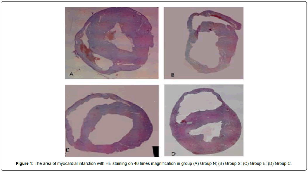
To determine the size of the area of myocardial infarction, Triphenyl Tetrazolium Chloride (TTC) was used to identify tissue that was still viable. The heart in the middle and bottom section was sliced 2-3 mm each. All sections were cleaned with TTC and then incubated for 30 minutes at 37°C and stored in 10% formaldehyde for further analysis. Viable heart tissue appeared brick red while infarcted tissue areas appeared pale white. Each section was photographed and the area of infarction was marked and counted in the image. The size of the infarction was expressed as the area and percentage of infarct area to the total left ventricular area Figure 1.
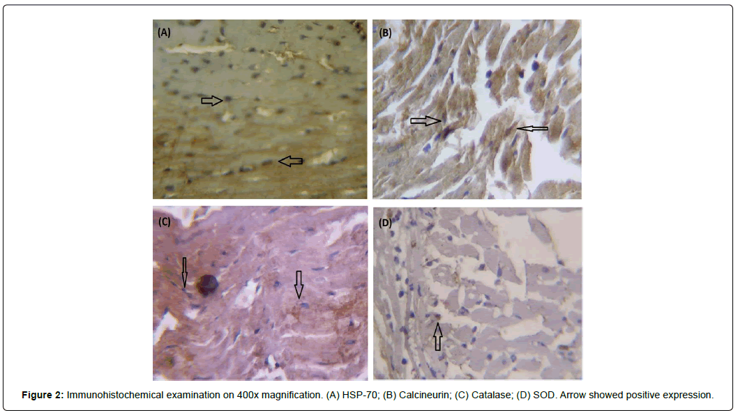
For immuno histochemical procedure, first, the slides are dipped in Xilol 1 and Xilol 2 solutions for 3-5 minutes each, followed by dipping in alcohol solution with gradual decreased concentrations, 99% to 70% alcohol respectively, respectively for 2 minutes. After rinsed with aquadest, the slides were added in 0.3% H2O2 for 5 minutes, and then rinsed with aquadest for 5 minutes. After rinsed with PBS 3 times, the slide was added a provision of 2 drops of Ab (1: 50) monoclonal Ab (spen insulin) and 1 ml of diluent for 25 minutes. After rinsed with PBS 3 times, polymer solution was added for 30 minutes. After rinsed with PBS 3 times, solution containing 1 drop of DAB chromogen and 49 drops of DAB substrate buffer was added for 3-10 minutes in dark places until the slides coloured brown. After rinsed with PBS 3 times and aquabidest 3 times, meyer hematoxilin was added for 6-15 minutes Figure 2.
The expressions of HSP-70, calcineurin, SOD, and catalase were examined as much as 10 times with an area width of 125 μmx125 μm. All data are expressed as means ± SD. Differences among groups were analysed using independent t-test. Correlation was analysed using Pearson correlation. The statistical analysis was conducted using SPSS 16. The α-level for statistical significance was set at 0.05.
Results
HSP-70 expression in the group E was higher than in other groups as shown in Table 1. Group C had the lowest HSP-70 expression. There was significant difference in HSP-70 expression between group N and S (Mean Difference (MD): -0.30; 95% CI -0.47 to -0.13; p<0.05).
| Group | HSP70 Mean ± SD |
Mean Difference (95%CI) | ||
|---|---|---|---|---|
| Group S | Group E | Group C | ||
| Group N | 0.38 ± 0.15 | -0.30 (-0.47 to -0.13)* | -1.27 (-1.65 to -0.89)* | 0.32 (0.16 to 0.47)* |
| Group S | 0.68 ± 0.12 | -0.97 (-1.34 to -0.60)* | 0.62 (0.49 to 0.75)* | |
| Group E | 1.57 ± 0.43 | 1.58 (1.22 to 1.94)* | ||
| Group C | 0.07 ± 0.08 | |||
Table 1: HSP-70 expression in each group.
There was significant difference in calcineurin expression between each group, except between N and C (MD=0.07; 95% CI -0.12 to 0.25; p=0.41) as shown in table 2. Calcineurin expression in group E was higher than other groups. Sham group had lower calcineurin expression compared to other groups. There was difference in calcineurin expression between group N and S (MD): -0.25; 95% CI -0.42 to -0.08; p<0.05).
| Group | Calcineurin Mean ± SD |
Mean Difference (95%CI) | ||
|---|---|---|---|---|
| Group S | Group E | Group C | ||
| Group N | 0.40 ± 0.06 | -0.25 (-0.42 to -0.08)* | -1.50 (-2.13 to -0.87)* | 0.07 (-0.12 to 0.25) |
| Group S | 0.65 ± 0.18 | -1.25 (-1.82 to -0.68)* | 0.32 (0.09 to 0.54)* | |
| Group E | 1.90 ± 0.95 | 1.57 (1.00 to 2.14)* | ||
| Group C | 0.33 ± 0.18 | |||
| Group | Catalase Mean ± SD |
Mean Difference 95%CI) | ||
| Group S | Group E | Group C | ||
| Group N | 0.62 ± 0.24 | 0.27 (0.01 to 0.52)* | -0.30 (-0.65 to 0.05) | -0.05 (-0.43 to 0.33) |
| Group S | 0.35 ± 0.14 | -0.57 (-0.88 to -0.25)* | -0.32 (-0.65 to 0.02) | |
| Group E | 0.92 ± 0.30 | 0.25 (-0.17 to 0.67) | ||
| Group C | 0.67 ± 0.34 | |||
| Group | SOD Mean ± SD |
Mean Difference 95%CI) | ||
| Group S | Group E | Group C | ||
| Group N | 0.40 ± 0.13 | -0.07 (-0.23 to 0.09) | -0.48 (-0.76 to -0.20)* | -1.10 (-1.37 to -0.83)* |
| Group S | 0.47 ± 0.12 | -0.42 (-0.69 to -0.14)* | -1.03 (-1.30 to -0.77)* | |
| Group E | 0.88 ± 0.28 | -0.62 (-0.97 to -0.26)* | ||
| Group C | 1.50 ± 0.27 | |||
Table 2: Calcineurin, catalase, SOD expression in each group.
There was no significant difference in catalase expression between each group, except between N and S; S and E as shown in Table 2. Group E had higher catalase expression compared to group S (MD= 0.57; 95% CI 0.27 to 0.87; p <0.05). There was significant difference in catalase expression between N and S (Mean difference (MD): 0.27; 95% CI 0.02 to 0.52; p<0.05).
There was significant difference in SOD expression between each group, except between N and S (MD= -0.07; 95% CI -0.23 to 0.09; p=0.37) as shown in Table 2. SOD expression in group E was higher than group N and S, but not group C. Sham group had higher SOD expression compared to other groups.
Correlation analysis showed that HSP had strong positive correlation with calcineurin (r=0.856, p=0.000). As shown in Table 3, there was no significant correlation between HSP-70 with catalase and SOD. However, HSP-70 was correlated with catalase and SOD when the analysis included only group who had intervention.
| Calcineurin | Catalase | SOD | |
|---|---|---|---|
| HSP 70a | r= 0.856 (p= 0.000) | r= 0.344 (p= 0.099) | r= –0.193 (p= 0.366) |
| HSP 70b | r= 0.781 (p= 0.003) | r= 0.801 (p= 0.002) | r= 0.646 (p= 0.023) |
b: analysis in group who had intervention (E and S)
Table 3: Correlation HSP-70 with calcineurin, catalase and SOD.
Discussion
In our study we found that HSP-70 in group E was higher than other groups. In reverse, sham group had lower HSP-70 expression compared to other groups. This indicated that HSP-70 will be expressed in cardiomyocytes experiencing acute myocardial infarction, and then will increase more after having mild exercises. This result was consistent with previous studies that exercise increased HSP-70 expression in heart [10-12].
HSP-70 expression may increase after exercise because of influence of extracellular HSP-70 caused by exocytosis of skeletal muscle cells and production of intracellular HSP-70 in the myocardial cells [13,14]. Extracellular HSP-70 will bind to TLR 4 which is widely found in the cell wall of cardiomyocytes [15]. The HSP-70- TLR 4 bonding process will help HSP-70 enter through the help of endolysosomes [16,17]. Increased intracellular HSP-70 will also be influenced by exercises that directly affect changes in myocardial cells such as increased intracellular calcium, changes in acidity that will activate HSF 1 in the intracellular and release bonds with HSP-70 [18,19].
No calcineurin expression difference between group N and C while it was increased in groups who had intervention indicated that calcineurin was not expressed shortly after infarction. Exercise increased calcineurin expression as shown in higher calcineurin expression in group E than Group S. It consistent with previous study that calcineurin expression peaked during ischemia [20]. There was a strong positive correlation between HSP-70 and calcineurin. This result confirmed the conclusion from previous study that HSP-70 directly interacted with calcineurin and stimulated an increase in calcineurin activity [21,22]
Higher catalase and SOD expression in sham group than group who had intervention suggested that endogenous catalase and SOD was depleted post-AMI and exercise cannot return the anti-oxidant enzymes back to normal level. However, there was statistically significant increase of catalase and SOD expression in group E compared to group S. Prolonged exercise training results in improvements in catalase and SOD activities [23-25]. However, they appear to conflict with another study [12]. Age of subjects and duration of exercise appeared to be possible explanation regarding conflicting results. Old subjects may have decline antioxidant enzymes because of an age-related decline in the HSP-70. There was adaptation response in increasing anti-oxidant enzymes after prolonged exercise [11] Changes in antioxidant enzymes occurred during the early phase of the exercise but returned to sedentary values over time as the animals adapted to the exercise [10].
ROS have an important role in the regulation of homeostasis. Excessive amounts of ROS can harm DNA, protein, and lipids. Exercise increases acute oxygen consumption in various organs and increases in ROS. However, the body has large amounts of enzymes (superoxide dismutase, catalase, glutathione peroxidase) and nonenzymatic antioxidants (vitamin E, vitamin A and vitamin C) to prevent the formation of ROS. Factors affecting the determination of oxidative stress and antioxidant capacity during excessive exercise are still unclear, and different results depend on the type, intensity and duration of the exercise [26].
There was strong correlation between HSP-70 and catalase and SOD in group who had intervention. It indicated that HSP-70 increased anti-oxidant activity in ischemic condition. It was shown that HSP-70 resulted in reduced ROS accumulation in ischemic hearts [27]. Through its chaperone activity, HPS-70 facilitates the translocation of newly synthesized SOD into the mitochondria and subsequently inhibits the generation of ROS within the mitochondria [27,28].
This study provided new findings of a mild exercise mechanism in inhibiting the expansion of the area of myocardial infarction after acute myocardial infarction through HSP-70 expression. This study also suggested the role of increased HSP-70 which induced in upregulation of calcineurin expression and increased the antioxidant enzymes of SOD and catalase in the post-acute myocardial infarction condition. However, as limitation in our study, the evaluations were carried out only on the first day and 6th week post-acute myocardial infarction, so we cannot know when the process of adaptation response in cells begins which it can affect subsequent processes. For further studies, it is necessary to evaluate the effect of mild aerobic exercise on inhibiting the expansion of the area of myocardial infarction in the human population by cardiac rehabilitation after acute myocardial infarction.
Conclusion
This study aimed to explain the relationship of HSP-70 on calcineurin, SOD, and catalase post-AMI. The result showed that HSP- 70 and calcineurin was increased, but catalase and SOD was depleted in post-AMI condition. Exercise improved HSP-70, calcineurin, catalase, and SOD activities post-AMI. HSP-70 expression increased directly calcineurin expression.
References
- Chen S, Brown IR (2007) Translocation of constitutively expressed heat shock protein Hsc70 to synapse-enriched areas of the cerebral cortex after hyperthermic stress. J Neurosci Res. 85: 402-409.
- Marber MS, Latchman DS, Walker JM, Yellon DM (1993) Cardiac stress protein elevation 24 hours after brief ischemia or heat stress is associated with resistance to myocardial infarction. Circulation. 88: 1264–1272.
- Okubo S, Wildner O, Shah MR, Chelliah JC, Hess ML, et al. (2001) Gene transfer of heat-shock protein 70 reduces infarct size in vivo after ischemia/reperfusion in the rabbit heart. Circulation. 103: 877-881.
- Li Q, Shi M, Li B (2013) Anandamide enhances expression of heat shock protein 72 to protect against ischemia-reperfusion injury in rat heart. J Physiol Sci. 63: 47-53.
- Mathur S, Walley KR, Wang Y, Indrambarya T, Boyd JH (2011) Extracellular heat shock protein 70 induces cardiomyocyte inflammation and contractile dysfunction via TLR2. Cric J. 75: 2445-2452.
- Someren JS1, Faber LE, Klein JD, Tumlin JA (1999) Heat shock proteins 70 and 90 increase calcineurin activity in vitro through calmodulin-dependent and independent mechanisms. Biochem Biophys Res Commun. 260: 619-625.
- Mustafi SB, Chakraborty PK, Dey RS, Raha S (2009) Heat stress upregulates chaperone heat shock protein 70 and antioxidant manganese superoxide dismutase through reactive oxygen species (ROS), p38MAPK, and Akt. Cell Stress Chaperon 14: 579-589.
- Currie RW, Karmazyn M, Kloc M, Mailer K (1988) Heat-shock response is associated with enhanced postischemic ventricular recovery. Circ Res. 63: 543-549.
- Karmazyn M, Mailer K, Currie WR (1990) Aquisition and decay of heat-shock-enhanced postischemic ventricular recovery. Am J Physiol. 259: 424-431.
- Harris MB, Starnes JW (2001) Effects of body temperature during exercise training on myocardial adaptations. Am J Physiol Heart Circ Physiol. 280: 2271-2280.
- Starnes JW, Taylor RP, Park Y (2003) Exercise improves postischemic function in aging hearts. Am J Physiol Heart Circ Physiol. 285: 347–351.
- Starnes JW, Choilawala AM, Taylor RP, Nelson MJ, Delp MD (2005) Myocardial heat shock protein 70 expression in young and old rats after identical exercise programs. J Gerontol A Biol Sci Med Sci. 60A: 963–969.
- Yamada P, Amorin F, Moseley P, Schneider S (2008) Heat shock protein 72 response to exercise in humans. Sport Med. 38: 715-733.
- Ogawa K, Seta R, Shimizu T, Shinkai S, Calderwood SK, et al. (2011) Plasma adenosine triphosphate and heat shock protein 72 concentrations after aerobic and eccentric exercise. Exerc Immunol Rev. 17: 136-149.
- Schmitt E, Gehrmann M, Brunet M, Multhoff G, Garido C (2007) Intracellular and extracellular function of heat shock protein: repercussion in cancer theraphy. J Leukoc Biol. 81: 15-27.
- Guzhova IV, Arnholdt ACV, Darieva ZA, Kinev AV, Lasunskaia EB, et al. (1998) Effects of exogenous stress protein 70 on the functional properties of human promonocytes through binding to cell surface and internationalization. Cell Stress Chaperones. 3: 67-77.
- Zou N, Ao L, Cleveland JC Jr, Yang X, Su X, et al. (2008) Critical role of extracellular heat shock cognate protein 70 in the myocardial inflammatory response and cardiac dysfunction after global ischemia-reperfusion. Am J Physiol Heart Circ Physiol 294: 2805–2813.
- Kiang JG, Tsokos GC. (1998) Heat Shock Protein 70 kDa: Molecular Biology, Biochemistry, and Physiology. Pharmacol Therapeut. 80: 183–201.
- Melling CWJ, Thorp DB, Milne KJ, Krause MP, Noble EG (2007) Exercise-mediated regulation of Hsp70 expression following aerobic exercise training. Am J Physiol Heart Circ Physiol. 293: 3692-3698.
- Parameswaran S, Sharma RK (2015) Expression of calcineurin, calpastatin and heat shock proteins during ischemia and reperfusion. Biochem Biophys Rep. 25: 207-214.
- Norman DAM, Yacoub MH, Barton PJR (1998) Nuclear factor NF-kB in myocardium: developmental expression of subunits and activation by interleukin-1b in cardiac myocytes in vitro. Cardiovasc Res. 39:434–441.
- Lakshmikuttyamma A, Selvakumar P, Sharma RK (2006) Interaction between heat shock protein 70 kDa and calcineurin in cardiovascular systems (Review). Int J Mol Med. 17: 419-423.
- Powers S, Demirel H, Vincent H, Coombes J, Naito H, et al. (1998) Exercise training improves myocardial tolerance to in vivo ischemia-reperfusion in the rat. Am J Physiol. 275: 1468–1477.
- Kim JD, Yu BP, McCarter RJ, Lee SY, Herlihy JT (1996) Exercise and diet modulate cardiac lipid peroxidation and antioxidant defenses. Free Radic Biol Med. 20: 83–88.
- Lawler JM, Kwak HB, Song W, Parker JL (2006) Exercise training reverses downregulation of HSP70 and antioxidant enzymes in porcine skeletal muscle after chronic coronary artery occlusion. Am J Physiol Regul Integr Comp Physiol. 291: 1756-1763.
- Golbidi S and Laher I (2011) Molecular mechanisms in exercise-induced cardioprotection. Cardiol Res Pract. 2011: 972807.
- Chen Z, Shen X, Shen F, Zhong W, Wu H., et al. (2013) TAK1 activates AMPK‐dependent cell death pathway in hydrogen peroxide‐treated cardiomyocytes, inhibited by heat shock protein‐70. Mol Cell Biochem. 377: 35–44.
- Suzuki K, Murtuza B, Sammut IA, Latif N, Jayakumar J, et al. (2002) Heat shock protein 72 enhances manganese superoxide dismutase activity during myocardial ischemia‐ reperfusion injury, associated with mitochondrial protection and apoptosis reduction. Circulation. 106: 270–276.
 Spanish
  Chinese
  Russian
  German
  French
  Japanese
  Portuguese
  Hindi
Hindi 


