Research Article, J Tra Rehalib Vol: 4 Issue: 1
Anatomical Classification of Flail Chest better than Numerical?
Rachel J Chubsey MMedSci, MRCS1, Manikandar Srinivas Cheruvu MRCS1, Christopher Michael R Satur MS, FRCS (C- Th)1,2
1Department of Cardiothoracic Surgery, University Hospitals of North Midlands, Stoke-on- Trent, United Kingdom, ST4 6QG
2Keele University, United Kingdom, ST5 5BG
*Corresponding author: Christopher M R Satur, University Hospital of North Midlands, Stoke on Trent, United Kingdom, ST4 6QG, Telephone Number +441782 675956, christopher.satur@uhnm.nhs.uk
Abstract
Objective: Objective Severity of chest wall trauma, commonly defined by the number of fractured ribs, fails to provide adequate definition of injuries. We describe an anatomical classification of flail chest and evaluate its predictive in comparison numerical and Injury Score characterisation. Methods Between September 2014 and December 2019, 156 (12.0%) of patients with major thoracic trauma, aged 57.6 years (SD 15.5) and 109 (69.9%) male, underwent surgical treatment chest wall injuries. We classified injuries according to our institutional classification of flail chest patterns, Types A – D or non-flail, to describe regional patterns of injury. The capacity to predict clinical outcome was compared to Abbreviated Injury Scores and the New Injury Severity Score.
Keywords: Flail chest, Trauma, Injury Score, Rib, Sternum Vertebral Fracture. Classification
Introduction
Thoracic injuries are the most severe of injuries, contributing to 57.5% of deaths following poly-trauma. Rib fractures have been identified in 93% of those seriously or fatally injured in vehicle collisions [1]. It is unarguable that mechanisms of trauma define the patterns and severity of chest wall injury, yet current clinical practices do not utilise anatomical classification to define patterns of injury. Clinical attention focusses on quantifying the number of ribs fractured, greater than six in number, indicating elevated demand for critical care and mortality expectation in excess of 30% [2 – 4]. Yet this descriptor does not account for injuries to the whole chest wall and does not provide description of patterns of injury.
Early in the twentieth century the importance of anatomical description of chest wall injuries and the relationship to outcome was documented. In 1940’s injury causing a “stove-in chest”, was reported to be associated with shock, compromised ventilation and cardiac function [5 – 7]. The term “Flail Chest” coined later in 1955 was used in conjunction with description three patterns of injury of disrupted chest wall structure, anterior and lateral and postero-lateral [8 – 10]. In recent years we have disregarded these apposite definitions and as a result may fail to provide holistic management of complex chest wall injuries [2, 3].
Since developing a chest wall reconstruction service at our institute that was designated one of 22 Major Trauma Centres in the United Kingdom in 2012, we found numerical description of rib fractures provided inadequate quality of distinction of injury characteristics [11]. It provided little prognostic value or structure to plan surgical strategy. We therefore developed, or more correctly rediscovered a classification of flail chest that has aided our assessment and management of chest wall injuries. We have evaluated this classification and provide this report to highlight the importance of anatomical classification of chest trauma.
Patients and Methods:
The University Hospital of North Midlands NHS Trust (UHNM) was designated a Major Trauma Centre (MTC) by the United Kingdom Government in 2012 and chest wall reconstruction (CWR) introduced in 2014 [11]. We have retrospectively analysed the patterns of chest wall injury of patients treated with chest wall reconstruction up to December 2019.
All patients were admitted to this institute as part of the major trauma protocol. Demographic and clinical data related to assessment, investigation and management were recorded prospectively on the national Trauma Audit Research Network (TARN) [12] by trained analysts. Patients included in this study were those with major thoracic trauma identified by the descriptor Abbreviated Injury Scale (AIS) score for chest, AIS Chest ≥3, and who had received treatment of chest wall injuries. Patient Data was cross checked with the departmental
database. The Abbreviated Injury Score (AIS), score 0 – 6, documented regional injury and New Injury Severity Score (NISS), scores 1-75, were derived from evaluation of total injury [12].
Between September 2014 and December 2019, of 1296 patients recorded with major chest wall injuries, 156 (12.0%) with a mean age 57.6 years, (SD, 15.5, range 19 – 89), and 109 (69.9%) male, underwent CWR. Two patients were excluded from statistical analysis due to inadequate data. Patients were primarily assessed by a general multi-disciplinary trauma team, and investigation protocol included whole body CT scan. Secondary referral to the Thoracic Surgical Trauma team for management of thoracic injuries was performed after relevant triage and resuscitation. Patients considered for surgical intervention underwent 3D reconstruction CT scan imaging and clinical assessment of cardiorespiratory status. Patients treated by reconstruction of chest wall injuries (CWR) underwent retrospective evaluation of clinical records and radiology, to characterise chest wall injuries and provide comparative analysis.
Definition of flail chest.
During evolution of our thoracic trauma service and management of flail chest we realised a need to redefine the term “Flail Chest”. This in turn required use of the comprehensive definition of the chest wall: The chest wall, a single cylinder, is constituted of ribs and costal cartilages united anteriorly by the sternum and posteriorly by the vertebral column, and joined by the clavicles [13]. A “flail segment” is a vertical series of fractures, situated parallel or diagonally in any part the cylinder, that allow a segment of the chest wall cylinder to move independently.
Diagnosis of a flail chest requires both radiological assessment and clinical examination. Clinical evaluation is designed to identify characteristics of a flail chest which include severity of pain on respiration, depression of the chest wall contour, paradoxical movement of the region, and compromised cardio-respiratory status. Four patterns of flail segment are described, types A – D. Depressed non-mobile segments causing significant distortion of the chest wall contour that were non-flail chest wall injuries also required CWR.
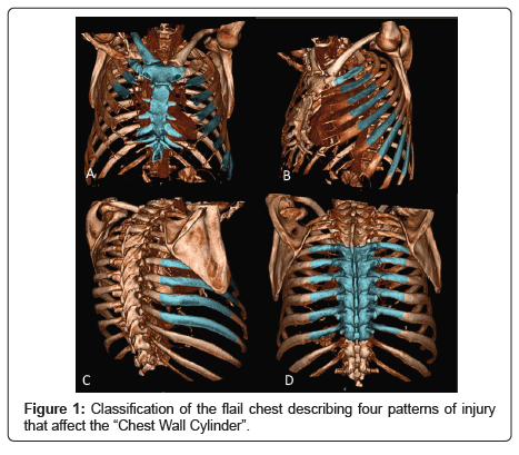
The following are descriptions of radiological characteristics of flail Types A – D, Figure 1:
Type A – Anterior flail segment; Transverse, oblique or comminuted fracture of sternum, associated with bilateral rib and costal cartilage fractures, and clavicular fracture. This pattern of injury is commonly caused by seat belt restraint, and less frequently the impact of the chest against the steering wheel following a road traffic collision. The orientation of injuries is often an oblique line of fractures that extends from a clavicle and includes ipsilateral ribs and costal cartilages, the manubrium or body of sternum, and contralateral costal cartilages and rib fractures extending from the second rib down to the costal margin. Care is required during examination to identify fractures of costal cartilages, as these may be missed.
The anterior central component of the chest is flail, moving paradoxically during respiration. Furthermore, respiration is compromised due to the inability of diaphragmatic attachments to sternum and ribs to provide traction during respiration (5, 6). Secondary injuries are caused by traction from the lower end of the seat belt and include, rupture of the diaphragm, liver and spleen.
Type B – Lateral flail segment; Sequential parallel fractures are situated unilaterally. They may extend from the clavicle and first rib down to the costal margin. Fractures may be situated within costal cartilages or medial bony end of ribs, the former being difficult to identify on CT scan. The lateral fractured ribs of the flail segment may extend as far lateral as the posterior axillary line. This flail, situated deep to the pectoralis muscles is commonly caused by direct impact; falls from low to medium to heights and in the elderly.
Type C – Postero-lateral flail segment; A pattern of fractures primarily caused by direct high velocity impact, such as motorbike accidents and falls from heights, causing compression fractures by impact of the scapular on the rib cage. Ribs are commonly fractured outside the margins of the scapula forming a V-shaped flail segment. Fractures anterior to the scapula, are situated on the posterior margin of the axilla. Posterior fractures most commonly follow a vertical line 1 – 2 cm lateral and parallel to the costo-transverse joint and may extend from rib one to rib ten. The scapula itself may be fractured, and is often comminuted. Secondary injuries are a result of the high- speed mechanism of injury, including transection of aorta, vertebral fractures, clavicular fractures and extra-thoracic injuries.
Type D – Posterior flail segment; An uncommon pattern of injury resultant of major compression of falling heavy objects and high-speed road traffic collisions. The epicentre of the injury pattern is an unstable thoracic vertebral column associated with bilateral rib fractures, the latter situated equidistant from the vertebra. The primary vertebral injuries demand neurosurgical attention, however due to concomitant instability of the rib fractures, thoracic surgical intervention may be required.
Statistical Analysis
Data obtained by retrospective evaluation of clinical and radiological characterisation were compared to demographic and injury stratification data recorded on the TARN database. Analysis used StataCorp, 2011. Release 12. College Station, Tx: StataCorp LP statistical software. Comparison of categorical and continuous data was achieved by use of Chi Square test and Student’s t-Test respectively. Statistical significance was considered p <0.05. Ethical approval was sought from the Ethical Committee of the University Hospital of North Midlands, and provided as the study constiuted an evaluation of clinical practice.
Results:
The distribution of flail types were evenly distributed between Types A – C and non-flail groups, type D occurring less frequently, Table 1. Demographic characteristics of Flail pattern types were similar, 84.6% of injuries were caused by Road Traffic Collisions (RTCs) and falls. RTCs was responsible for the majority of Type A and D flail patterns, Table 2. Type B flail patterns were caused equally by RTCs and Falls, as were non-Flail patterns.
Table 1: Showing the incidence of chest wall injuries according to the
classification of fail chest.
| Flail InjuryClassification | Incidence | Percent (%) |
|---|---|---|
| Type A | 30 | 19.48 |
| Type B | 41 | 26.62 |
| Type C | 38 | 24.68 |
| Type D | 10 | 6.49 |
| Non-Flail | 35 | 22.73 |
Fracture of elements of the thoracic cylinder, other than ribs, showed a statistical relationship to injury pattern. Type A had a 73.3% incidence of sternal fractures, and Type D 90% incidence of vertebral injuries, p<0.001, Rupture of the diaphragm was an infrequent injury, identified in 4 (8.9%) and 2 (6.5%) following RTC and falls respectively and predominated in Types A-C flail chest.
Table 2: Showing the mechanisms of injury responsible for each group of chest wall injuries. Percentage values are the proportion of the injury types. Comparison of frequencies demonstrated that the patterns showed a relationship to mechanism of injury, Chi square p <0.001.
| Injury Pattern | Mechanism of Injury | |||
|---|---|---|---|---|
| Flail Type; n (%) | RTCn = 71 (46.7%) | Falln = 62 (40.8%) | Blunt Trauman = 11 (7.2%) | Crushn = 8 (5.3%) |
| A | 24 (80%) | 3 (10.0%.) | 1 (3.3%) | 2 (6.7%) |
| B | 17 (41.5%) | 20 (48.7%) | 1 (2.4%) | 3 (7.3%) |
| C | 8 (22.9%) | 21 (58.3 %) | 5 (14.3%) | 1 (24.4%) |
| D | 7 (70%) | 2 (20%) | 0 | 1 (10%) |
| Non-Flail | 14 (40.0%) | 16 (45.7%) | 4 (11.4%) | 1 (2.9%) |
AIS scores, AIS Chest and AIS Rib, were not found to be different across flail types, yet Type A and Type D exhibited the highest NISS scores, Type A had greatest number of rib fractures and Types C and Non-Flail, the lowest numbers, P<0.001. The incidence of polytrauma recorded in injury subtypes and compared with the population was found to be greater when Type A injuries were recorded, p = 0.022, . Comparison of the incidence of extra-abdominal injuries showed no significant differences between patients of the Flail Types and non- Flail groups, except for vertebral injury that had a preponderance in type D, p <0.001,
Types A required the longest period of mechanical ventilation, treatment in the Critical Care Unit and treatment in hospital, p < 0.001, whilst patients with non-flail injuries required significantly reduced investment of intensive treatments, The mortality of the study population was 6 (3.85%).
Comment
Patients who have suffered major thoracic trauma experience increased demand for intensive care treatments and exhibit increased mortality that is related to severity of injury. Of patients aged between 18 – 50 years and aged over 60 years, mortality was 30% and 76% respectively, was attributable to thoracic injuries [2]. Severity of chest wall trauma is most commonly assessed by number of fractured ribs, more than 8 ribs fractured demonstrating increased mortality [3]. Furthermore,
the number of fractured ribs is utilised to define characteristics of the injury severity score, where ISS > 30 being is associated with a mortality in excess of 40%, [14, 15]. Yet there remains debate as to the merit of CWR in management of thoracic trauma, a fact that we propose is related to inadequate characterisation of chest wall injuries.
Sillar and Proctor, in an era before mechanical ventilation, documented that there was differential mortality related to different patterns of chest wall injury [9, 10]. Anterior flail chest injury, commonly caused by motor vehicle accidents was associated with an untreated mortality of 85%. The cause, severe respiratory distress, resulted from paradoxical movement of the flail segment and diaphragmatic dysfunction. The latter was attributed to loss of a rigid anterior chest wall to which the diaphragm was attached causing ineffective contraction. The impact on cardiovascular function of flail segments was also recognised as contributory to mortality associated with these injuries. Surgical treatment of anterior flail segments was reported to reduced mortality by 75% [9].
Current medical literature fails to discriminate anatomical patterns of chest wall injury. There is common focus on the numerical extent of rib trauma and assumption that flail chest is determined by parallel fracture of regions of the rib structure [4, 14 – 16]. Other than early reports, literature does not consider the chest wall a single structure unified by component parts, that when disrupted in any part of the circumference causes a flail segment. There is therefore limited consideration of anatomical subtypes of chest wall injury.
We sought to emphasise of the importance of considering the chest wall as integrated structure, a cylinder. Various parts of that cylinder, have differing structures and functions, and therefore cause differential pathophysiological effects when subjected to trauma. We have undertaken evaluation of the utility of an anatomical classification of flail chest. The results demonstrated, not surprisingly, that chest wall injury patterns described by the classification bore regional relationship to mechanisms of trauma, severity of injury and to outcome. Most notable was Type A, that was distinguished by a close relationship to RTC as the mechanism of trauma, the greatest number of fractured ribs, demand for ventilation and care in the critical care unit. By contrast non-flail chest wall injuries, which despite having a large number of fractured ribs, exhibited reduced respiratory compromise and demand for critical care. The differences between groups were not however distinguished by AIS Chest or AIS Rib subclasses of the internationally recognised NISS scoring system.
Determination of accurate anatomical description remains the essence of surgical practice, the core principle upon which this classification was founded. Treatment of Type A flail has required bilateral rib/ costal cartilage and sternal fixation through subpectoral incisions, and clavicular fixation by orthopaedic surgeons. Treatment of Type B flail required access to cartilaginous and bony factures by elevation of the pectoralis major. Type C flail have required dual incision to access the paravertebral rib fractures and anterior fractures situated along the posterior axillary line. Treatment of Type D flail, has exemplified treatment of chest trauma in the setting of poly-trauma and a collaborative approach to surgical treatment.
The study has demonstrated that despite a high NISS value of the population treated by CWR, comprehensive operative treatment has resulted in a low hospital mortality. This statistic, <4% mortality, was significantly below estimated mortality statistics of >11% and
>34% when 6 and 8 ribs were fractured respectively [3, 4, 14]. It was also below an estimated mortality of 20% determined by values of NISS > 30. Our mortality statistic however consistent with other reports of surgical treatment of flail chest, [17, 18]. Whilst the study is a retrospective descriptive analysis without a comparative non- treatment population, we believe the findings support the premise that a comprehensive understanding of the patterns of injury has facilitated successful surgical strategy and excellent outcome.
Conclusion
We have undertaken retrospective analysis of an anatomical classification of chest all injuries that defines flail injuries of the chest wall. This study demonstrated that the classification delineates injury patterns and the Types A-D corresponds to differing pathophysiological patterns and related to clinical outcomes. We would recommend the classification is used in clinical practice and also receives further evaluation. We recommend that future studies, that seek to evaluate treatment methods and outcomes of chest wall trauma, should classify injuries anatomically. We commend the current classification for these purposes.
Acknowledgment:
We offer our thanks to clinicians who have provided collaborative care leading to development of current clinical pathways, and thank administrative staff who have undertaken data management.
References
- Arajavi E, Sanavirta S. Chest injuries sustained in severe traffic accidents by seatbelt wearers. J Trauma 1989; 29: 37 â?? 41
- Kent R, Woods W, Bostrom O. Fatality risk and the presence of rib
- Ann Adv Automot Med 2008; 52: 73 â?? 84
- Flagel Et al. Half-a-dozen ribs: The breakpoint for mortality. Surgery 2005; 138: 717-25
- Crandall J, Kent R, Patrie J, Fertile J, Martin Rib fracture patterns and
- radiologic detection â?? A restraint-based
- Annu Proc Assoc Adv Automot 2000; 44:235-60 Brewer LA. Wounds of the chest in war and peace: 1943â??1968.
- Ann Thorac Surg. 1969;7(5): 387â??408. Knoepp Fractures of the ribs: A review of 386 cases. Am J Surg. 1941;52(3): 405â??14.
- Hagen Multiple rib fractures treated with a drinker respirator: a case report.
- JBJS 1945;27: 330- 334
- Cohen EA. Treatment of the fail chest by towel clip traction. Am J Surg 1965;109:604 -10
- Sillar Management of the flail anterior segment. J Bone Joint Surg. 1961;43B(4): 738â??45
- Proctor , London PS. The -Stove-in chest with paradoxical respiration. BJS 1955:42:622- 633
- Fisher A, Ross C, Henderson C, Kirk S, Feroze M, Richmond D, Bett L, et Major trauma care in England. National Audit Office. 2010; 1-37.
- Lecky F, Woodford M, Edwards A, Bouamra O, Coats Trauma scoring systems and databases. Brit J Anaesa 2014;113: 286-94
- Shields TW, Locicero J, Ponn RB. Anatomy of the Thorax. General Thoracic Surgery, Fifth Edition; Page 3. Lipincott Williams & Wilkins
- Lin FCF, Li RY, Tung YW, Jeng KC, Tsai Morbidity, mortality, associated injuries, and management of traumatic rib fractures. J Chin Med Assoc. 2016;79(6): 329â??34.
- Jamulitrat S, Sangkerd P, Thongpiyapoom S, Narong MNA. Comparison of mortality predictive abilities between NISS and ISS in Trauma J Med Assoc Thai 2001;84:1416 -21
- Naidoo K, Hanbali L, Bates The natural history of flail chest injuries. Chin J Trauma 2017; 20: 293-6
- Marasco SF, Martin K, Niggemeyer L, Summerhayes R, Fitzgerald M, Bailey
- Impact of rib fixation on quality of life after major trauma with multiple rib fractures. Injury, Int J Care 2019;50:119-24
- Leinicke JA, Elmore L, Freeman BD, Colditz GA. Operative management of rib fractures in the setting of flail chest: A systematic review and meta- analysis. Ann Surg. 2013;258(6): 914â??21.
 Spanish
  Chinese
  Russian
  German
  French
  Japanese
  Portuguese
  Hindi
Hindi 
