Case Report, J Spine Neurosurg Vol: 8 Issue: 1
C1 Arch Reconstruction: A Novel Alternative to Fusion in Maintaining Mobility of the Atlanto-Axial Joint
Sibhi Ganapathy1*, Ramakrishna TV2 and Swaroop Gopal2
1Manipal Hospital, Whitefield, Bangalore, India
2Sakra World Hospital, Bangalore, India
*Corresponding Author : Dr. Sibhi Ganapathy
Consultant Neurosurgeon, Manipal Hospital, Whitefield, Bangalore, India
Tel: 8041694039
E-mail: sibhig@yahoo.co.uk
Received: October 1, 2018 Accepted: November 06, 2018 Published: February 14, 2019
Citation: Ganapathy S, Ramakrishna TV, Gopal S (2019) C1 Arch Reconstruction: A Novel Alternative to Fusion in Maintaining Mobility of the Atlanto-Axial Joint. J Spine Neurosurg 8:1. doi: 10.4172/2325-9701.1000310
Abstract
First decribed by Sir Goeffrey Jefferson in London in 1924, the term Jefferson’s fracture has become an eponym for all fractures of the Atlas. Complex Jefferson fractures and unstable Jefferson fractures require surgical fusion to maintain stability of the spinal axis. This however results in debilitating loss of movement at the crucial C1C2 joint, the most versatile of spinal joints where stability and mobility are intricately balanced into a complex yet supremely agile structure. The desire to attempt in some way to preserve a degree of movement and to spare the patient the crippling loss of movement at this joint has led to many attempts at innovation, both surgical and technological. This has led to the discovery of C1 osteosynthesis. This revolutionary technique enables the anatomical fixation of the defect thus allowing motion preservation without compromising on stability, which is a major advantage given the extreme importance of both to the proper functioning of the C1C2 and C1C0 joints. We present a case report of a 54 year old man with an isolated Jefferson’s fracture which was treated successfully by this technique. We also present an overview of the biomechanics of C1 injury and the relevant review of literature regarding options for management of complex C1C2 injuries.
Keywords: Surgical fusion; Anatomical fixation; Osteosynthesis
Introduction
Jefferson fractures are uncommon and traditionally depict the 4 section break of the map book. They make up around 7% of cervical spine fractures, be that as it may, are nearly connected with C2 breaks or fractures (right around half). As the danger of neurological damage is low, (around 4%-17%), these fractures are regularly missed in crisis rooms [1].
Although initially described by Sir Goeffrey Jefferson in London in 1924, the term has turned into an eponym for all fractures of the Atlas. Albeit Complex Jefferson cracks and temperamental Jefferson fractures require careful surgical fusion, it brings about crippling loss of development and movement at the urgent C1C2 joint. This has driven numerous to consider movement protecting choices in medical procedure, in this manner keeping up soundness yet saving a level of movement and development at the CVJ. This has prompted the revelation of C1 osteosynthesis. This revolutionary technique enables the anatomical fixation of the defect thus allowing motion preservation without compromising on stability, which is a major advantage given the extreme importance of both to the proper functioning of the C1C2 and C1C0 joints.
There are approximately 67 cases reported of this study in the world. We are proud to add another successful case report to this ever increasing number of surgeries in the hope that it one day becomes a standard of care for C1 fractures with instability but intact transverse ligament and facets.
Case Report
A 54 year old male had a fall from a height of approximately 6 metres. He complained of severe neck pain but was otherwise deficit free. On examination he had tenderness over the back of the neck, but otherwise no clinical findings.
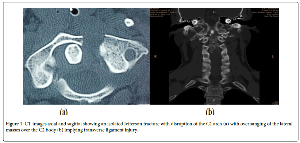
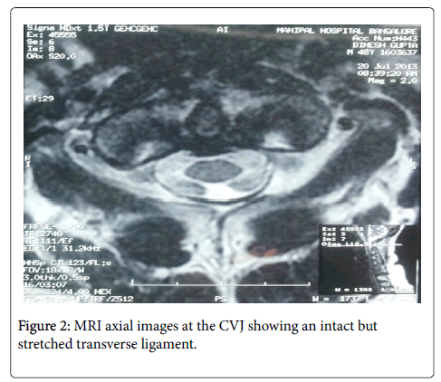
His CT of the cervical spine revealed an isolated Jefferson fracture with a primarily posterior arch pathology (Figure 1). The MRI further revealed that the transverse ligament although stretched and frayed was intact, allowing the odontoid to stay within the articular anterior C1 compartment, thereby preventing it from compressing the cervico-medullary junction (Figure 2).
The prescribed treatment of this injury is a Harm Goel fusion whereby, the C1lateral mass screws are connected to C2 pedicle or pars screws fusion the C1C2 joint and ending movement at the most mobile and critically active joints of the spine. The other option was to preserve movements with a C1 osteosynthesis.
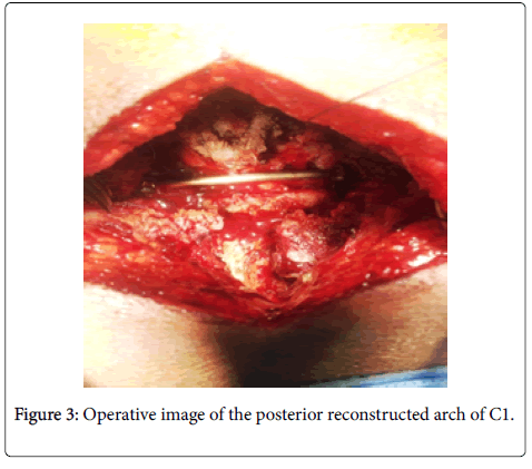
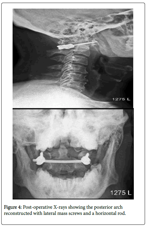
Posterior arch reconstruction was done by placing lateral mass screws into the C1 and placing a horizontal curved rod into place. Compressing the construct under flouroscopic guidance to ensure the overhang of the lateral masses over the C2 body was achieved carefully and slowly ensuring that there were no changes in SSEP or MEP action potentials. After final tightening of the screws (Figure 3), the wound was closed and the patient mobilized on a Philadelphia collar for 6 weeks. His Follow up imaging (Figure 4) bear testimony to an excellent reduction and successful osteosynthesis. His range of movements remains normal despite his injury.
Discussion
Operative management in Jefferson’s fracture
Jefferson fractures are a frequently ignored part of cranio-vertebral intersection injury. Most don't require careful mediation and a substantial dominant part are without neurological pressure. Decent information of life systems and biomechanics helps in the administration of this burden.
Sir Goeffrey Jefferson proposed in 1920, that because of the wedge state of the parallel masses, a compressive stacking pressure was changed over into a decompressive power prompting the crack being referred [2]. Agent treatment is once in a while required as the fractures only from time to time cause precariousness or neural pressure. Signs for medical procedure in Jefferson Breaks incorporate:
Transverse Ligament Disruption and Associated wounds, for example, AAD, Hangman's cracks or Odontoid breaks [3].
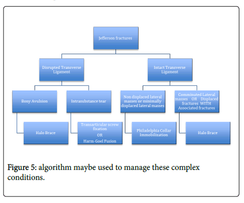
Complex C1C2 fractures require unique consideration. The accompanying calculation possibly used to deal with these unpredictable conditions. The favored kind of medical procedure is reliant upon the intricacy present and additionally the nearness or nonappearance of transverse tendon burst (Figure 5) [4].
Biomechanics
In light of injury, the CVJ shows unsurprising examples of disappointment dependent on the system of damage. The C1 goes about as a washer between the occipital condoyles and the C2 beffering the power transmitted down. In this way the C1 is to a great degree helpless against pivotal powers which while squeezing down cause the burst fracture average to the sidelong masses.
The sidelong mass on C1, being taller, is coordinated along the side. In this way vertical powers are compacting the sidelong masses between the occipital condyles and the hub divide them, fracturing either of the front or back curves. The effect power cause an outward spread of the sidelong masses of C1 [5].
A Jefferson fracture doesn't generally result in spinal line damage, as the measurements of the hard ring increment, prompting spinal trench auto decompression.
Spinal cord damage is more expected if the transverse Atlanta ligament (TAL) is burst due to atlanto-hub shakiness. An ADI 3mm demonstrates AAD while an ADI of 5mm shows alar ligament also, TAL damage [5].
In creature models especially animals, fracture examples have been replicated in axial compressive powers and distractive shear weights on the horizontal masses [6,7].
Ligamentous damage is generally uncommon in great Jefferson cracks or fractures, yet at the point when present is normally happens as an osteoperiosteal separation of the transverse tendon or an entire separation of the tubercle of the map book. This prompts relocation of the parallel masses which can be picked up on radiology. It very well may be analyzed by recognizing bony goad separation on CT, by TAL burst on MRI and additionally by distinguishing the widenning of the parallel masses on a CV intersection Xray (open mouth AP view) [8].
C1 arch reconstruction
The method takes into account versatility safeguarding in spite of adjustment utilizing screws and poles. The methodology perhaps foremost or back contingent on the idea of the sections, related odontoid fractures and in addition deficients presents [9].
The front methodology is regularly not favored because of the grimness related with transoral dismemberment, danger of meningitis and CSF release and risk, Figure of dysphagia and odynophagia. This Procedure licenses development and diminishes the spreading of the horizontal masses (due to TAL harm), in this way conveying soundness to a precarious break or fracture [9-12].
Conclusion
C1 arch osteosynthesis is a novel technique that can allow the patient to have his proverbial cake and eat it as well!! A Robust knowledge of anatomy and biomechanics of the spine, excellent prowess on the surgical stage are definitely helpful, in ensuring optimal results.
References
- Levine AM, Edwards CC (1991) Fractures of the atlas. J Bone Joint Surg Am 73: 680-691.
- Jefferson G (1920) Fractures of the atlas vertebra: Report of four cases and a review of those previously reported. Br J Surg 7: 407-422.
- Fielding JW, Cochran GB, Lawsing JF, Hohl M (1974) Tears of the transverse ligament of the atlas: a clinical and biomechanical study. J Bone Joint Surg Am 56:1683-1691.
- Spence KF Jr, Decker S, Sell KW (1970) Bursting atlantal fracture associated with rupture of the transverse ligament. J Bone Joint Surg Am 52: 543-549.
- Heller JG, Viroslav S, Hudson T (1993) Jefferson fractures: The role of magnification artifact in assessing transverse ligament integrity. J Spinal Disord 6: 392-396.
- Dickman CA, Greene KA, Sonntag VK (1996) Injuries involving the transverse atlantal ligament: classification and treatment guidelines based upon experience with 39 injuries. Neurosurgery 38: 44-50.
- Dickman CA, Sonntag VK (1997) Injuries involving the transverse atlantal ligament: Classification and treatment guidelines based upon experience with 39 injuries. Neurosurgery 40: 886-887.
- Greene KA, Dickman CA, Marciano FF, Drabier J, Drayer BP, et al. (1994) Transverse atlantal ligament disruption associated with odontoid fractures. Spine (Phila Pa 1976) 19: 2307-2314.
- Fowler JL, Sandhu A, Fraser RD (1990) Are view of fractures of the atlas vertebra. J Spinal Disord 3: 19-24.
- Lee TT, Green BA, Petrin DR (1998) Treatment of stable burst fracture of the atlas (Jefferson fracture) with rigid cervical collar. Spine (Phila Pa 1976) 23: 1963-1967.
- Kesterson L, Benzel E, Orrison W, Coleman J (1991) Evaluation and treatment of atlas burst fractures (Jefferson fractures). J Neurosurg 75: 213-220.
- Landells CD, Van Peteghem PK (1988) Fractures of the atlas: Classification, treatment and morbidity. Spine (Phila Pa 1976) 13: 450-452.
 Spanish
  Chinese
  Russian
  German
  French
  Japanese
  Portuguese
  Hindi
Hindi 