Clinical Image, J Clin Image Case Rep Vol: 4 Issue: 1
Clinical Images: Extrapleural Hemorrhage - An Unrecognized Cause of Sudden Chest Pain
Takeaki Sato* and Shigeki Kushimoto
Department of Emergency Medicine, Tohoku University Graduate School of Medicine, Sendai, Miyagi, Japan
*Corresponding Author: Takeaki Sato
Tohoku University Hospital Emergency Center, 981-0872, Miyagi-pref, Sendai-shi, Aoba-ku, Seiryo-cho 1-1, Miyagi, Japan
Tel: +81-022-717-7489
Fax: +81-022-717-7492
E-mail: takeakisato@gmail.com; takesato@hkg.odn.ne.jp
Received: November 21, 2019 Accepted: December 12, 2019 Published: December 19, 2019
Citation: Sato T, Kushimoto S (2019) Clinical Images: Extrapleural Hemorrhage – An Unrecognized Cause of Sudden Chest Pain. J Clin Image Case Rep 3:2.
Abstract
A 53-year-old man presented with sudden posterior chest pain without any preceding causative events. He had no significant medical history, including conditions requiring antithrombotic medication. His consciousness was clear, blood pressure was 110/80 mmHg, heart rate was 70 beats per minute (bpm), respiratory rate was 14 per minute, and SpO2 was 98% in room air.
Keywords: Sudden Chest Pain, Extrapleural Hemorrhage
Clinical Image
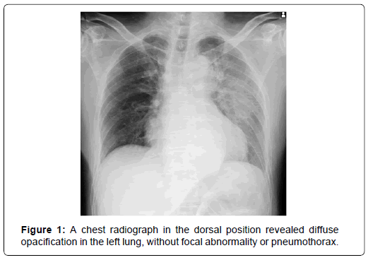
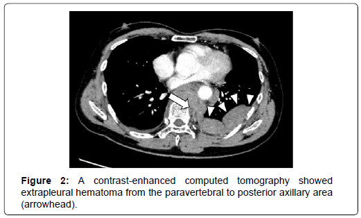
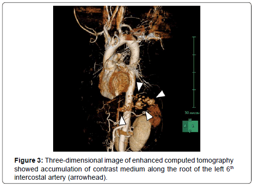
A 53-year-old man presented with sudden posterior chest pain without any preceding causative events. He had no significant medical history, including conditions requiring antithrombotic medication. His consciousness was clear, blood pressure was 110/80 mmHg, heart rate was 70 beats per minute (bpm), respiratory rate was 14 per minute, and SpO2 was 98% in room air. He reported a sharp continuous pain that was unrelated to respiration and physical exertion. There was no episode of coughing and hemoptysis. The electrocardiogram findings were within normal limits. A chest radiograph in the dorsal position revealed diffuse opacification in the left lung, without focal abnormality or pneumothorax (Figure 1). A contrast-enhanced computed tomography showed extrapleural hematoma from the paravertebral to posterior axillary area (Figure 2, arrowhead), with extravasation from the intercostal artery (arrow). No pseudo aneurysm was observed. Three-dimensional image of enhanced computed tomography showed accumulation of contrast medium along the root of the left 6th intercostal artery (Figure 3, arrowhead). Based on his stable physiologic status with normal hemoglobin level and coagulation test results, we observed it as a conservative therapy without angioembolization. The patient has been followed up for 6 months, without recurrent hemorrhage.
Extrapleural hemorrhage without a preceding traumatic event is an extremely rare condition; it has never been considered as a cause of chest pain [1]. Hemorrhage from the intercostal artery may be considered an etiological factor, which may require intervention [2]. In the present case, as the bleeding was limited to the tight extrapleural space, pseudoaneurysm formation was not observed. Idiopathic extrapleural hemorrhage may be an important unrecognized cause of sudden chest pain.
References
- Bosners S, Becker A, Haasenritter J, Abu Hani M, Keller H, et al. (2009) Chest pain in primary care: epidemiology and pre-work probabilities. Eur J Gen Pract 15: 141-146.
- Chemelli AP, Thauerer M, Wiedermann F, Strasak A, Klocker J, et al. (2009) Transcatheter arterial embolization for the management of iatrogenic and blunt traumatic intercostal artery injuries. J Vasc Surg 49: 1505-1513.
 Spanish
  Chinese
  Russian
  German
  French
  Japanese
  Portuguese
  Hindi
Hindi 


