Spanish
  Chinese
  Russian
  German
  French
  Japanese
  Portuguese
  Hindi
Hindi Research Article, J Regen Med Med Vol: 15 Issue: 1
Collagen Hydrolysates, from Beauty Supplements to Supporting Wound Healing: How Solid is the Rationale? An Overview and Insights for Regenerative Medicine Practitioners
Emanuele Salvatore Aragona1, Maurizio Cavallini2, George Christopoulos3, Marco Mantoan4 and Mauro Raichi5*
1Complex Operative Unit Diabetic Foot, Melzo, Milan, Italy
2Department of Plastic Surgery, Centro Diagnostico Italiano, Milan, Italy
3The Ghanem Clinic, London, UK & College of Medicine and Dentistry, Ulster University, Birmingham, UK
4Department of Diagnostics and Public Health, Section of Hygiene and Preventive, Environmental and Occupational Medicine, University of Verona, 37134 Verona, Italy
5Clinical Pharmacology Consultant and Medical Director, Astéria Pharma Srl, Milan, Italy
*Corresponding Author: Mauro Raichi
Clinical Pharmacology Consultant and Medical Director, Astéria Pharma Srl, Milan, Italy; Email: mauro.raichi@gmail.com
E-mail: mauro.raichi@gmail.com
Received date: 28 April, 2026, Manuscript No. JRGM-26-189113; Editor assigned date: 01 May, 2026, PreQC No. JRGM-26-189113 (PQ); Reviewed date: 15 May, 2026, QC No. JRGM-26-189113; Revised date: 04 June, 2026, Manuscript No. JRGM-26-189113 (R); Published date: 11 June, 2026, DOI: 10.4172/2325-9620.1000378
Citation:Aragona ES, Cavallini M, Christopoulos G, Mantoan M, Raichi M (2026) Collagen Hydrolysates, from Beauty Supplements to Supporting Wound Healing: How Solid is the Rationale? An Overview and Insights for Regenerative Medicine Practitioners. J Regen Med Med 15:1.
Abstract
Introduction and purpose: Aquatic organisms, including invertebrates such as sponges, mollusks, and jellyfish, are sources of environmentally friendly marine collagen and lowmolecular-weight bioactive oligopeptides, which are purified using advanced technologies. Since the early 2010’s, numerous high-quality experimental and human studies have explored the properties of hydrolyzed marine collagen fragments as systemic functional ingredients in regenerative medicine. This review discusses these properties and the rationale behind them, with a focus on wound healing. Mechanisms underlying chronic wound healing may provide a strong foundation for the stillmissing, high-level clinical studies, particularly in patients with diabetic and pressure ulcers.
Methods: This review examines only academically significant studies published in PubMed-indexed journals with significant impact factors, supplemented by a few contributions from Google Scholar for methodologically sound in vitro and animal studies.
Results: Activation of skin fibroblasts and other mesenchymal cells underpins the systemic regenerative properties of highly purified hydrolyzed marine collagens. For example, 50 µg/mL of hydrolyzed marine collagen peptides nearly replicated in vitro the accelerated cell migration induced by 10 µg/mL of recombinant human epidermal growth factor. Faster wound healing, associated with increased collagen neosynthesis, is accompanied by increased immunohistochemical expression of platelet-endothelial cell adhesion molecule-1, basic fibroblast growth factor, and transforming growth factor β-1. The potential supportive role of collagen hydrolysates in managing insulin resistance could benefit the treatment of chronic diabetic and pressure ulcers.
Conclusion: An increasing number of preclinical and human studies highlight the systemic regenerative properties of hydrolyzed marine collagens and their excellent safety profile. The evidence for their regenerative properties in aesthetic skin rejuvenation appears solid. Pre-clinical evidence supporting wound healing is also growing. Unfortunately, sound clinical studies confirming the experimental evidence in everyday wound care practice are still lacking, and long-term safety remains a primary concern.
Keywords: Collagen hydrolysates; Diabetic foot care; Fibroblast activation; Hydrolyzed marine collagen; Regenerative medicine; Wound care
Keywords
Collagen hydrolysates; Diabetic foot care; Fibroblast activation; Hydrolyzed marine collagen; Regenerative medicine; Wound care
Introduction
Marine collagen hydrolysates and their growing role as beauty supplements
Collagen derivatives from cowhide and other land mammals, along with more recent marine collagen supplements, have gained prominence in the global beauty supplement industry, despite the limited number of well-designed studies. The decline in native collagen with age after early adulthood, which contributes to visible signs of skin aging, such as wrinkles and sagging, especially among women after menopause, has fueled the recent widespread acclaim for collagen derivative supplements [1].
Seawater and freshwater organisms including invertebrates like sponges, mollusks, echinoderms, and jellyfish are sources of marine collagen [2-4]. Unlike bovine, porcine, or human recombinant Type 1 collagens, all marine collagens are naturally less cross-linked, making them biophysically unique. They exhibit lower tensile strength, which is less critical in the low-gravity aquatic environment, but show greater flexibility [4]. Collagen from aquatic sources addresses religious concerns and avoids the risks of prion disease transmitted by landmammal collagens; additionally, it avoids the risks of contamination with heavy metals and marine pollutants [2,5]. Highly purified marine collagens are also environmentally friendly because they originate from the estimated 20 million tons of waste organic residues produced annually by the global industrial fishing industry [2,5]. Moreover, published studies and a recent review of their regenerative potential indicate that marine collagen hydrolysates have safety profiles similar to placebo, with only occasional mild nausea and negligible risks of Type 1 immediate hypersensitivity [2].
The high bioavailability of hydrolyzed marine collagens after oral intake explains their widespread distribution throughout the body [2]. Several studies discuss their skin-rejuvenating effects and potential to reactivate functions in joints and other organs. However, these studies are outside the scope of this review, which focuses on the potential benefits of marine collagen fragments for wound healing, especially in slow-healing wounds.
Because they were introduced only recently, the number of published papers on hydrolyzed marine collagens indexed in PubMed is limited. This is especially true in high-impact, peer-reviewed journals. A PubMed search conducted in early January 2026, using the search string “marine collagen peptides” and limited to Englishlanguage articles, returned only 30 citations, including a review in aesthetic regenerative medicine by two authors of this manuscript [2]. Many of these papers mainly focus on purification procedures and other technical, non-medical issues.
Wound care dressings often use hydrolyzed collagen-based matrices, and a substantial body of literature supports their use. Still, as of early January 2026, no paper in PubMed-indexed journals appeared to have reviewed the effectiveness of orally administered marine collagen hydrolysates for wound care in human patients.
A role for marine collagen oligopeptides in chronic wound healing is soundly based
Despite the lack of clinical studies on marine collagen bioactive fragments in wound care, especially for chronic, difficult-to-heal wounds such as diabetic and pressure ulcers, the rationale for their widespread use appears solid. Fibroblast dysfunction and abnormally high levels of Matrix Metalloproteinases (MMPs) disrupt extracellular matrix remodeling by reducing collagen content [6-8]. Histochemical studies in experimental diabetic models confirm this disruption, showing delayed appearance of sparse, poorly stained precollagen (reticulin) fibers and weak staining for acid and neutral mucopolysaccharides [6]. Histologically, scar formation is delayed, with abnormal persistence of early vascularization and cellular activity. In fibroblasts, wound-healing mediators such as COL16A1 are poorly expressed. Likewise, several intracellular signaling pathways, including the transcription factors PLAGL1, RUNX2, and ZKSCAN7, which appear downregulated in poorly healing lesions, respond favorably to regenerative treatments such as platelet-rich plasma [9,10].
The purpose of this review is to discuss the properties of bioactive hydrolyzed marine collagen oligopeptides, with a particular focus on wound healing, and to present the rationale for their potential effectiveness in this indication. The mechanisms underlying chronic wound healing, as demonstrated preclinically and discussed herein, may provide a strong foundation for the high-level clinical studies that remain warranted.
The rationale appears especially well documented for diabetic ulcers, but may extend to other hard-to-heal chronic wounds, as suggested, in patients with chronic tissue ischemia and pressure ulcers, usually over bony prominences, through the oral administration of bioactive oligopeptides derived from gelatin or heat-denatured collagen from land mammals [11].
Materials and Methods
The authors adopted selective criteria for their review of published papers on hydrolyzed marine collagens in wound management:
• Inclusion only of well-described, methodologically sound, and reproducible studies, published in PubMed-indexed journals with notable impact factors.
• Exclusion of all studies of questionable quality published in nonindexed, non-peer-reviewed journals.
These selection criteria aimed to minimize the risk of promotional bias. The early 2010’s mark the period when the first indexed, academically credible papers on hydrolyzed marine collagens appeared in the literature; this period was set as the search cutoff for the consulted databases. The authors made the final decision about which studies to include in their review during an online consensus meeting held in mid-November 2025. They adhered as closely as possible to the PRISMA guidelines for reviews while maintaining a general narrative style to make their manuscript easier to read.
Results and Discussion
Even if still limited, evidence from the literature on marine collagenderived oligopeptides indicates a notable oral bioavailability and systemic distribution (Figure 1) [12]. After oral intake and systemic distribution, the low-molecular-weight 14C-labelled fragments persist in target tissues for more than two weeks-skin, muscles, and mesenchymal tissues involved in slow-healing wounds [12,13].
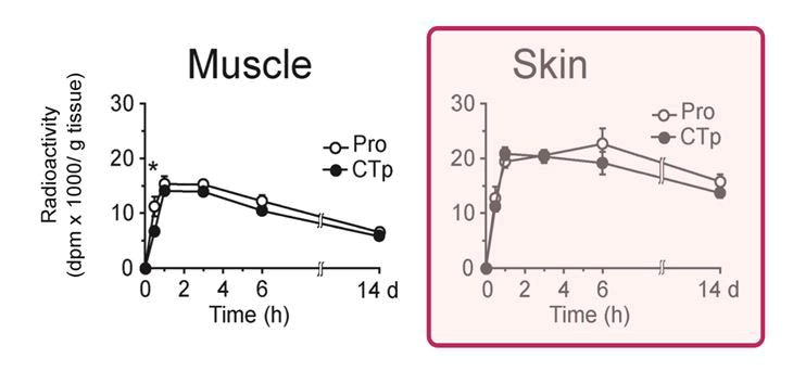
Figure 1: Distribution of radioactivity in the skin and muscle, normalized to 1 g of wet tissue, in laboratory Wistar rats during the first six hours and after 14 days following oral administration of 14Clabeled hydrolyzed collagen fragments (CTp) and proline (Pro); mean ± standard error. Reproduced and adapted from Sibilla S, et al. under a Creative Commons CC BY-NC 4.0 License [12].
Efficient absorption, mediated by transporters in the intestinal brush border membrane, such as the proton-coupled oligopeptide transporter 1 (PepT1), prevents digestion of marine collagen fragments into individual amino acids [2]. The high systemic bioavailability of marine collagen oligopeptides, which is indistinguishable from that of the amino acid proline, sets hydrolyzed marine collagens apart from collagen fragments derived from land mammals. Absorption is up to 50% more efficient compared to mammalian collagens, likely owing to the higher content of glycine, serine, and threonine and the lower levels of proline and hydroxyproline [14,15].
Safety and oral-route bioavailability of hydrolyzed marine collagens
In preclinical investigations, marine collagen oligopeptide hydrolysates showed negligible impact on pro-inflammatory mediators in animal studies [2]. The low histidine content, a histamine precursor, indicates the low antigenic potential of marine fragments and reduced risk of type-1 immediate hypersensitivity [16]. These hydrolysates also act as potent antioxidants, with free radical scavenging activity comparable to E321, a widely used food preservative (Figure 2) [5].
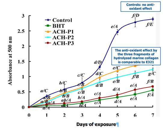
Figure 2: The decrease in spectrophotometric absorbance by cell membranes over seven days directly correlates with the oxidative damage caused by free radicals. The antioxidant effectiveness of the positive control E321 and three peptides from the same batch of marine collagen is comparable. Control: inert negative control. Reproduced and adapted from Geahchan S, et al. under a Creative Commons CC BY-NC 4.0 License [5].
The biological properties of hydrolyzed marine collagens
Activation of skin fibroblasts underpins the regenerative properties of marine collagen hydrolysates after oral intake, both in the skin and systemically. These properties are unique to marine breakdown fragments, compared with dermally infiltrated intact collagen [2,16-19]. Fibroblast activation requires a polypeptide structure, as exposure to proline and hydroxyproline, the amino acids typical of all collagens, fails to exhibit the same biological behavior (Figure 3) [12].
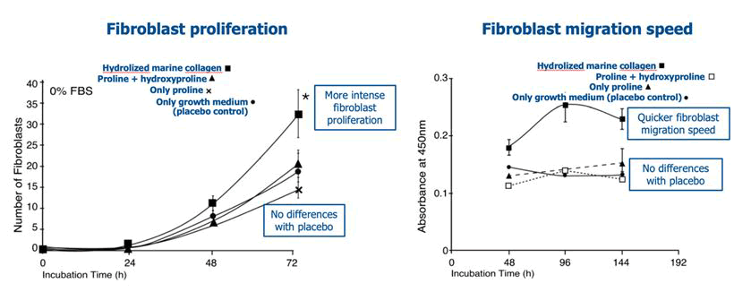
Figure 3: Effects on in vitro proliferation and migration speed of human fibroblasts after exposure to marine collagen fragments for 72 hours, compared with the growth medium (inert placebo), the amino acid proline, and a proline-hydroxyproline mixture; *p<0.05 versus placebo. Reproduced from Sibilla S, et al. under a Creative Commons CC BY-NC 4.0 License [12].
The effect on fibroblasts appears direct. For example, the hydrophobic peptide F5 from Mozambique tilapia (Oreochromis mossambicus) fish scales not only scavenges reactive oxygen species and prevents oxidative stress-induced damage to fibroblasts, but also docks to the active sites of matrix metalloproteinases MMP-1, MMP-3, and MMP-12, which break down collagen and elastin [3]. Furthermore, in laboratory animals supplemented with oral marine collagen hydrolysate for 24 months, there is an increase in the expression of Type I and III collagen compared to aged untreated controls. This increase in collagen deposition occurs alongside a decrease in Metalloproteinase-1 (MMP-1) expression and higher tissue levels of the MMP-1 inhibitor, as revealed by immunohistochemical and Western blot analysis [20].
These changes are linked to the activation of the SMAD signaling pathway mediated by the SMAD group of related proteins acting as the primary signal transducers for receptors of the Transforming Growth Factor beta (TGF-ß) superfamily and the up-regulated expression of the SMAD activator TGF-βRII (TβRII), supporting the direct effect on fibroblasts [21]. The activation of the Hyaluronan Synthase 2 (HAS2) transcription pathway stimulates the synthesis of high-molecularweight hyaluronan [20].
The rationale behind these properties is sound and unsurprising, aligning with the innovative concept of transitioning from inflammatory repair to non-inflammatory tissue regeneration (Anti-Inflammatory Regenerative Medicine, AIMED) [22]. The small peptides, known as matrikines, released during the breakdown of extracellular matrix collagen, are highly homologous to marine collagen fragments and act as good models of the biological effects for marine collagen hydrolysates (Figure 4). Molecular docking studies demonstrate the high affinity of several matrikines for receptors for chemokines, cytokines, ion channels, and growth factors [2,23]. Examples include the collagen matrikine α1 C-1158/59, which promotes wound healing and cell migration, and the glycine- and proline-containing matrikine PGP, which binds at the chemokine receptors C-X-C motif chemokine receptors 1 and 2 (CXCR1 and CXCR2), thereby regulating both the inflammatory and epithelialization phases of wound healing [23,24].
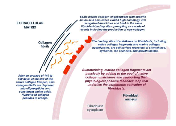
Figure 4: The effects of hydrolyzed marine collagens on fibroblasts resemble and complement those of physiological matrikines as native collagen reaches the end of its lifespan in the dermis and subdermal tissues. Reproduced and adapted from Bartoletti E, et al. under a Creative Commons CC BY-NC 4.0 License [2].
The promising rationale for hydrolyzed marine collagens in wound care
A recent review examined the emerging benefits of orally administered marine collagen hydrolysates in aesthetic medicine and plastic surgery [2]. Building on those findings, the authors present some ideas about the rationale and expected advantages of marine collagen hydrolysates in wound care.
Experimental in vitro exposure to marine collagen peptides promotes wound healing in a dose-dependent manner, as demonstrated in several early preclinical studies from the mid-2010’s. In a 2015 study based on the in vitro scratch closure assay, with marine collagen proteolysate concentrations ranging from 6.25 to 50 μg/mL, there was no difference in the rate of cell migration and repair ability of the injured cell monolayer between fibroblasts exposed to 50 μg/mL of marine collagen fragments and those exposed to 10 μg/mL of recombinant human Epidermal Growth Factor (rhEGF), used as positive controls [25]. The same year, a study in young male Balb/c mice injected in the tails (test solutions: 80 μL) with Acid-Soluble Collagen (ASC) from haddock (Melanogrammus aeglefinus) skin and four ASC hydrolysates with different average molecular weights H1 - (53.8 kDa), H2 (23 kDa), H3 (14.4 kDa), and H4 (6.5 kDa) - found that ASC and the hydrolysates with higher molecular weights (H1 and H2) shortened clotting and bleeding times compared to controls (saline) [26]. Moreover, during experimental burn wound healing, skin cellularity and maturity increased in both the epidermal and dermal layers compared to the control group. After a week, blood capillaries appeared in scalded skin treated with hydrolysates but not in controls (Figure 5) [26].
In another similar study conducted two years later, chum salmon skin collagen fragments accelerated wound healing after cesarean section. This was associated with increased immunohistochemical expression of platelet-endothelial cell adhesion molecule-1, basic fibroblast growth factor, and transforming growth factor ß-1 [27]. The dose-dependent increase in fibroblast proliferation following exposure to marine collagen peptides is also chronologically connected to the dose-dependent activation of the fibroblast intracellular NF-κB p65 signaling pathway, followed by enhanced signaling along other secondary intracellular pathways [28].
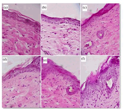
Figure 5: Increased cellularity and maturation of cells in the epidermis and dermis of scalded mouse skin (×400) after treatment with (a) saline (control group); (b) haddock skin ASC; (c) H1; (d) H2; (e) H3; (f) H4. Reproduced from Dang QF, et al. with permission from the publisher [26].
Monolayer scratch wounds close quickly. In a previously discussed wound-healing scratch assay, the repair rate of the scarified monolayer of Human Umbilical Vein Endothelial Cells (HUVEC) after 48 hours was about 74-75% following exposure to 6.25 μg/mL of two slightly different formulations of marine collagen peptides from the jellyfish Rhopilema esculentum. The repair rate of vehicle-treated, scarified control HUVEC cells was less than 50%. Starting at 18 hours, the difference in wound closure was significant compared to controls (Figures 6 and 7) [19].
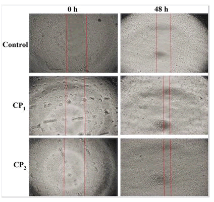
Figure 6: Migration of HUVEC cells in a wound-healing scratch assay after exposure to two slightly different formulations of jellyfish collagen peptides (6.25 μg/mL) compared with migration after exposure to vehicle (control) at baseline and after 48 hours. Red lines mark the edges of the wound gap. Reproduced from Hu Z, et al. under a Creative Commons CC BY-NC 4.0 License [19].
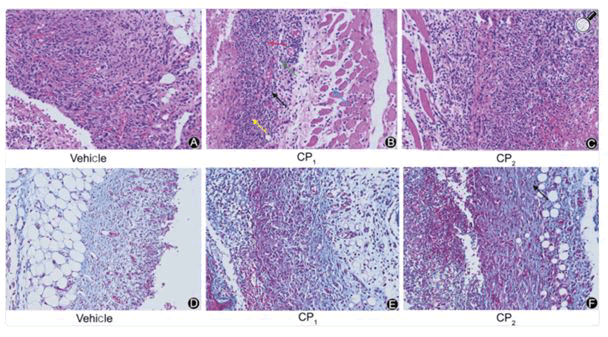
Figure 7: Skin sections from mice treated with 0.9 g/kg body weight collagen peptides or vehicle on day 6; magnification 200X. A to C micrographs: Skin sections showing fibroblasts (black arrows), fiber cells (red arrow), new blood vessels (yellow arrow), inflammatory cells (green arrow), and interstitial connective tissue around muscle cells (blue arrow). Hematoxylin and eosin stain. D to F micrographs: Blue-stained collagen (black arrow). Masson trichrome stain. Reproduced from Hu Z, et al. under a Creative Commons CC BY-NC 4.0 License [19].
Most of the limited number of published papers on hydrolyzed marine collagens indexed in PubMed are experimental, conducted in vitro and in laboratory models, including studies on wound healing.
Surprisingly, despite the sound rationale for marine collagen hydrolysates, there are no studies specifically focused on pressure injuries in laboratory models, nor have human studies examined the potential of hydrolyzed marine collagen in wound management. The results of the only (as far as the authors know) double-blind placebocontrolled study investigating the effectiveness of non-marine collagen hydrolysates could approximate the expected positive outcomes of the still-absent human studies [11]. Patients with pressure ulcers receiving standard care were randomized to twice-daily, high-dose oral intake of low-molecular-weight hydrolysates from heat-denatured pork skin gelatin containing either low or high levels of the prolylhydroxyproline (Pro-Hyp) and hydroxyprolylglycine (Hyp-Gly) dipeptides or placebo (maltodextrin TK-16). The primary efficacy parameters, the two scoring scales Pressure Ulcer Scale for Healing (PUSH) and Pressure Sore Status Tool (PSST), as well as the wound area, were significantly lower at the end of 16 weeks compared to placebo, with only minor differences between the two formulations. The PUSH scores for the slightly more effective formulation were 6.46 ± 0.98 and 9.26 ± 2.09 for the placebo (p<0.01), while the PSST scores were 19.71 ± 3.08 and 23.38 ± 3.85 (p = 0.01), and the wound areas were 3.19 ± 2.88 and 5.00 ± 3.88 (p=0.027). Over 71% of individuals showed improvements in their PUSH and PSST scores. According to the authors [11], the significant improvements in PUSH and PSST scores and wound area could be attributed to the physiological activity of the systemically absorbed oligopeptides.
Conversely, evidence is accumulating on the potential role of collagen hydrolysates in modulating insulin resistance, an interesting finding given the rising prevalence of diabetic ulcers [29-33]. Helping control insulin resistance and regulate glycemic metabolism could be beneficial, as the prevalence of diabetic foot, a serious complication of diabetes, is estimated at 4-10%. It primarily affects older patients, with a lifetime risk of about 15% for diabetic individuals to develop this complication [34].
Although not strictly derived from an aquatic organism, exposure to the skin collagen polypeptide GPAGAP from Andrias davidianus, a giant, critically endangered amphibious salamander from the Yangtze River basin in central China, improved insulin resistance in the HepG2 cell line through multiple-target, multi-pathway mechanisms. Molecular docking studies revealed interference with several intracellular signaling pathways involved in type 2 diabetes mellitus and its complications, including the PI3K-Akt and AGE-RAGE pathways, as well as α- glucosidase inhibition [29]. Marine collagen polypeptides from the bones of Thunnus albacares, a yellowfin tuna from tropical and subtropical oceans, also demonstrated competitive inhibition of α- glucosidase [30]. Even more convincing is the evidence that marine collagen hydrolysates inhibit Dipeptidyl Peptidase IV (DPP-IV), which degrades the intestinal incretin hormones GLP-1 (Glucagon-Like Peptide-1) and GIP (Glucose-Dependent Insulinotropic polypeptide), thereby promoting insulin secretion, inhibiting glucagon release, and delaying gastric emptying [30]. Already in 2014, the tripeptide GPA from fish scale collagen proved to be a competitive inhibitor (Ki=4.5 mM) of DPP-IV; in contrast, collagen hydrolysates from pig and cattle skin and chicken feet showed no inhibitory activity against DPP-IV [31]. Collagen polypeptides from the Far East fish Selar crumenophthalmus also increased GLP-1 expression [32]. Molecular docking analysis identified nine collagen oligopeptides from bighead carp skin with potential DPP-IV inhibitory activity, with the fragment PPGF exhibiting the highest potency (IC50=4.63 nM) [33]. Emerging clinical evidence from well-designed, double-blind, actively controlled trials indicates that patients with type 2 diabetes treated with fish collagen fragments experience significantly better short- and medium-term control, including reductions in fasting blood glucose and glycosylated Hemoglobin (HbA1c) over a three-month period [34].
Building on this preclinical evidence, one of the authors, M.R., is currently coordinating a pilot study sponsored by his company that uses a patented hydrolyzed marine collagen supplement (NUXIDIO, Asteria Pharma Srl, Milan, Italy) to explore the potential of oral marine collagen hydrolysates in the early stages of diabetic foot ulcers as an adjunct to standard diabetic ulcer treatment in the early stages of diabetic foot ulcers [35-40].
Conclusion
The rationale for the value of collagen oligopeptides, particularly their potential to support wound healing via oral intake, appears trustworthy. This conclusion is primarily based on methodologically sound, reproducible preclinical studies. However, there are no clinical studies specifically addressing wound care. This lack of human studies hinders a balanced discussion of the advantages and disadvantages of hydrolyzed marine collagens in wound management. It also prevents drawing definitive conclusions beyond a cautious acknowledgment of the consistent preclinical rationale and the potential for clinical benefits in support of standard conservative wound management, such as surgical debridement, correction of peripheral arterial insufficiency, offloading the wound, daily saline or similar dressings to maintain a moist wound environment, and infection control.
Conversely, clinical investigations for the indication of skin rejuvenation for aesthetic purposes are available and discussed in systematic reviews and meta-analyses. However, even in that indication, acceptance of generally favorable outcomes is not universal, as suggested by subgroup meta-analyses by funding source corporate-dependent or corporate-independent.
Although persuasive, experimental data on wound management from in vitro and animal models cannot escape debate and doubts about their predictive value for humans. These considerations led the Corresponding Author to oversee an academic clinical research program on early diabetic foot care with medium-term follow-up. Based on the described preclinical evidence, short-term benefits are likely; however, only long-term studies will confirm their sustainability and prevent overestimating benefits. Verifying the longterm safety of marine collagen fragments is another critical issue. Chronic wound care is a prolonged medical and surgical endeavor, and currently, there is limited information on long-term safety, including for the anti-aging and skin rejuvenation indications.
In conclusion, although it may still be too early to provide definitive recommendations to wound care specialists about adjunctive biologically active marine collagen hydrolyzed oligopeptides, pending confirmation from the well-designed clinical trials currently underway, the substantial experimental evidence to date suggests potential benefits in clinical settings. Clinicians may consider the existing evidence when making decisions on a case-by-case basis.
Ethical Statement
The submitted review covers previously published information, thereby waiving any formal preliminary requirement for Institutional Review Board approval.
Data Sharing Statement
The minutes of board discussions are archived and can be provided by the Corresponding Author upon reasonable request.
Funding
The corporate sponsor Asteria Pharma Srl, Milan, Italy, will be the sole provider of funding to support the article processing charges required by the Journal of Regenerative Medicine.
Disclosure
The authors declare no conflicts of interest. No author received funding or other benefits. Dr. Mauro Raichi, the corresponding author, is the medical director of a company that sells hydrolyzed marine collagen nutritional supplements. However, he acted solely as an expert in the field and as a clinical pharmacology consultant, making every effort to ensure that his position did not influence his judgments. Dr. Maurizio Cavallini received research grants as a member of R&D steering boards and as a lecturer and tutor in continuing medical education activities from several companies that may be only occasionally connected to the issues discussed in the manuscript. Similarly, Dr. Emanuele S. Aragona, Dr. George Christopoulos, and Dr. Mauro Raichi received fees over the past year and a half from various producers of plastic and aesthetic surgery products for their roles as R&D consultants, editors of educational materials, and lecturers at continuing medical education and academic meetings.
Authors’ Contribution
All authors made significant contributions to the manuscript, whether in conception, study design, execution, data acquisition, analysis, and interpretation, or across all these areas; participated in drafting, revising, or critically reviewing the article; gave final approval of the manuscript for submission; and agreed to be accountable for all aspects of the work. Dr. Mauro Raichi was the primary medical writer.
Acknowledgements
The authors thank Asteria Pharma Srl, Milan, Italy (https:// asteriapharma.com/), for their unrestricted support towards openaccess publication costs.
References
- Global Beauty Supplements Market Outlook (2024) Research and Markets, Dublin, Ireland.
- Bartoletti E, Cavallini C, Klinger MEA, Lim TS, Llorca Perez VM, et al. (2025) Hydrolyzed marine collagen: Emerging evidence of benefits via the oral route-Review and insights for medical aesthetics practitioners. Clin Cosmet Investig Dermatol 18: 3339–3356.
[Crossref] [Google Scholar] [PubMed]
- Ma X, Chuang PH, Tseng YH, Wang X, Ma Z, et al. (2025) Progress in research on animal collagen peptides: Preparation, bioactivity, and application. Molecules 30: 3061.
[Crossref] [Google Scholar] [PubMed]
- Coppola D, Oliviero M, Vitale GA, Lauritano C, D'Ambra I, et al. (2020) Marine collagen from alternative and sustainable sources: Extraction, processing and applications. Mar Drugs 18: 214.
[Crossref] [Google Scholar] [PubMed]
- Geahchan S, Baharlouei P, Rahman A (2022) Marine collagen: A promising biomaterial for wound healing and skin anti-aging. Mar Drugs 20: 61.
[Crossref] [Google Scholar] [PubMed]
- Ekmektzoglou KA, Zografos GC (2006) A concomitant review of the effects of diabetes mellitus and hypothyroidism in wound healing. World J Gastroenterol 12: 2721–2729.
[Crossref] [Google Scholar] [PubMed]
- Goulding V (2015) The effects of diabetes on collagen within wound healing. Diabetic Foot J 18: 75–80.
- Huang Y, Kyriakides TR (2020) The role of extracellular matrix in the pathophysiology of diabetic wounds. Matrix Biol Plus 6–7: 100037.
[Crossref] [Google Scholar] [PubMed]
- Wang P, Sun J, Wang Y, Ren B, Liu Y, et al. (2026) Promotion of DFU wound healing via BRG1-COL16A1 axis in fibroblasts. Adv Sci (Weinh): e16687.
[Crossref] [Google Scholar] [PubMed]
- Li X, He Z, Long C, Chen M, Chen M, et al. (2025) Single-cell dissection of fibroblast heterogeneity in diabetic ulcers: Platelet-rich plasma therapy activates core regenerative programs via PLAGL1/RUNX2/ZKSCAN7 networks. Front Biosci (Landmark Ed) 30: 47450.
[Crossref] [Google Scholar] [PubMed]
- Sugihara F, Inoue N, Venkateswarathirukumara S (2018) Ingestion of bioactive collagen hydrolysates enhanced pressure ulcer healing in a randomized double-blind placebo-controlled clinical study. Sci Rep 8: 11403.
[Crossref] [Google Scholar] [PubMed]
- Sibilla S, Godfrey M, Brewer S, Budh-Raja A, Genovese L (2015) An overview of the beneficial effects of hydrolyzed collagen as a nutraceutical on skin properties: Scientific background and clinical studies. Open Nutraceut J 8: 29–42.
- Watanabe-Kamiyama M, Shimizu M, Kamiyama S, Taguchi Y, Sone H, et al. (2010) Absorption and effectiveness of orally administered low-molecular-weight collagen hydrolysate in rats. J Agric Food Chem 58: 835–841.
[Crossref] [Google Scholar] [PubMed]
- Jafari H, Lista A, Siekapen MM, Ghaffari-Bohlouli P, Nie L, et al. (2020) Fish collagen: Extraction, characterisation, and applications for biomaterials engineering. Polymers (Basel) 12: 2230.
[Crossref] [Google Scholar] [PubMed]
- Islam J, Solval KEM (2025) Recent advancements in marine collagen: Exploring new sources, processing approaches, and nutritional applications. Mar Drugs 23: 190.
[Crossref] [Google Scholar] [PubMed]
- Salvatore L, Gallo N, Natali ML, Campa L, Lunetti P, et al. (2020) Marine collagen and its derivatives: Versatile and sustainable bio-resources for healthcare. Mater Sci Eng C Mater Biol Appl 113: 110963.
[Crossref] [Google Scholar] [PubMed]
- Ricard-Blum S, Vallet SD (2019) Fragments generated upon extracellular matrix remodeling: Biological regulators and potential drugs. Matrix Biol 75–76: 170–189.
[Crossref] [Google Scholar] [PubMed]
- Kisling A, Lust RM, Katwa LC (2019) What is the role of peptide fragments of collagen I and IV in health and disease? Life Sci 228: 30–34.
[Crossref] [Google Scholar] [PubMed]
- Hu Z, Yang P, Zhou C, Li S, Hong P (2017) Marine collagen peptides from the skin of Nile tilapia (Oreochromis niloticus): Characterization and wound healing evaluation. Mar Drugs 15: 102.
[Crossref] [Google Scholar] [PubMed]
- Liang J, Pei X, Zhang Z, Wang N, Wang J, et al. (2010) The protective effects of long-term oral administration of marine collagen hydrolysate from chum salmon on collagen matrix homeostasis in the chronologically aged skin of Sprague-Dawley male rats. J Food Sci 75: H230–H238.
[Crossref] [Google Scholar] [PubMed]
- Kim DU, Chung HC, Choi J, Sakai Y, Lee BY (2018) Oral intake of low-molecular-weight collagen peptide improves hydration, elasticity, and wrinkling in human skin: A randomized, double-blind, placebo-controlled study. Nutrients 10: 826.
[Crossref] [Google Scholar] [PubMed]
- Aragona SE, Mereghetti G, Ciprandi G (2022) LumiHeal® and anti-inflammatory regenerative medicine (AIMED): A new management strategy for shortening wound healing and effective outcomes, from repair 1.0 to wound regeneration 4.0. J Biol Regul Homeost Agents 36: 87–95.
- Jariwala N, Ozols M, Bell M, Bradley E, Gilmore A, et al. (2022) Matrikines as mediators of tissue remodelling. Adv Drug Deliv Rev 185: 114240.
[Crossref] [Google Scholar] [PubMed]
- Ma Y, Kleinbeck K, Kao WJ (2011) Extracellular matrix-derived tripeptide proline-glycine-proline inhibits keratinocyte proliferation and migration. Wound Repair Regen 19: 718–726.
[Crossref] [Google Scholar] [PubMed]
- Kwon YW, Heo SC, Lee TW, Park GT, Yoon JW, et al. (2017) N-acetylated proline-glycine-proline accelerates cutaneous wound healing and neovascularisation by human endothelial progenitor cells. Sci Rep 7: 43057.
[Crossref] [Google Scholar] [PubMed]
- Dang QF, Liu H, Yan JQ, Liu CS, Liu Y, et al. (2015) Characterisation of collagen from haddock skin and wound healing properties of its hydrolysates. Biomed Mater 10: 015022.
[Crossref] [Google Scholar] [PubMed]
- Wang J, Xu M, Liang R, Zhao M, Zhang Z, et al. (2015) Oral administration of marine collagen peptides prepared from chum salmon (Oncorhynchus keta) improves wound healing following cesarean section in rats. Food Nutr Res 59: 26411.
[Crossref] [Google Scholar] [PubMed]
- Yang F, Jin S, Tang Y (2019) Marine collagen peptides promote cell proliferation of NIH-3T3 fibroblasts via NF-κB signaling pathway. Molecules 24: 4201.
[Crossref] [Google Scholar] [PubMed]
- Zhou M, Ren G, Zhang B, Ma F, Fan J, et al. (2022) Screening and identification of a novel antidiabetic peptide from collagen hydrolysates of Chinese giant salamander skin: Network pharmacology, inhibition kinetics and protection of IR-HepG2 cells. Food Funct 13: 3329–3342.
[Crossref] [Google Scholar] [PubMed]
- Natsir H, Dali S, Arif AR, Sartika, Leliani (2019) Activity and kinetics of α-glucosidase inhibition by collagen hydrolysate from Thunnus albacares bone. J Phys Conf Ser 1341: 032015.
- Hatanaka T, Kawakami K, Uraji M (2014) Inhibitory effect of collagen-derived tripeptides on dipeptidyl peptidase-IV activity. J Enzyme Inhib Med Chem 29: 823–828.
[Crossref] [Google Scholar] [PubMed]
- Kusuma RJ, Ermamilia A, Halimah SN, Pradani NF, Sholikha I (2020) Selar (Selar crumenophthalmus) fish protein hydrolysate has antidiabetic properties possibly through GLP-1. Curr Nutr Food Sci 17: 516–522.
- Zheng H, Zhao L, Xie Y, Tan Y (2024) Purification and identification of novel dipeptidyl peptidase IV inhibitory peptides derived from bighead carp (Hypophthalmichthys nobilis). Foods 13: 2644.
[Crossref] [Google Scholar] [PubMed]
- Devasia S, Kumar S, Stephena PS, Inoue N, Sugihara F, et al. (2019) A double-blind, randomized, four-arm clinical study to evaluate the safety, efficacy, and tolerability of collagen peptide as a nutraceutical therapy in the management of type II diabetes mellitus. J Diabetes Metab 10: 1–7.
- Alexiadou K, Doupis J (2012) Management of diabetic foot ulcers. Diabetes Ther 3: 4.
- Dewi DAR, Arimuko A, Norawati L, Yenny SW, Setiasih NL, et al. (2023) Exploring the impact of hydrolyzed collagen oral supplementation on skin rejuvenation: A systematic review and meta-analysis. Cureus 15: e50231.
[Crossref] [Google Scholar] [PubMed]
- Mokari-Yamchi A, Davoodi SH (2020) Collagen supplementation for skin health: A mechanistic systematic review. J Cosmet Dermatol 19: 2820–2829.
[Crossref] [Google Scholar] [PubMed]
- Ravindran R, Pizzol D, Lopez-Gil JF, Rahmati M, Boyer L, et al. (2026) Collagen supplementation for skin and musculoskeletal health: An umbrella review of meta-analyses on elasticity, hydration, and structural outcomes. Aesthet Surg J Open Forum 8: ojag018.
- Myung SK, Park Y (2025) Effects of collagen supplements on skin aging: A systematic review and meta-analysis of randomized controlled trials. Am J Med 138: 1264–1277.
[Crossref] [Google Scholar] [PubMed]
- Shanks N, Greek R, Greek J (2009) Are animal models predictive for humans? Philos Ethics Humanit Med 4: 2.
[Crossref] [Google Scholar] [PubMed]