Research Article, J Trauma Stress Disor Treat Vol: 6 Issue: 4
EMDR Therapy Mechanisms Explained by the Theory of Neural Cognition
Stephanie Khalfa1* and Touzet CF2
1Department of Médecine Timone, Institut de Neuroscience de la Timone, France
2Aix Marseille Université, Laboratoire de Neurosciences Intégratives et Adaptatives, France
*Corresponding Author : Stéphanie Khalfa
Aix Marseille Université, CNRS UMR 7289, Institut de Neuroscience de la Timone, Faculté de Médecine Timone, 27 Bd Jean Moulin 13385 Marseille cedex 05, France
Tel: +(33) 689 990 712
Fax:+(33) 491 324 056
E-mail: stephanie.khalfa@gmail.com
Received: October 23, 2017 Accepted: November 14, 2017 Published: November 21, 2017
Citation: Khalfa S, Touzet CF (2017) EMDR Therapy Mechanisms Explained by the Theory of Neural Cognition. J Trauma Stress Disor Treat 6:4. doi: 10.4172/2324-8947.1000179
Abstract
Eye Movement Desensitization and Reprocessing (EMDR) is a therapy of choice for post-traumatic stress disorder (PTSD). The mechanism of EMDR therapy is still unknown but it is hypothesized to favor memory reconsolidation. A new learning occurs relieved from the emotional load. Based on the Theory of neural Cognition
(TnC), we propose an explanation of this phenomenon that implicates hebbian synaptic plasticity, i.e., long-term potentiation (LTP) and long-term depression (LTD). The new learning is mediated by the bilateral alternating stimulations (BAS) that are essential to the EMDR therapy. These repeated BAS modify the neural traces of a traumatic memory through the incorporation of newly activated cortical columns. These activated columns form a sparse coding representation of the situation called the global state of activation (GSA). Some of these added cortical activities will eventually crystallize in a column’s activation that is able to join the current GSA, making a new GSA, i.e., a stable network of activity. This process (trauma recall and BAS) is repeated several times, and each time, the activity of new columns is being added to the current GSA, until a GSAn totally cleared of its emotional content is obtained. Each GSA is a stable network of activity which gets
reinforced thanks to LTP. Each time, a lessened traumatic memory is experienced. These modifications end up with a shift from the amygdalae’s involvement in the traumatic memory towards a more cognitive representation of the traumatic event, exempt from the previously associated strong negative feeling.
Keywords: Post-traumatic stress disorder; EMDR; Bilateral alternating stimulations; Global state of activation; Reconsolidation
Introduction
Eye Movement Desensitization and Reprocessing (EMDR) therapy is an eight-phase treatment approach intended to solve the consequences of traumatic memories [1]. It has been proved to be an especially efficient and recommended therapy for post-traumatic stress disorder (PTSD) [2-4]. PTSD occurs in the aftermath of a traumatic event such as accidents, natural disasters, and aggressions [5]. The life prevalence varies according to countries but is important, i.e., about 7% to 11% [6-8]. Prevalence increases in war and terrorist attacks from 13% to 20% [9-11]. PTSD is characterized by intrusive thoughts (flashbacks, nightmares…), remembering avoidance of the traumatic event, negative thoughts and feelings, and hyperarousal [4]. These symptoms are worsened in most of PTSD cases by psychiatric comorbidities such as depression, anxiety, addiction [12]. PTSD mechanisms are known to involve disruption of the neuronal network of fear with the amygdala being too activated and the medial prefrontal cortex being less activated than in control subjects which accounts for the difficulty of the prefrontal cortex to inhibit the fear response [13,14]. However, other cerebral dysfunctions have been characterized especially those of the motivation and reward system [15,16], and the resting state networks [17,18]. To sum up, many networks appear to be disrupted in PTSD demonstrating the complexity of this pathology.
The EMDR therapy includes associations of cognitive, emotional and physical assessments of actual distress to the traumatic scenery, as well as imagined exposure during bilateral alternating stimulations (BAS) (auditory, visual, tactile stimuli alternating between the two sides of the body) [19]. The major therapeutic action of EMDR therapy is through the association of the patient’s traumatic memory with these BAS [20]. This exposure results in the extremely fast extinction of emotional responses elicited by the traumatic memory [1]. 77% to 100% of PTSD patients had symptoms’ remission after 3 to 10 hours of treatment [21], and the effect seems to be long-lasting [22]. Given its swiftness and efficiency, many researches attempt to understand its underlying neural mechanisms.
State of the Art
To date, the most cited formalization work is the Adaptive Information Processing (AIP) model of the founder of EMDR’s therapy Francine Shapiro [23]. In a nutshell, the AIP model hypothesizes that the memories are processed and stored in an adaptive form. A particularly distressing incident may be unprocessed and stored in state-specific form (“frozen in time”) with its own neural network, unable to connect to other neural networks. The EMDR protocol is able to transmute the “unprocessed event” to an adaptive resolution. It is important to note that the AIP model is a functional model, which does not provide, nor rely on neuronal explanations of the involved mechanisms.
The Theory of neural Cognition (TnC) [24-26] is a general framework that seeks to explain all the cognitive processes at the neural network’s level. In this paper, we aimed at using the TnC in order to explain the EMDR protocol’s efficiency using only properties of normal functioning neurons and neural networks.
We sought to explain how negative emotions can disappear from the traumatic memory, why there is inter-individual variability in the number of EMDR sessions for a single trauma (i.e., from 1 to more), and why BAS are more efficient than bilateral non-intermittent stimulations or unilateral intermittent stimulations [19,27].
Theory of Neural Cognition
A few dozens of repeated depolarizations (spikes) are sufficient to exhaust any neuron. Therefore, in order to maintain a certain level of activity, a population of neurons must be involved. In the human cortex, such populations are functional sets of 110,000 neurons, occupying 1 square millimeter of the cortex surface (cortical thickness is 2 millimeters): the cortical columns [28,29]. For the TnC, the cortical column is the unit of information processing able to code continuous values, while the neuron is only able to code transient binary values [30]. The total number of brain neurons is estimated to 82 billion, but the cortex accounts only for 20% of the total number of neurons in the brain [31]. It follows that the number of cortical columns is close to 160,000. Careful recent analysis of the cortical architecture has shown that the cortex is made of 360 areas (or cortical maps) [32]. Knowing that, the average number of columns per map is 450 (e.g., a square of 22 x 22 columns).
Each map is devoted to a specific dimension of the event [33]. Cortical maps receive the sensory inputs form the visual, auditory, olfactory and proprioceptive cortices (or primary cortex [34]). The secondary cortex is made of the maps that receive inputs from the primary cortex’ maps, making associations such as between form and color. The associative cortex is made of the maps that are left – about 65% of the whole cortex [35].
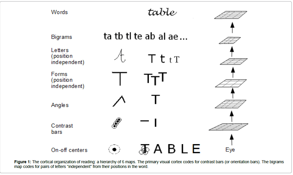
For example, the cognitive ability of reading requires a hierarchy of 6 successive maps. Its neural organization has been modeled by Dehaene in 2005 [36], and others [37-39]. The hierarchy (Figure 1) starts with the oriented edges of the primary visual cortex, up to letters, pairs of letters (bigrams) and words [29,40,41].
Figure 1 about here of the 360 anatomically-identified maps, 80 has already been assigned a functional role [42]. Their role is to code for a specific dimension of reality. It may be a high level representation such as faces, digits, tools you can size, body parts, animals, etc.
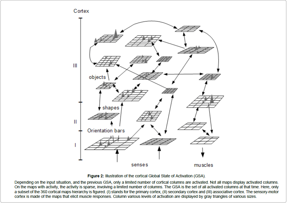
Thus, the cortex is a hierarchy of maps, each one coding for a specific dimension of the situation (or event), and each map being organized respectively to the subject’s personal experience. On a given map, at any time, due to local inhibition between the columns, there is an inter-column competition, each one inhibiting the others, but also being inhibited by them [33]. It follows that, over the whole hierarchy of cortical maps, only a small number of columns are fully activated, depending on the situation experienced at that time. These activated columns form a sparse coding representation of the situation called the global state of activation (GSA, Figure 2) [24].
Memories are traces of experienced events, i.e., traces of GSAs. They allow us to recognize the event which is currently experienced as either identical (or similar) to a situation that has already occurred (i.e., an existing GSA), or as similar in a number of aspects (i.e., a partly similar GSA), or as quite different from everything that had already been experienced during the personal life (i.e., no match with any GSA). If the current event is identical to an already memorized item, then the prediction of what is about to happen is easy. Since the world is continuous, there is a high probability that the next event will be identical to the event that followed the old memorized item [24].
How does the brain retrieve such information? The functioning of cortical maps does preserve the topology of the input’s space. It means that if a map’s input activates a given cortical column, then a similar input will activate a neighbor column, and a very different input will activate a far distant column of the map. Therefore, by activating the neighbor columns as soon as the dimension of an event is identified (by the “winner” cortical column on a map), it is possible to predict what could be the future value of the next event in this dimension. By taking into account all the maps of the cortical hierarchy (i.e., all of the event’s dimensions), the brain automatically makes predictions and acts accordingly [24].
Synaptic Update by LTP and LTD Mechanisms
Long-term potentiation (LTP) intervenes in order to enhance the matching between all of the activated cortical columns (the “winner” ones), strengthening their connections’ efficacy [43]. At the same time, long-term depression (LTD) erodes the connections between all columns that are not activated [44]. This synergy between LTP, LTD and the cortical neural architecture is sufficient to organize the maps’ hierarchy and to achieve precise representations of all experienced situations [24].
TnC’s main statement is that “the brain does not process the information, but only represents the information”. This is best demonstrated by the fact that the successive GSAs do represent the succession of experienced events. However, there is a side effect to the GSA representation of events. Since GSAs are attractors, they dynamically modify the representation of information (i.e., process the information) [33].
Figure 2 about here LTP and LTD not only concern cortical neurons, but all neurons. Therefore, the neurons providing inputs to the cortex, and the neurons receiving outputs from the cortex do also experience synaptic efficiency adjustments depending on their use. GSA must not be seen as restricted to the cortex, but as implicating neurons belonging to all brain structures such as the hippocampus, thalamus, amygdala, etc.
All neural activities of a GSA are coherent, i.e., they are part of an attractor which bends the activities’ dynamics towards the memorized GSA. If the neural activities have not yet reached a GSA, they will evolve toward a coherent network of activities, i.e., a memorized GSA. There is no dichotomy between neurons which belong to high-level cortical maps (and code for abstractions such as ideas) and neurons which belong to other structures, such as the amygdala (an important member of the fear network). The GSA is unique and includes the whole brain activities.
Dangerous situations elicit improved body efficiency in order to fight-or-flight with maximum chances of success. The physiological modifications associated to these (potentially dangerous) situations have received the name of stress [45]. Improved strength and faster processing are all adaptive solutions. In order to get the shortest delay, the current situation must be recognized and categorized as dangerous as quickly as possible. The time delay increases by 10 ms at each implicated cortical step [46]. Therefore, recognition able to offer a result in only one cortical step would be advantageous, even if the recognition was cruder. It is exactly the function of the amygdala.
Amygdala
The amygdala acts as an early warning system able to start fightor- flight behaviors. At the same time, in parallel, the cortex analyses the situation in details, and may (a little later) stop the reflex behavior (in case of false alarm), or complete the already started action (in case of an identified danger). The amygdala may be seen as a “one-level cortex” whose activation by sensory inputs only necessitates 10 ms [46,47].
The two amygdalae are almond-shaped groups of 20 nuclei located deep and medially below the cortex. They are well connected to the sensory system (including the olfactory bulb), and send information to many locations, including the cortex (via the thalamus and hypothalamus) and motor neuron areas [48]. The amygdala’s neural organization is cortical-like. The Amygdala’s principal neurons are large cells that resemble pyramidal neurons [49]. The huge inhibition allowing for a sparse final activation of the amygdalae is provided by GABAergic interneurons which represent about 20% of the neuronal population [50].
The amygdala is a key structure in the acquisition and expression of fear conditioning, and its extinction [46,47]. It has strong connections with the medial prefrontal cortex (mPFC). However, the mPFC does not play a role during fear acquisition but is required for the expression of learnt fear, and the consolidation of extinction memory [48,51]. In addition to the fight-or-flight behavior actions, the amygdalae start a number of other actions targeting the autonomic regulation system [52]. These actions improve physiological capacities, therefore helping the individual to survive the dangerous event. Each one of these “improvements” is deleterious on the long run as in PTSD [53], but may account for avoiding short-term death.
Among the various physiological responses to stress, there is an improved neural memorization [54] – quite useful since it allows a one-time learning (instead of the usual requirement for multiple presentations). The GSA associated to the stressful situation is then automatically reinforced in a manner that is much more efficient than usual. This GSA can prevail, which means that it will easily get recalled, and each recall will generates the same physiological stress answer. The person subjected to such condition is experiencing acute stress disorder or PTSD, and the stressful situation is a “traumatic memory”.
Traumatic Memory
A traumatic memory is part of the episodic memory, and in PTSD patients as compared to controls, it appears to abnormally elicit the amygdalae and the prefrontal cortex [55]. The amygdala (early warning system) has recognized the traumatic event; the latter has also been processed by the cortex. Synaptic LTP guarantees that the memorization of the traumatic event implies neural connections between the amygdalae [56] and the cortex (involving catecholamine and glucocorticoid release) [57,58]). The GSA of the traumatic event includes an activation of the amygdalae additionally to its cortical representation.
Each time the event is recalled; a part of the amygdalae is also activated and starts its automatic stress response inducing what is labeled as a negative emotion. Each recall reinforces the association between the cortex and amygdalae. Not surprisingly, it has been observed that chronic stress exposure physically increases dendritic branching in the basolateral amygdala nuclei (BLA), therefore reinforcing the stress response of the amygdalae [59]. In PTSD, intrusive flashbacks provoke repetitive recalls of the traumatic events which maintain, or even may reinforce the stress response (PTSD). However, the memorization of a traumatic event does not automatically lead to a PTSD. According to the TnC, the traumatic event’s effect will depend upon pre-existing cortical and amygdala connections for similar GSAs. The more a set of GSAs have been reinforced by several traumatic or deleterious events, the more a person could be at risk for developing PTSD.
Making a PTSD patient relive his traumatic event in association with BAS in the EMDR therapy is able to decrease the emotional content of this traumatic event’s memorization and efficiently decrease the PTSD symptoms [1].
Bilateral Alternating Stimulations
BAS are repeated alternating stimuli. They can be auditory broad band beeps, or horizontal eye movements or tactile vibrations [19]. They are perceived by sensory neurons which relay the information to their target neurons, which propagate the information, and so on. Since the practitioner has elicited the patient’s traumatic memory, the current GSA is the one representing the traumatic memory (GSAo). The additional information builds up, and – after a certain amount of time – adds new column activations to GSAo. The new GSA – let’s called it GSA1 – is not any GSA. It is a stable GSA, i.e., one for which the added columns make sense (respectively to the patient’s personal experience). This addition can de facto be verbalized.
BAS activity percolates through the brain, and adds nonspecific activations to the current GSA. These added activations have little chance to correlate with any amygdalae’s activation (the ratio of traumatic memories versus non-traumatic memories is quite small). GSA1 is larger than GSAo, but no new connections with the amygdala are involved. The relative weight of the amygdala compared to the cortical activation is shifting in favor of the cortex. The process (trauma recall and BAS) is repeated several times, and each time new column activities are added to the current GSA. After n repetitions, the current GSA (GSAn) is no more able to specifically elicit the amygdala (the additional columns do not have amygdala connections). The stress response is therefore unable to happen. The patient is not able to experience negative emotions related to his traumatic memory anymore.
The additional columns code for aspects that are new in the context of GSAo. It could be interpreted as the learning of new associations [59,60], or interpreted as memory “reconsolidation” [23].
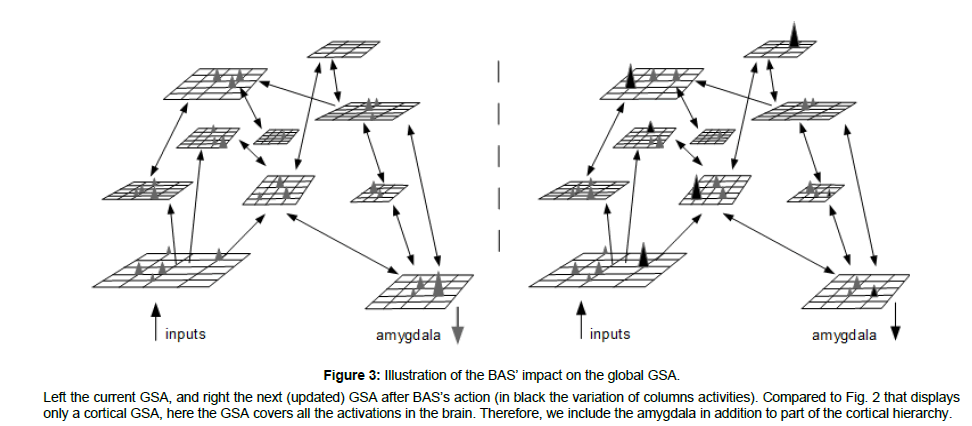
To sum up, an EMDR session must be understood as a recall of the traumatic memory in order to identify GSAo, associated with several BAS sessions whose activities will percolate through the brain, and add non-specific activations to the GSA. Some of these added cortical activities will eventually crystallize in a column’s activation that is able to join the current GSA, making a new GSA, i.e., a stable network of activity. This process (trauma recall and BAS) is repeated several times, and each time, the activity of new columns is being added to the current GSA, until a GSAn totally cleared of its emotional content is obtained. Each GSA is a stable network of activity which gets reinforced thanks to LTP. Each time, a lessened traumatic memory is experienced (Figure 3).
Figure 3: Illustration of the BAS’ impact on the global GSA.
Left the current GSA, and right the next (updated) GSA after BAS’s action (in black the variation of columns activities). Compared to Fig. 2 that displays only a cortical GSA, here the GSA covers all the activations in the brain. Therefore, we include the amygdala in addition to part of the cortical hierarchy.
The New Global State of Activation (Gsan)
After n BAS sessions, the initial GSAo has been replaced by a new larger GSAn. GSAn sends to the amygdalae a pattern of activity that is so different from GSAo that the amygdalae’s activation is strongly lessened. In addition, the prefrontal cortex is more involved in the new GSAn. This is indeed what is found following EMDR therapy in PTSD patients [61-63]. Initially, it starts by an over-activation of the amygdala (GSAo), as demonstrated by increased amygdala functioning when simply using BAS in parallel to negative emotion elicitation [64]. This finally ends with greater prefrontal cortex activity and less amygdala activity. It is important to understand that the series of GSAs experienced by the subject imply a change in stress (i.e., emotion) that may not be a continuous decrease. It may be that a particular GSA evokes more amygdala activity than the previous one. However, on the long run, the GSA activity pattern arriving on the amygdalae is going to be so different from the initial GSAo that it will not activate the amygdalae anymore. The discovery of this GSAn may take some time, but it is quite certain that the EMDR’s BAS will be successful at building it most of the time, in the case of single traumatic events for instance; most of the PTSD patients are symptoms free after an EMDR therapy [65]. The cortical connections of GSAn linking with the amygdalae are now susceptible to LTD. After each recall devoid of a stress response, these connections are depressed. They will finally be erased. Therefore, as soon as the subject experiences his traumatic memory without stress once, stress should not step in anymore. The person is free from the traumatic part of his memory.
Considering our explanatory model, it is important to note that the new GSAn is the old GSAo plus a few elements that the subject considers to be related. The subject has not been “brainwashed”. On the contrary, his memory of the traumatic event is larger, more accurate, and released from the automatic stress response.
EMDR’s BAS action cannot be avoided. Inhibitory mechanisms have not been trained, and are therefore not able to avoid the impact of the BAS. In case of an auditory BAS, each second, some neurons of the auditory cortex are depolarized because of the audio stimuli. They “pass” this information to their target neurons, whose activations may not be immediate, but will add-up until a depolarization occurs that is sent to the target’s target neurons, etc. This explains why even if patients do not believe in the efficiency of EMDR therapy, it works anyway. This cognitive neuronal explanation is line with the efficiency of the EMDR even in mice [27] where BAS are effective in improving fear extinction learning.
Discussion
Based on validated and well-known neuronal mechanisms such as LTP and LTD, the TnC is able to explain how the emotional load can disappear from the memory of the traumatic event in PTSD patients. It is also able to explain why the EMDR therapy for a single trauma can be very short from a few minutes to a few hours [65] depending on the number of steps required to reach the final GSAn. Thus, our explanations can also account for the inter-individual variability in the response to EMDR therapy.
The fast effect to treat traumatic memories that have impeded the patient’ quality of life (sometimes) for years or decades can be explained by LTP and LTD’s learning mechanisms. The stabilization (memorization) of new GSAs is able to explain why it is impossible to step back: as soon as a new learning is made which does not elicit emotional reactions through the amygdalae’s activation, the negative emotion is gone forever. Any recall is just a re-learning. If the patient does remember the event, he reinforces its absence of connection with the amygdalae. LTD occurs and lessens whatever connections with the amygdala still exist. This is in line with the long-time effect of the EMDR therapy in PTSD (at least 35 months) [22].
Our explanations of EMDR therapy’s underlying mechanisms are in accordance with previous studies showing a shift in PTSD patients’ brain activity after EMDR therapy, from limbic regions including the amygdalae to frontal regions according to both EEG and BOLD signals [61,62]. Neuroanatomical experiments also showed increased gray matter density in the prefrontal cortex after a decrease in symptoms following EMDR therapy [63]. All these results could corroborate the idea of a new GSAn emergence after the EMDR therapy sustaining an enhanced prefrontal cortex activity and decreased amygdalae activities. TnC is therefore able to explain the emotional load decrease in traumatic event memory after EMDR therapy and the inter-individual variability response to this therapy.
In addition, TnC can also outline why BAS are more efficient than bilateral non-alternated stimulations or unilateral ones [19,27]. Bilateral stimuli have a largest recruitment area compared to unilateral stimuli. The ability of the stimulations to recruit a large area depends upon predictions at map level that elicit inhibition processes [24]. Alternation and intermittency are discontinuities that do not favor predictions. Since predictions authorize inhibition, less predictable BAS are more efficient than bilateral non alternated or bilateral non intermittent stimuli.
BAS’s nature was initially visual with patients performing horizontal saccadic eye movements following a visual target [1, 21]. However, BAS can also be efficient using tactile or auditory stimulations. Indeed, in these three cases, neuronal activation spread across the brain forcing the emergence of new neuronal associations and therefore of new GSAs. The important quality that BAS must respect is to be repetitive in order to build-up neural activations and discontinuous in order to avoid inhibition.
In addition to these evidences, future experiments should further confirm our explanations by continuously monitoring EMDR sessions using functional Magnetic Resonance Imagery recordings (fMRI) to show how the BAS induced activities percolate through the cortex, slowly building-up activations on cortical maps until – from time to time – this activity resumes into a local activation of a cortical column. Immediate questioning of the patient may help label this columnar activity as related to a particular dimension of the souvenir.
Given that mice proved to be a good model for EMDR therapy [27], experiments could be led in mice with localized intracranial electrodes recording within the amygdalae and the prefrontal cortex. This technique will benefit from both high spatial and temporal resolutions in order to follow the emotional desensitization accompanying the BAS.
During EMDR therapy, patients frequently describe that the target image (traumatic event) disappears or becomes distant throughout the successive BAS. Depending on the GSAs experienced by the patient, in particular if the ratio of “old” (GSAo) versus “new” associations (GSAn / GSAo) is small, it may be that the new GSAn does not allow the clear visualization of the target image anymore.
During the therapy, old traumatic memories often rush back, and again our explanations account for that since the wide spread activation for cortical columns by BAS may bring back memories that are related with the traumatic one.
Regarding the AIP model [21], our explanations agree with the observations that traumatic events are stored with their original emotions, physical sensations and beliefs, and thereafter are reconsolidated [23,1]. Our explanations target the underlying neural mechanisms of reconsolidation and claim that this reconsolidation is both learning of new associations, and forgetting of old ones.
Conclusion
A better understanding of the cortex’s functioning (LTP, LTD), cortex function (i.e., the representation of the events in order to predict what will happen next) and amygdalae function (early warning system) allows explaining PTSD. At the same time, knowing the inter-relations between the cortex and the amygdalae, and based on the TnC framework, we develop the hypothesis that EMDR BAS forces further learning (re-learning) of the traumatic event, in which the amygdalae are not implicated anymore. These modifications could induce a shift from the amygdalae’s involvement in the traumatic memory to a more cognitive representation of the traumatic event without the strong negative feeling previously associated. Our explanation proposal still awaits further proof, but it is able to account for many aspects of the EMDR therapy, including results of EMDR therapy imaging study.
References
- Foa EB, Keane TM, Friedman MJ, and Cohen JA (2009) Effective Treatments for PTSD: Practice Guidelines of the International Society for Traumatic Stress Studies. New York, NY: Guilford Press.
- Shapiro F (2014) The role of eye movement desensitization and reprocessing (EMDR) therapy in medicine: addressing the psychological and physical symptoms stemming from adverse life experiences. The Permanente Journal 18: 71-77.
- World Health Organization [WHO] (2013) Guidelines for the Management of Conditions Specifically Related to Stress. Geneva: World Health Organization.
- American Psychiatric Association [APA] (2004) Practice guideline for the treatment of patients with acute stress disorder and posttraumatic stress disorder. Am J Psychiatry 161(Suppl. 11): 3-31.
- de Vries GJ, Olff M (2009) The lifetime prevalence of traumatic events and posttraumatic stress disorder in the Netherlands. J Trauma Stress 22: 259-67.
- Alonso J, Angermeyer MC, Bernert S, Bruffaerts R, Brugha TS, et al. (2004) Prevalence of mental disorders in Europe: results from the European Study of the Epidemiology of Mental Disorders (ESEMeD) project. Acta psychiatry Scand Suppl 2004: 21-27.
- American Psychiatric Association [APA] (2013) Diagnostic and Statistical Manual of Mental Disorders, 5th Edn. Washington, DC: American Psychiatric Association.
- de Vries GJ, Olff M (2009) The lifetime prevalence of traumatic events and posttraumatic stress disorder in the Netherlands. J Trauma Stress 22: 259-67.
- Alonso J, Angermeyer MC, Bernert S, Bruffaerts R, Brugha TS, et al. (2004) Prevalence of mental disorders in Europe: results from the European Study of the Epidemiology of Mental Disorders (ESEMeD) project. Acta psychiatry Scand Suppl 2004: 21-27.
- Kessler RC, McGonagle KA, Zhao S, Nelson CB, Hughes M, Eshleman S, Wittchen HU, Kendler KS (1994) Lifetime and 12-month prevalence of DSM-III-R psychiatric disorders in the United States. Results from the National Comorbidity Survey. Arch gen psychiatry 51: 8-19.
- Jolly A (2003) Epidémiologie des PTSD. Int J Victimology 2.
- Magruder K, Serpi T, Kimerling R, Kilbourne AM, Collins JF, et al. (2015) Prevalence of Posttraumatic Stress Disorder in Vietnam-Era Women Veterans: The Health of Vietnam-Era Women's Study (HealthVIEWS). JAMA Psychiatry 72: 1127-34.
- Hoge CW, Riviere LA, Wilk JE, Herrell RK, Weathers FW (2014) The prevalence of post-traumatic stress disorder (PTSD) in US combat soldiers: a head-to-head comparison of DSM-5 versus DSM-IV-TR symptom criteria with the PTSD checklist. Lancet Psychiatry 1: 269-77.
- Ginzburg K, Ein-Dor T, Solomon Z (2010) Comorbidity of posttraumatic stress disorder, anxiety and depression: a 20-year longitudinal study of war veterans. J Affect Disord 123: 249-257.
- Etkin A, Wager TD (2007) Functional neuroimaging of anxiety: a meta-analysis of emotional processing in PTSD, social anxiety disorder, and specific phobia. Am J Psychiatry 164: 1476-1488.
- Patel R, Spreng RN, Shin LM, Girard TA (2012) Neurocircuitry models of posttraumatic stress disorder and beyond: a meta-analysis of functional neuroimaging studies. Neurosci Biobehav Rev 36: 2130-2142.
- Sailer U, Robinson S, Fischmeister FPS, König D, Oppenauer C, et al. (2008) Altered reward processing in the nucleus accumbens and mesial prefrontal cortex of patients with posttraumatic stress disorder. Neuropsychologia 46: 2836-2844.
- Felmingham K L, Falconer E M, Williams, L, Kemp A H, Allen A, Peduto A, & Bryant R A (2014) Reduced amygdala and ventral striatal activity to happy faces in PTSD is associated with emotional numbing. PloS One 9: e103653.
- Bluhm RL, Williamson PC, Osuch EA, Frewen PA, Stevens TK et al. (2009) Alterations in default network connectivity in posttraumatic stress disorder related to early-life trauma. JPN 34: 187-94.
- Sripada RK, King AP, Garfinkel SN, Wang X, Sripada CS, et al. (2012) Altered resting-state amygdala functional connectivity in men with posttraumatic stress disorder. JPN 37: 241-249.
- Servan-Schreiber D, Schooler J, Dew MA, Carter C, Bartone P (2006) Eye movement desensitization and reprocessing for posttraumatic stress disorder: a pilot blinded, randomized study of stimulation type. Psychother and psychosom 75: 290-297.
- Shapiro F (1996) Eye movement desensitization and reprocessing (EMDR): evaluation of controlled PTSD research. J behav ther exp psychiatry 27: 209-218.
- Shapiro F (2001) Eye movement desensitization and reprocessing (EMDR): basic principles, protocols and procedures. 2nd ed. New York, NY: The Guilford Press.
- Högberg G, Pagani M, Sundin O, Soares J, Aberg-Wistedt A, et al. (2008) Treatment of post-traumatic stress disorder with eye movement desensitization and reprocessing: outcome is stable in 35-month follow-up. Psychiatry Res 159: 101-108.
- Solomon R M, Shapiro F (2008) EMDR and the adaptive information processing model: potential mechanisms of change. J EMDR Pract Res 2: 315-325.
- Touzet C (2015) The Theory of neural Cognition applied to Robotics. Int J Adv Robot Syst 12: 74.
- Touzet, C (2014) Hypnose, sommeil, placebo? Les réponses de la Théorie neuronale de la Cognition – Volume 2.
- Touzet C (2010) Conscience, intelligence, libre-arbitre ? Les réponses de la Théorie neuronale de la Cognition – Volume 1.
- Wurtz H, El-Khoury-Malhame M, Wilhelm F H H, Michael T, Beetz E M, et al. (2016) Preventing long-lasting fear recovery using bilateral alternating sensory stimulation: a translational study. Neurosci 321: 222-235.
- Mountcastle VB (1997) The columnar organization of the neocortex, Brain 120: 701-722.
- Hubel D (1995) Eye, Brain, and Vision. Freeman.
- Touzet C (2013) Why neurons are not the right level of abstraction for implementing cognition (edtn.) Biologically Inspired Cognitive Architectures 2012: Proceedings of the Third Annual meeting of the BICA Society, Advances in Intelligent Systems and Computing 196, Springer 317-318.
- Herculano-Houzel S (2009) The human brain in numbers: a linearly scaled-up primate brain, Front hum neurosci 3: 31.
- Glasser M F, Coalson, T, Robinson E, Hacker C, Harwell J, et al. (2016) A Multi-modal parcellation of human cerebral cortex. Nature 536: 171-178.
- Kohonen T (2001) Self-Organizing Maps, (3rd edtn.) Springer Series in Information Sciences.
- Penfield W, Rasmussen T (1950) The cerebral cortex of man. Macmillan.
- Yeo BT, Krienen FM, Sepulcre J, Sabuncu MR, Lashkari D, Hollinshead M, et al. (2011) The organization of the human cerebral cortex estimated by intrinsic functional connectivity. J Neurophysiol 106: 1125-1165.
- Dehaene S, Cohen L, Sigman M, Vinckier F (2005) The neural code for written words: A proposal. Tr Cogn Sci 9: 335-341.
- Dufau S, Lété B, Touzet C, Glotin H, Ziegler J et al. (2010) Developmental Perspective on Visual Word Recognition: New Evidence and a Self-Organizing Model. Eur J Cogn Psychol 22: 669-694.
- Whitney C, Bertrand D, Grainger J (2012) On coding the position of letters in words: A test of two models. Exp Psychol 59: 109-114.
- Touzet C, Kermorvant C, Glotin H (2014) A Biologically Plausible SOM Representation of the Orthographic Form of 50,000 French Words. (Edtn.) Advances in Self-Organizing Maps and Learning Vector Quantization, Springer AISC 295: 303-312.
- Vinckier F, Qiao E, Pallier C, Dehaene S & Cohen L (2011) The impact of letter spacing on reading: A test of the bigram coding hypothesis. J Vis 11: 8-8.
- Grainger J, Hannagan T (2014) What is special about orthographic processing? Wr Lang Liter 17: 225-252.
- Ledoux J (1998) The emotional brain, Weidenfeld & Nicolson.
- Silver M, Kastner S (2009) Topographic maps in human frontal and parietal cortex. Tr Cogn Sci 13: 488-495.
- Frégnac Y, Pananceau M, René A, Huguet N, Marre O, et al (2010) A re-examination of Hebbian-covariance rules and spike timing-dependent plasticity in cat visual cortex in vivo. Spike-timing dependent plasticity. Front Synaptic Neurosci 2: 147.
- Selye H (1998) A syndrome produced by diverse nocuous agents. Nature 138: 32.
- Malenka R C & Bear M F (2004) LTP and LTD: an embarrassment of riches. Neuron 44: 5-21.
- Quirk GJ, Repa C, LeDoux JE (1995) Fear conditioning enhances short-latency auditory responses of lateral amygdala neurons: parallel recordings in the freely behaving rat. Neuron 15: 1029-39.
- Marek R, Strobel C, Bredy TW, Sah P (2013) The amygdala and medial prefrontal cortex: partners in the fear circuit. J physiol 591: 2381-2391.
- Bedi US, Arora R (2007) Cardiovascular manifestations of posttraumatic stress disorder. J Natl Med Assoc 99: 642-649.
- Faber ESL, Callister RJ & Sah P (2001) Morphological and electrophysiological properties of principal neurons in the rat lateral amygdala in vitro. J Neurophysiol 85: 714-723.
- Spampanato J, Polepalli J & Sah P (2011) Interneurons in the basolateral amygdala. Neuropharmacology 60: 765-773.
- Quirk GJ, Garcia R, González-Lima F (2006) Prefrontal mechanisms in extinction of conditioned fear. Biol Psychiatry 60: 337-43.
- Ulrich-Lai Y M, Herman J P (2009) Neural Regulation of Endocrine and Autonomic Stress Responses. Nat Rev Neurosci 10: 397-409.
- McEwen B S (2007) Physiology and neurobiology of stress and adaptation: central role of the brain. Physiol rev 87: 873-904.
- Shin LM, Orr SP, Carson MA, Rauch SL, Macklin ML, et al. (2004) Regional cerebral blood flow in the amygdala and medial prefrontal cortex during traumatic imagery in male and female Vietnam veterans with PTSD. Arch Gen Psychiatry 61: 168-76.
- Maren S (1999) Long-term potentiation in the amygdala: a mechanism for emotional learning and memory. Tr neurosci 22: 561-567.
- Sigurdsson T, Doyère V, Cain C K, LeDoux J E (2007) Long-term potentiation in the amygdala: a cellular mechanism of fear learning and memory. Neuropharmacology 52: 215-227.
- Xiong H & Krugers H J (2015) Tuning hippocampal synapses by stress-hormones: relevance for emotional memory formation. Brain res 1621: 114-120.
- Vyas A, Mitra R, Shankaranarayana Rao BS, Chattarji S (2002) Chronic stress induces contrasting patterns of dendritic remodeling in hippocampal and amygdaloid neurons. J Neurosci 22: 6810–6818.
- Bevilaqua LR, Medina JH, Izquierdo I, Cammarota M (2008) Reconsolidation and the fate of consolidated memories. Neurotox Res 14: 353-358.
 Spanish
  Chinese
  Russian
  German
  French
  Japanese
  Portuguese
  Hindi
Hindi 


