Research Article, J Regen Med Vol: 11 Issue: 6
Evaluation of 18 Protocols Using Single and Double Spin Techniques for the Production of Platelet-Rich Plasma
Carlos De la Hoz1, Philippe Wuilleumier1, Amy Riumbau1, Carla Cerra2
1Regenerative Medicine, The Neomedicine Institute, Doral, FL 33172, USA
2Biological Sciences, Florida International University, Miami, FL 33199, USA
*Corresponding Author: Carlos De la Hoz
Regenerative Medicine, The Neomedicine Institute Doral
FL 33172, USA
Tel: 7866789590
E-mail: delahoz. carlos@gmail.com
Received: 07-Oct-2022, Manuscript No. JRGM-22-76747;
Editor assigned: 11-Oct-2022, PreQC No. JRGM-22-76747 (PQ);
Reviewed: 27-Oct-2022, QC No. JRGM-22-76747;
Revised: 01-Nov-2022, Manuscript No. JRGM-22-76747 (R);
Published: 07-Nov-2022, DOI:10.4172/2325-9620.1000230
Citation: Riumbau A, Wuilleumier P, De la Hoz C, Cerra C (2022) Evaluation of 18 Protocols Using Single and Double Spin Techniques for the Production of Platelet-Rich Plasma. J Regen Med 11:6.
Abstract
Platelet-Rich plasma (PRP) is an autologous biologic product derived from the centrifugation of whole blood that produces a liquid fraction containing platelet concentrate above its baseline along with multiple growth factors. PRP has been used in different musculoskeletal pathologies promoting angiogenesis, tissue regeneration and repair. In order to inject good quality of PRP, it is important to evaluate and understand how different protocols affect the collection and production of good quality PRP. This study demonstrates and compares different existing protocols and their results regarding pure platelet rich plasma concentration. After performing 18 different protocols with different centrifugal force and times, it was found that the single spin technique of 600g for 5 minutes had the greatest concentration of platelets with a concentration factor of 2.64 greater than the baseline. The double spin technique showed to concentrate the platelets even more than the single spin technique. The double spin technique with the protocol of 600g for 5 minutes showed a concentration factor of 5.76 greater than the baseline. This study offers a guide for the production and collection best concentrated PRP.
Keywords: Platelet-Rich plasma; Concentration; Single spin technique; Double spin technique.
Keywords
Platelet-Rich plasma; Concentration; Single spin technique; Double spin technique.
Introductıon
Platelet-rich plasma (PRP) is a processed liquid fraction derived from centrifuged blood containing platelet concentrate above its baseline [1]. PRP is an autologous biologic product that has been used for numerous indications in the past years, particularly in regenerative medicine and sports medicine fields. Different studies have shown the positive outcomes that PRP has on different musculoskeletal conditions such as tendons, muscles, and cartilage rupture and degeneration. These results are a result of the properties that PRP has on tissue healing and repair. PRP contains alpha granules, dense granules, and lysosomes in addition to growth factors and fibrin. The combination of such factors stimulates cell proliferation and migration, synthesis, and deposition of extracellular matrix components all of which promote angiogenesis, tissue regeneration, and repair [2,3]. The clinical use of PRP therapies in regenerative medicine has gained increasing popularity as they have proven effective in initiating tissue repair via the release of previously mentioned growth factors. Such growth factors are listed in Table 1. Therefore, through the release of growth stimulants, PRP therapy can accelerate the healing process of chronic and acute injuries.
| Name of Growth Factors | Functions |
|---|---|
| Transforming growth factors beta- 1; TGFB1 | Proliferation, differentiation, in many cell types |
| Platelet-derived growth factor, alpha polypeptide; PDGFA | Potent mitogen for connective tissue cells |
| Platelet-derived growth factor, beta polypeptide; PDGFB | Promotes cellular proliferation, inhibits apoptosis |
| Platelet-derived growth factor C; PDGFC | Increase in the motility of different cells (mesenchymal cells, fibroblasts, smooth muscle cells, capillary endothelial cells, neurons) |
| Platelet-derived growth factor D; PDGFD | Plays a role in developmental and physiological processes and in a disease like cancer, fibrotic diseases, and arteriosclerosis |
| Insulin-like growth factor I; IGF1 | Mediates growth-promoting effects of growth hormone |
| Fibroblast growth factor I; FGF1 | Induces liver gene expression, angiogenesis, and fibroblast proliferation |
| Epidermal growth factor; EGF | Induces differentiation of specific cells, act as a potent mitogenic factor for a variety of cultured cells |
| Vascular endothelial growth factor A; VEGFA | Primarily a mitogen for vascular endothelial cells induces angiogenesis |
| Vascular endothelial growth factor B; VEGFB | Regulator of blood vessel physiology, a role in endothelial targeting of lipids to peripheral tissues |
| Vascular endothelial growth factor C; VEGFC | Angiogenesis and endothelial cell growth, and also affect the permeability of blood vessels. |
Table 1. List of growth factors present in platelet-rich plasma (PRP) along with their respective functions [11].
As stated above, PRP has been used in multiple conditions, in the case of knee osteoarthritis different studies have shown statistically significant benefits. When PRP is injected intraosseous, growth factors get access to this area and to the deep layers of the cartilage which is otherwise non-accessible in intra-articular infiltration. This stimulates the synthesis of hyaluronic acid and lubricin by synoviocytes and chondrocytes. Meanwhile, it also helps in homing and chondrogenic differentiation of mesenchymal stem cells of subchondral bone and synovial fluids. It also suppresses NF-KB pathway activation [2]. However, a lack of consistency in the reporting and usage of PRP preparation protocols has made it difficult to compare PRP therapy outcomes and procedures [1]. Due to such inconsistencies, more research must be done to investigate the different PRP protocols to create a consensus for optimal PRP preparation and therapeutic procedures. There are different factors to consider at the time of testing different protocols to obtain PRP. One of them is the type of centrifugation, either horizontal centrifugation or fixed-angle centrifugation. It was reported that in horizontal centrifugation the PRP was superior at accumulating platelets and leukocytes when compared to the results from standard fixed-angle centrifuges [4]. This also relies on the fact that one of the advantages of a horizontal centrifuge is that it separates layers based on density. A horizontal centrifuge tube produced from a swing-out bucket allows for a better differential between the minimum and maximum radius found within a centrifugation tube [5]. This increases the ability to separate cells based on the disparities between the RCF-min and RCF-max. Additionally, fixed-angle centrifugation results in more trauma to the cells because it pushes cells outward and downward to the wall of the tube. Therefore, to avoid cell trauma separation of layers is always observed in an angulated fashion. Moreover, horizontal centrifugation also allows cells to separate and migrate freely throughout the blood layers. Based on this data, this study was designed to evaluate all 18 protocols by using horizontal centrifugation. Other factors that affect the processing and obtention of PRP are volume of the sample of whole blood, time period of centrifugation, range of centrifugal acceleration, type of material of the centrifuged tubes, different size of platelets, and hematocrit variability among others.
A study done on the effects test tube material has on obtaining PRF concentration growth factors concluded that plastic tubes are better at platelet distribution than glass tubing. The results of the study demonstrated that with increased centrifugal speed, the PRF matrix stuck onto one surface region of the glass test tubes while in plastic test tubes the PRF matrix remained uniform throughout the sample [6]. Based on the results of this study, plastic test tubes were used to ensure the uniform distribution of PRP in testing the various protocols.
PRP is obtained by centrifugation varying the relative centrifugal force, temperature, and time. It also could be obtained by single or double spin centrifugation. There are numerous protocols in current literature describing optimal conditions of centrifugation. As previously mentioned there are many factors affecting this process, therefore all of them must be addressed first in order to get more realistic and reproducible results. In one study in which 22 healthy male and female volunteer donors participated, 4.5 mL of whole blood was obtained and mixed with 0.5 mL of citrate solution. Blood samples were centrifuged at a range of 240-400 g, ranging from 8-19 minutes. The best performance was obtained by using the parameters of 300 g for 5 minutes at 12 °C and 240 g for 8 minutes at 16°C for 1 single spin [7]. Another study determined a centrifugal acceleration of 3731 g for 4 minutes was the optimal condition for obtaining the highest platelet concentration from 478 ml of whole blood [8]. Another protocol was determined by a study that showed PRP samples that concentrated platelets at 3.2 times the concentration of the whole blood baseline. The centrifugation consisted of 5 mL of whole blood for two spins at 200 g for 10 minutes per spin [9].
Another study conducted by [10,11] consisted of a one-step centrifugation for the collection of PRP directly above the erythrocyte layer. The sample was collected in sterile tubes containing 4.5 mL of whole blood and 3.8% of trisodium citrate 0.5 mL. The sample was centrifuged at 460 g for 8 minutes. This protocol was the most efficient in concentrating platelets by a factor of 2.67 above the baseline value. Considering all these different studies suggesting different protocols, this case study produced a study in which the best protocols were evaluated to determine which one best concentrated platelets. All protocols were performed with horizontal centrifugation to avoid cell trauma seen in fixed-angle centrifugation. In addition, all PRP samples were taken from the exact same layer for all protocols by the same physician to limit variations among protocols.
Materials and Methods
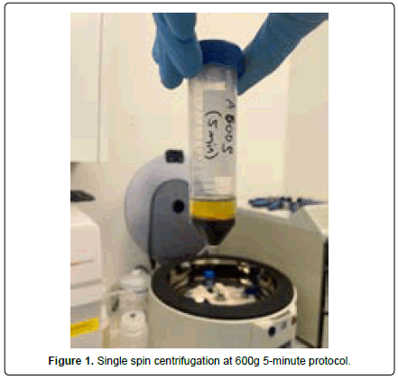
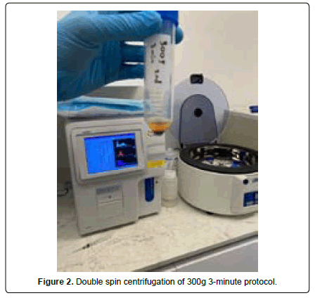
A total of 18 protocols were conducted: 300g, 500g, 600g, 700g, 800g, and 900g each at 3 minutes, 5 minutes, and 8 minutes respectively. Three volunteers were used to obtain the blood samples, each donating a total of 60cc of whole blood (WB) for a total of 180cc. One of the donors donated an extra 40cc of WB for the double spin technique. Each of the 60cc of WB was combined with 12 ccs of anticoagulant and then divided into 10cc samples and randomly assigned to a protocol. In order to limit variability, each of the three volunteers were randomly assigned to 2 centrifugal force protocols at 3 minutes, 5 minutes, and 8 minutes respectively. For example, volunteer A was assigned to protocols 300g and 500g each at 3 minutes, 5 minutes, and 8 minutes. To create a baseline for comparing the PRP concentration after the tested protocol, the concentration of each volunteer’s platelets was measured beforehand. After recording the baselines, 10 ccs of blood were drawn for each protocol and centrifuged horizontally in a plastic tube to allow for better cell separation. After each sample was centrifuged, the buffy coat and a little quantity exactly below and above the buffy coat was extracted using a syringe and dispensed into another plastic tube for further analysis. The buffy coat and the exact layers above and below contain the highest concentration of platelets of the layers of centrifuged blood: plasma, buffy coat, and erythrocytes. After obtaining the mentioned sample, the platelet concentration was measured using a CBC machine. After all protocols were analyzed, the four protocols that created the highest concentration of PRP were reassessed with a double-spin technique using the same protocol. For the double spin, Donor A provided an additional 40 ccs of WB for the four different selected protocols to undergo a double spin analysis. The double spin centrifugation consisted of collecting the poor platelet rich plasma and the buffy coat and transferring it to another sterile plastic tube. After that manual separation, the sample containing the buffy coat was centrifuged for a second time in order to concentrate the platelets in a second round creating a platelet pellet. Those platelet pellets were further analyzed in the CBC machine to determine the concentration of platelets (Figure 1 and 2).
Results and Discussion
Current studies suggest different protocols and lack an overall consensus on standardizing a particular group of protocols for the obtention of PRP. In this study, several protocols were evaluated and compared to determine which were most successful in concentrating platelets at a greater concentration. A study performed by [4] analyzed the total volume of plasma along with the total yield and concentration of platelets and leukocytes of various protocols. The results demonstrated that the best yield of leukocytes was achieved after centrifugation for 8 min utilizing 700g, 1000g, and 1200g protocols. From those, the highest concentration was achieved at 700g for 8 min. They determined that the protocol for best-distributing platelets and leukocytes within the upper 3-5 ml plasma layer was at 700g for 8 min. Meanwhile, the protocol which demonstrated the highest concentration of platelets and leukocytes was observed at 200g for 5 min. Noting that while the total yield of platelets and leukocytes was lower in the 200g for 5 min protocol when compared to 700g for 8 min, the overall concentration was higher. In 2014 lower centrifugation speeds were proposed as a method to achieve a better accumulation of growth factors and cells within the upper plasma layers by modifying the protocols from 700g down to 200g [12]. Lowering the centrifugation speed showed an increase of approximately 20% in platelet concentration. In this case study, the current protocols were evaluated to determine which specific protocols concentrated the greatest number of platelets in the buffy coat layer and layers immediately under and above it. This study allowed for a better understanding of how different centrifugation forces and speeds affect platelet concentration. Note that every protocol was performed under the same circumstances, analyzing and extracting the samples from the same exact location for every single protocol.
According to the results of the single spin seen in Table 2, using a centrifugal force greater than 800g does not significantly concentrate platelets. As a matter of fact, at 900g the concentration of platelets decreased in comparison to the baseline. Such protocols seem to be too aggressive to adequately distribute platelets along layers. The centrifugal force that showed the most consistent increased concentration of platelets regardless of the speed time was at 600 g. The four protocols that had the best outcomes concentrating platelets were 600g at 5 min, 500g at 3 min, 300g at 3 min and 600g at 8 min. As seen in Table 3, these four protocols with the highest effectiveness concentrating platelets were reevaluated for a double spin. The double spin technique allows for the concentration of platelets at the bottom of the tube to form into platelet pellets. The most successful protocol in concentrating platelets after a double spin centrifugation was 600g for 5 min. The protocol concentrated the platelets with a concentration factor of 5.76 greater than the baseline. Taking into consideration that the concentration of platelets is dependent on various factors, the replication of this study may result in variability among the results due to different resources. Therefore, this case study functions as a guide for successful protocols proven in our particular institution that should be further tested according to different resources and materials.
| Centrifugal Force (RCF) | Baseline PLT (uL) | Time (min) | PLT (uL) | Concentration Factor | |
|---|---|---|---|---|---|
| Donor A | 300 g | 215,000 | 3 min | 504,000 | Increased 2.34 |
| 215,000 | 5 min | 317,000 | Increased 1.47 | ||
| 215,000 | 8 min | 366,000 | Increased 1.70 | ||
| Donor B | 500 g | 332,000 | 3 min | 873,000 | Increased 2.62 |
| 332,000 | 5 min | 425,000 | Increased 1.28 | ||
| 332,000 | 8 min | 309,000 | Decreased 0.93 | ||
| Donor B | 600 g | 280,000 | 3 min | 625,000 | Increased 2.32 |
| 280,000 | 5 min | 740,000 | Increased 2.64 | ||
| 280,000 | 8 min | 652,000 | Increased 2.34 | ||
| Donor A | 700 g | 273,000 | 3 min | 623,000 | Increased 2.28 |
| 273,000 | 5 min | 307,000 | Increased 1.12 | ||
| 273,000 | 8 min | 377,000 | Increased 1.38 | ||
| Donor C | 800 g | 356,000 | 3 min | 710,000 | Increased 1.99 |
| 356,000 | 5 min | 429,000 | Increased 1.21 | ||
| 356,000 | 8 min | 362,000 | Increased 1.02 | ||
| Donor C | 900 g | 356,000 | 3 min | 196,000 | Decreased 1.82 |
| 356,000 | 5 min | 273,000 | Decreased 1.30 | ||
| 356,000 | 8 min | 103,000 | Decreased 3.46 |
Table 2.Results of the single spin of 18 protocols at different centrifugal forces and times.
| Donor A | Baseline PLT (uL) |
Protocol | Double Spin | Concentration Factor |
|---|---|---|---|---|
| 257,000 | 600g for 5min | 1,482,000 | 5.76 | |
| 257,000 | 500g for 3 min | 1,339,000 | 5.21 | |
| 257,000 | 300g for 3 min | 951,000 | 3.70 | |
| 257,000 | 600g for 8min | 317,000 | 1.23 |
Table 3. Results of the reevaluation of the best protocols in single spin centrifugation having undergone a double spin centrifugation.
Conclusion
This study showed that the centrifugal force of 600g showed the highest and most consistent concentration of platelets regardless of time. The single spin protocol of 600g at 5 min was the most effective protocol concentrating platelets with a concentration factor of 2.64 times greater than the baseline. As the results showed, centrifugal forces greater than 800g are not ideal for concentrating platelets. Such results could be due to the inability high centrifugal force have in adequately distributing platelets among the upper PRP layers. Regarding the double spin technique, it showed an overall greater concentration factor with respect to the single spin technique. The double spin technique with the protocol of 600g for 5 min allowed for the highest concentration factor of platelets overall. This study is an essential guide in determining the most efficient protocol in concentrating platelets for procedures such as PRP injections.
References
- Everts P, Onishi K, Jayaram P, Lana JF, Mautner K (2020) Platelet-rich plasma: New performance understandings and therapeutic considerations in 2020. Int J Mol Sci, 21(20):7794.
- Milano G, Sánchez M, Jo CH, Saccomanno MF, Thampatty BP, et al. (2019) Platelet-rich plasma in orthopaedic sports medicine: state of the art . J ISAKOS, 4(4): 188-95.
- Reed GL (2004) Platelet secretory mechanisms. Semin Thromb Hemost, 30(4):441-50.
- Miron RJ, Chai J, Zheng S, Feng M, Sculean A, et al. (2019) A novel method for evaluating and quantifying cell types in platelet-rich fibrin and an introduction to horizontal centrifugation. J Biomed Mater Res A, 107(10):2257-71.
- Lourenço ES, Mourão CFAB, Leite PEC, Granjeiro JM, Calasans-Maia MD, et al. (2018) The in vitro release of cytokines and growth factors from fibrin membranes produced through horizontal centrifugation. J Biomed Mater Res A, 106(5):1373-80.
- Tsujino T, Masuki H, Nakamura M, Isobe K, Kawabata H, et al. (2019) Striking differences in platelet distribution between advanced-platelet-rich fibrin and concentrated growth factors: Effects of silica-containing plastic tubes. J Funct Biomater, 10(3):43.
- Amable PR, Carias RB, Teixeira MV, da Cruz Pacheco I, Corrêa do Amaral RJ, et al. (2013) Platelet-rich plasma preparation for regenerative medicine: Optimization and quantification of cytokines and growth factors. Stem Cell Res Ther, 4(3):67.
- Kahn RA, Cossette I, Friedman LI (1976) Optimum centrifugation conditions for the preparation of platelet and plasma products. Transfusion, 16:162-5.
- Landesberg R, Roy M, Glickman RS (2000) Quantification of growth factor levels using a simplified method of platelet-rich plasma gel preparation. J Oral Maxillofac Surg, 58:297-300.
- Anitua E, Aguirre JJ, Algorta J, Ayerdi E, Cabezas AI, et al. (2008) Effectiveness of autologous preparation rich in growth factors for the treatment of chronic cutaneous ulcers. J Biomed Mater Res B Appl Biomater, 84:415-21.
- Trebinjac S, Nair MK (2020) Regenerative Injections in Sports Medicine.
- Ghanaati S, Booms P, Orlowska A, Kubesch A, Lorenz J, et al. (2014) Advanced platelet‐rich fibrin: a new concept for cell‐based tissue engineering by means of inflammatory cells. J Oral Implantol, 40(6):679-89.
Indexed at, Google Scholar, Cross Ref
Indexed at, Google Scholar, Cross Ref
Indexed at, Google Scholar, Cross Ref
Indexed at, Google Scholar, Cross Ref
Indexed at, Google Scholar, Cross Ref
Indexed at, Google Scholar, Cross Ref
Indexed at, Google Scholar, Cross Ref
Indexed at, Google Scholar, Cross Ref
Indexed at, Google Scholar, Cross Ref
 Spanish
  Chinese
  Russian
  German
  French
  Japanese
  Portuguese
  Hindi
Hindi 

