Research Article, Arch Med Biotechnol Vol: 3 Issue: 3
Evaluation of an Intragastric Model for Clinically Isolated Pathogenic Shigella Species-Induced Diarrhea in Albino Wistar Rats
Ashajyothi C1*, Prabhurajeshwar C2, Navya HM2, and Kelmani Chandrakanth R3
1Department of Biotechnology, Vijayanagara Sri Krishnadevaraya University, Ballari, Karnataka, India
2Department of Biotechnology, Davangere University, Davangere, Karnataka, India
3Department of Biotechnology, Gulbarga University, Gulbarga, India
*Corresponding Author: Ashajyothi C, Department of Biotechnology, Vijayanagara Sri Krishnadevaraya University, Ballari, Karnataka, India, E-mail: ashajyothi1988@gmail.com
Received date: 21 December, 2021, Manuscript No. AMB-21-50325;Editor assigned date: 23 December, 2021, PreQC No. AMB-21-50325 (PQ); Reviewed date: 07 January, 2022, QC No. AMB-21-50325; Revised date: 21 February, 2022, Manuscript No. AMB-21-50325 (R); Published date: 02 March, 2022, DOI:10.4172/amb.1000027
Citation: Ashajyothi C, Prabhurajeshwar C, Navya HM, Kelmani Chandrakanth R (2022) Evaluation of an Intragastric Model for Clinically Isolated Pathogenic Shigella Species-Induced Diarrhea in Albino Wistar Rats. Arch Med Biotechnol 3:3.
Abstract
Shigella is a main reason of dysentery all through the arena and is liable for 5-10 percentage of diarrheal contamination in many areas and also constitutes one of the developing numbers of antimicrobial-resistance micro-organism in growing countries. Diarrhea is a condition where there is stomach pain, fever, vomiting and excessive passage of very watery stools in initial stages are common. However, in serious cases, there can be passage of blood and mucus-streaked stools as well. For the pre-clinical evaluation of pathogenesis, screening the therapeutics and evaluating vaccines, wistar rats and different primates are the handiest animals which can be evidently vulnerable to shigellosis. In this study, wistar rats were divided into various groups and through oral and intragastrical route of administration, a dose of Shigella strains (S. dysentriae-07 and S. flexneri-277) were injected with varying numbers of Shigella strain. Based on better survival rate and real induction of diarrhea, intragastric administration was considered the most effective with a dose of 12 × 108 cfu/ml of Shigella strains. Further, diarrheal induction was associated with abdominal ailment; progressive increase in stool weight and increase in bacterial populations were observed. This study concludes that the Shigella induced rat model of shigellosis which might be helpful for physiopathological, pharmaceutical studies, for evaluation of vaccine and test the efficacy of microbial agents.
Keywords: Shigella, Shigellosis, Diarrhea, Dysentriae, Rat model, Histopathology
Keywords
Shigella; Shigellosis; Diarrhea; Dysentriae; Rat model; Histopathology
Introduction
Shigellosis is an infectious disease caused by a variety of species of Shigella;S. dysentriae, S. flexneri, S. sonnei and S. boydii. of these, S. dysentriae and S. flexneri are found worldwide and live in an intent overpopulated area, where malnutrition or starvation are out of control, have insufficient waste management facilities and shortage of safe drinking-water supplies [1].Shigella species-induced diarrhea is precise to mankind as well as affects various primate species. The natural reservoir for this strain is the digestive tract and in particular the colon [2]. Though S. dysentriae type 1 was formerly found to be the source of dysentery in Japan as early as in 1893, there has been neither a qualified vaccine for this pathogen nor consent on the mechanism of its pathology [3]. The outcomes have been an increase in shigellosis all over the world. It is concurrent with 5–15% of cases of diarrhoea and 30–50% of cases of dysentery globally [4]. Shigellosis may frequently be treated with antibiotics. Antibiotics usually used to control it are ampicillin, sulfonthoxazole, nalidixic acid, fluoroquinolone, and ciprofloxacin. SomeShigellaspecies have become resistant to antibiotics and their unsuitable usage to deal with shigellosis would possibly make the organisms extra resistant. Resistance to fluoroquinolone antibiotics have been reported forShigella species, which rapidly develop resistance to current therapy [5-7]. This situation has led the scientists to look for new sources of antimicrobial substances from diverse sources similar to alternative drugs [8,9]. The mechanisms by which Shigella organisms cause intestinal damage and bloody mucoid dysentery in humans are poorly understood. One reason is perhaps is the need of an appropriate animal model in which the disease can be reproduced and studied. To date, only a few species of laboratory animals have been reported to be experimentally infected. Additionally, most of these experiments were intended to examine a specific characteristic of the host or the pathogen rather than the pathogenic mechanisms of the disease. The guinea pig corneal epithelium [10] was used to examine the invasive characteristics of Shigella species as also the small intestine of rabbits [11,12] to study local antibody production and immune protection. Humans, chimpanzees and monkeys are the normal hosts for Shigella spp, whereas rats, guinea pigs and rabbits are normally resistant to both natural and experimental infections. Though these animals can be made susceptible to Shigella infection through various forms of pre-treatment manipulations, including starvation, administration of antimicrobial, antimotility and toxic agents (carbon tetrachloride), neutralization of gastric acid before oral challenge, and intraperitoneal administration of opium [13-15]. Some of the aspects hindering the improvement of a vaccine against Shigella infection require of an appropriate animal model for studying the pathology and examining of possible vaccines. However, only some limited animal models have been set up, for example, mouse and rat by other strains of bacteria or else rabbit for the study of verocytotoxins mostly on the anxious structure [16]. We believe that shigellosis induced in this way in starved and physiologically influenced animals represents an artificial infection and is of little practical value unless it imitates the disease process as it naturally occurs in the normal susceptible host. The present study is intended to describe an easy and superior animal model involving rats, which do not require starvation or elementary treatment, permits easy infection with virulent Shigella strains and imitates the characteristics of the disease. This report highlights an animal model that can develop Shigella strain-induced diarrhea, which could recover our perceptive of the pathological mechanism to develop alternative agents next to this kind of diarrhea.
Materials and Methods
Experimental live model
Animal care and management followed the procedure of the Committee for the Purpose of Control and Supervision of Experiments on Animals (CPCSEA), Government of India, and the protocols were permitted by the Institutional Animal Ethical Committee (IAEC) (Approval number: 346/CPCSEA). The specific pathogen-free, colony-bred, virgin adult female albino rats of Wistar strain, weighing around 100–120 g were acquired from the experimental animal facility of Mahaveer Enterprises, Hyderabad, India. Animals were fed a standard pellet rat chow (VRK Nutrition Solutions, Sangli, Maharashtra, India) and water ad-libidum and had open admittance to drinking water during the experimental period.
Bacterial strains
The strains used in this study are multi-drug resistant Shigella dysentriae-07 and Shigella flexneri-277 isolates isolated from the stools of infants with diarrhoea [7]. The isolates were routinely grown on Tryptic soya broth at 37℃ under aerobic environment for 18–20 h before to use.
Induction of diarrhoea
The turbidity of different S. dysentriae-07 and S. flexneri-277 inocula was matched spectrophotometrically at 450 nm to the 0.5, 1, 2, 3, 4, 5 and 6 McFarland standard values. One of the different saline diluted inocula was administered orally or intraperotonially to each cluster/group of four rats.
Assessment of diarrheal parameters
The prevalence and mass/weight of stools had been assessed each day over three successive days previous to diarrheal induction and over the six consequent days. Stools have been collected with a white cloth fixed to underside of the bars supporting the animals. Animals were observed for 5-6 days from the day of induction used for lethality. S. dysentriae-07 and S. flexneri-277 were found in stools prior to their administration (intraperotonially) and at 2, 26, 50 and 98 h after the diarrhea. The bacteria in the faces of rats were counted according to the modified method of Kamgang, 2005 [16].
Viability and detection of bacterial cultures in feces of rats
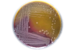
For determining the viability and to detect bacterial cultures in feces of rats, fresh void fecal materials were collected during the experimental period and pooled from all test and the control group. The samples have been homogenised in usual saline (0.85% cold NaCl) and gradually diluted. The diluted homogenates (0.1 ml) were spread on Xylose-lysine deoxycholate citrate agar plates to enumerate enterobacteria, especially Shigella species. The plates are incubated at 37℃ for 24 hours, the number of colonies was counted based on the colony's formation and documented consequently. A control group of unprocessed/untreated rat was also analyzed [17].
Singleplex polymerase chain reaction of faecal samples
Using the Genei Genomic DNA Extraction Manual Kit (Cat No. KT28) the genomic DNA was extracted from the faecal samples with minor modifications. The extracted DNA became amplified by conventional PCR using primers specific to the genes of interest to identify Shigella species. A separate PCRs reaction mixture was employed for the amplification of the DNA fragments. To identify the Shigella sps, the IpaB-F and IpaB-R primer set with forward and reverse primers, respectively, was used to amplify the 120 bp ipaB gene of Shigella sps,. The PCR amplification was performed according to Layton et al. [18] with some modifications. The amplification was carried out using a 20 µl reaction cocktail in Thermal Cycler (Eppendorf) (Table 1); the PCR program was standardized for 30 cycles and operated using modified conditions as in Tables 2 and 3 shows the nucleotide sequence of the primer pairs used for the PCR and the expected amplicon sizes.
| Constitutes | Quantity (µl) | Final Concentrations |
|---|---|---|
| Template DNA | 2 | 25 ng |
| 10X Taq DNA assay buffer | 2 | 1X |
| dNTPs | 0.4 | 200 µM each dNTPs |
| Primer | 2 | 20 picomoles |
| Taq DNA polymerase | 0.3 | 1 Unit |
| Sterile Water | 13.3 | 20 µl |
Table 1: The components of Polymerase Chain Reaction (PCR) mixture.
| Process | Temperature (°c) | Time | Cycles |
|---|---|---|---|
| Initial Denaturation | 95 | 3 min | 1 |
| Denaturation | 95 | 10 sec | - |
| Annealing | 60 | 20 sec | 30 |
| Extension | 72 | 30 sec | - |
| Final Extension | 72 | 7 min | 1 |
| Storage | 4 | - | - |
Table 2: Polymerase Chain Reaction (PCR) amplification programme condition for ipaB gene in Shigella species form stool samples.
| Pathogen | Primer | Sequence (51-31) | Amplicon Size (bp) |
|---|---|---|---|
| Shigella species | IPAB- F | 5’ GACGCCCAAGCCTTCGAGCA 3’ | 120 |
| IPAB-R | 5’ AGCAGCGACCGGCAATTCCT 3’ |
Table 3: Primers used for the PCR detection of IPA-B gene for shigella species.
Haematology and biochemical studies
At the end of the experimentation, all the existing animals were fasted for the night prior to anesthetization and necropsy. Blood samples were collected in EDTA-2K tube as anticoagulants. The haematological factors resoluted with an automatic programmed haematology analyzer, K-4500 (Sysmex Corp. Japan) incorporated White Blood Cells (WBCs), Red Blood Cells (RBCs), Haemoglobin (HB), Platelets Count (PLT) and differential leukocytes count. The non-EDTA-2K-treated blood samples were utilized for biochemical tests of the serum. These samples were used to resolve the levels of glucose, total cholesterol, creatinine and for the activity of Aspartate Aminotransferase (AST), Alkaline Phosphatise (ALP), and Alanine Aminotransferase (ALT). After the animals have been sacrificed, the intestine/gut was also separated, slight longitudinally, weighed and cleaned with 0.9% NaCl. Mucosae have been scrapped off, homogenised in 10 mM sodium phosphate buffer at pH 7.4 (1:10 w/v) and centrifuged at 3000 g for 10 minutes at 4℃. The supernatant was taken for the evaluation of protein, assay of alkaline phosphatise, reduced glutathione (GSH), Super Oxide Dismutase (SOD) and Catalase Activity (CAT) [19].
Relative organ weights and histopathological study
Coarse examination was completed by necropsy and recorded. Prior to further histopathological examination, small intestine, liver, kidney, spleen, lungs, and heart were weighed up. The comparative organ weights had been calculated based on the ultimate Body Weight (BW) of the rats. At necropsy, the essential organs had been surgically separated from the rats, rinsed with normal saline, fixed and sealed in 10% formalin. Collected tissues were repellently and microscopically observed during histopathological examination as per standard OECD guidelines [20].
Results
Selection of bacterial strains to induce diarrhoea in rats
Among the 32 clinical isolates of Shigella isolates; Shigella dysentriae-07 and Shigella flexneri-277 have been selected on the basis of highest multiple drug resistance and their susceptibility for clinical antibiotics and antimicrobial agents [7]. Hence, isolates S. dysentriae-07 and S. flexneri-277 were selected for the induction of diarrhoea.
Induction of diarrhoea in diarrhoeagenic rats with Shigella strains
A dose of Shigella strains (S. dysentriae-07 and S. flexneri-277) was determined by injecting the diarrheal rats with varying numbers of Shigella strain 12 × 106, 12 × 107 and 12 × 108 colony-forming unit per ml (cfu/ml) [19] of the oral and intragastric modes of administration, intragastric administration was considered the most effective. A dose of intragastric injection of 12 × 106 to 12 × 108 cfu/ml were injected to the rats; a real dysenteric diarrhea was induced in rats with 12 × 108 cfu/ml of Shigella strains, which showed better survival rate; therefore, this dose was considered optimal and was fixed for the entire experiment.
Evaluation of diarrheal parameters
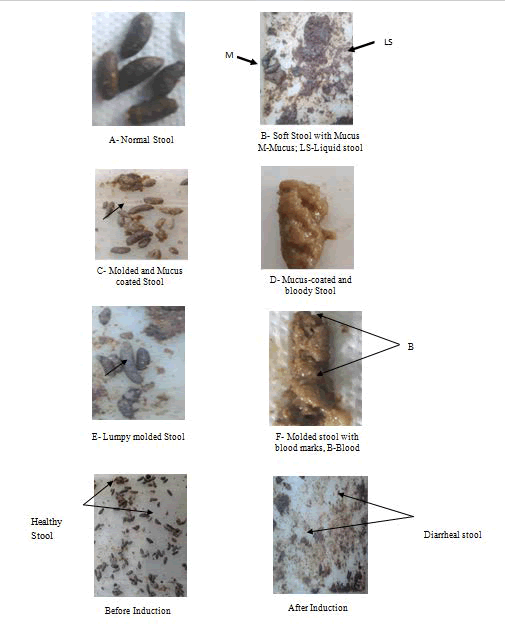
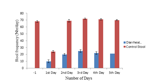
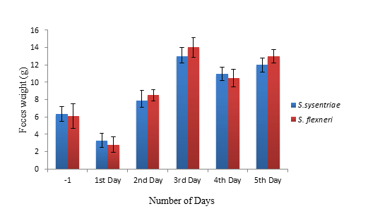
Prior to induction of diarrhea, animals having regular faeces, they were hard/solid, molded, brown or darkish and rough (Figure 1A). Some primary diarrheal appearances had been cited in rats with inoculum 1 × 10⁸ cfu/ml of Shigella strains (the 3 McFarland standards). We then perceived few soft faeces (unmolded) or liquid and with blood at the anal orifice. Actual dysenteric diarrhoea was attained in rats with inoculum of 12 × 108 cfu/ml. Diarrhoeal stools observed within 24 hours after induction. Stools were either soft or liquid, containing mucus (Figure 1B) or molded and smooth but mucus coated, frequently with mucus-linked molded faeces (Figure 1C). At times, the faeces as well appeared longer, dark and shiny because of blood and mucus (Figure 1D). These stools were molded and lumpy but brown and brittle (Figure 1E) or existing blood mark (Figure 1F). After 2-4 hours of Shigella strain induction, behavioural alterations like weakness, less mobile, curled up and strong aggressiveness were observed. The frequency of diarrheal feces (in case of both S. dysentriae-07 and S. flexneri-277) increased steadily from the first to the fifth day following induction, whereas the occurrence of total faeces decreased considerably after the first day following induction (12 ± 1.41 and 10 ± 1.21) and increased on each of the other days (Figure 2). The weight/mass of the stool decreased drastically after the first day after induction and increased on each of the following days, including a significant increase on the 3rd to 5th day, as shown in Figure 3 [16].
Figure 2: Stool frequency (number of feces per day: Nbr/day) before and after diarrheal induction in rats with S. dysentriae-07 and S. flexneri-277 (12 × 108 cfu/ml) [Where: DS: Diarrheal stool; CS: Control stool; -1: mean of feces frequency the day before diarrheal induction; 1-5: days after diarrheal induction (n=6 and Statistical significant at p<0.005)]
Feed, water intake and body weight of the rats
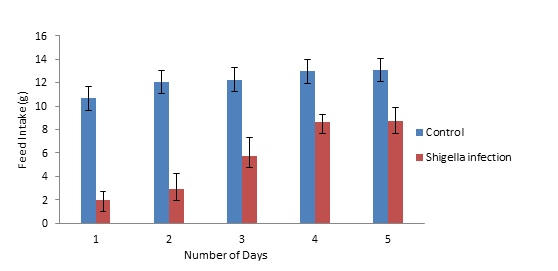
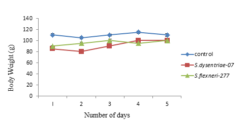
Table 4 illustrates the daily feed eating of the rats during the study period. The rat group fed with a test dose of 12 × 108 cfu/ml for Shigella strains showed considerably higher feed intake at the end of the experimental phase (Figure 4). In all the other cases, the feed intake was as usual for rats with no major differentiations among the control and experimental clusters, whereas in Shigella infection, the body weight of the rats was significantly decreased by 1st and 2nd day, but subsequently recovered on the 3rd and 5th days. There is no significant decrease in the body weight; rats were healthy and their physiological condition was as good as those in the control group (Figure 5). The water intake was as usual for all the rat groups during the complete experimental phase.
| Daily Feed Intake (g) | ||
|---|---|---|
| No. of days | Control | Shigella infection ( 12 × 108 cfu/ml) |
| 1 | 10.66 ± 0.42 | 2.01 ± 0.70 |
| 2 | 12.05 ± 0.24 | 2.9 ± 1.34 |
| 3 | 12.26 ± 1.33 | 5.76 ± 1.60 |
| 4 | 12.96 ± 0.67 | 8.66 ± 0.62 |
| 5 | 13.07 ± 1.12 | 8.69 ± 1.19 |
Table 4: Daily feed intake of rats fed with Shigella strains for experimental period values are means ± SEM, n=6.
Viability and detection of bacterial cultures in faeces of rats
The capability of the isolates to defend the gastro-intestinal tract against Shigella infection can be established (Figure 6) by examining the count of enerobactericiae, particularly Shigella species, on XLDA agar media, in rat faeces [17].
Polymerase chain reaction for confirmation of Shigella
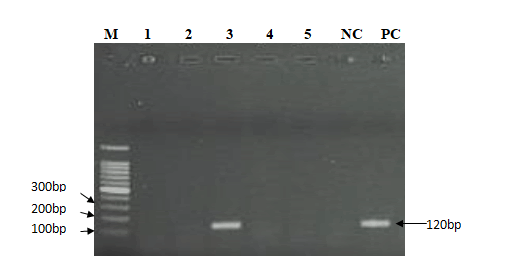
Further identification of Shigella from faecal samples of the dose-induced rats was made by amplifying ipaB gene using primer pairs described in Table 3. The amplicons were neatly separated on the 1.5% agarose gel. The amplification was positive, asserts the presence of ipaB gene of size 120 bp (Figure 7).
Figure 7: PCR amplification for ipaB gene of Shigella where: Lane M: contain 100 bp molecular weight marker; Lane 1-5: contained amplicons of samples from PCR; Lane 3: shows a band of 120 bp indicates the presence of ipaB gene; Lane PC: contained positive control purified Shigella DNA; Lane NC: contained negative control (No DNA).
Haematology and Biochemical Studies
Haematology study
To measure the toxicity of Shigella dysentriae-07 and Shigella flexneri-277 strain- treated rats in control and experimental groups, the estimation of standard haematological parameters, such as red blood cell count, white blood cell count, haemoglobin level and platelet count is an important aspect. Concentration-dependent haematology results for treatment of rats, this study found no significant difference in any analysed aspects in haematology; haematology values of all the parameters analysed were not extensively influenced by the induction of Shigella dysentriae-07 and Shigella flexneri-277 strains in both the control and experimental groups (Table 5). These findings suggest that the test article Shigella strains values were in the reference range and may not be toxic as they do not significantly affect the movement of haematological parameters. Additionally, the normal metabolism of the treated animals was not involved by the test article Shigella strain reinforcement.
| Experimental Group | Haematology Parameters | |||
|---|---|---|---|---|
| WBC | RBC | Haemoglobin | Platelets | |
| S. dysentriae- 07 induced | 7.8 ± 0.23 | 4.51 ± 0.04 | 120 ± 3.0 | 230 ± 4.7 |
| S. flexneri-277 induced | 9.7 ± 0.56 | 3.35 ± 0.02 | 13.6 ± 3.2 | 120 ± 2.3 |
| Control | 7.5 ± 0.54 | 5.20 ± 0.21 | 128 ± 1.02 | 210 ± 4.21 |
| Reference Range | 44838 | 3.50-5.50 | 110-160 | 100-300 |
Table 5: Haematology study of Shigella species treated rat after in vivo feeding trials in control and experimental groups. WBC: white Blood Cells (SI unit: 109/L), RBC: Red Blood Cells (SI unit: 109/L), Haemoglobin in g/L, Platelets in 109/L. All data are expressed in mean ± SD, n=6.
Blood serum biochemical tests: All the experimental groups in the present study were considered for the evaluation of different biochemical tests.
Alkaline Phosphatase (ALP) activity
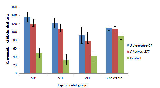
Alkaline phosphatase test measures the amount of the enzyme ALP in the blood stream of the humans; ALP analyses the serum infection mostly in the liver and in bone, with some made in the intestine and kidney. In the treatments, a marked elevation was seen in the activity of ALP in S. dysentriae-07- and S. flexneri-277-infected rats (135.5 ± 11.12, 119.8 ± 12.13 and 49.57 ± 12.52) in both the control and experimental groups (Figure 8).
Aspartate aminotransferase (AST) activity
AST is an enzyme that augments in activity in diseases, such as relentless bacterial infections, malaria, pneumonia, pulmonary infarcts and tumours of organs for example heart and muscle. In the treatment ofShigella strains, a noticeable elevation in the activity of ALT was observed in S. dysentriae-07- and S. flexneri-277-infected rats (121.5 ± 11.12, 106.21 ± 12.13 and 33.31 ± 12.15) among the control and experimental groups as shown in Figure 8.
Alanine aminotransferase (ALT) activity
Alanine Aminotransferase (ALT) is primarily found in the liver and is considered as being more precise than alanine aminotransferase for noticing liver cell damage. In Shigella strain treatment, a noticeable elevation was observed in the activity of ALT in S. dysentriae 07- and S. flexneri-277-infected rats (92.33 ± 12.15, 78.66 ± 20.18 and 41.66 ± 12.12) in both the control and experimental groups, as described in Figure 8.
Cholesterol activity
In cholesterol activity of the serum, no considerable differentiations were found in any of the analyzed phases in the experimental groups; in biochemical analysis all the experimental groups were within the standard range compared with control (Figure 8).
Intestinal Serum Biochemical Tests
Alkaline phosphatase activity
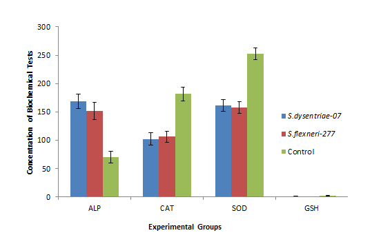
Intestinal ALP, a membrane-bound enzyme, is there in huge quantity in the intestine and occupied in fat assimilation in the intestine. It is a marker enzyme that enhances in the period of colonic injury [19]. Figure 9 illustrates the level of ALP among the control and experimental groups of S. dysenriae-07- and S. flexneri-277-treated rats. A noticeable elevation in the activity of ALP was noticed in S. dysentriae-07- and S. flexneri-277 infected rats (168.5 ± 13.12, 151.3 ± 15.17), whereas the control group showed significant decrease (70.17 ± 10.12).
Catalase activity
Catalase is an awfully significant enzyme in defensive the cells from oxidative injuries by reactive oxygen species. In this study, the S. dysenriae-07- and S-flexneri 277-treated rats had shown noticeably high oxygenic stress and damage in the experimental groups (102.36 ± 10.72 and 106.21 ± 9.75) when compared with the control (182 ± 12.12), as shown in Figure 9.
Super Oxide Dismutase (SOD) activity
SOD is an antioxidant enzyme produced in our bodies to neutralize a specific oxygen free radical called superoxide. In the treated experimental groups of rats, a noticeable result in the activity of SOD was noticed in S. dysentriae-07- and S. flexneri-277-infected rats (161 ± 10.41, 158 ± 10.31), when compared with the control (252.83 ± 10.31) as shown in Figure 9.
Reduced Glutathione (GSH) activity
In the treated experimental cluster of rats, a noticeable decline in the GSH activity was noticed in S. dysentriae-07- and S. flexneri-277-infected rats (0.76 ± 0.51, 0.68 ± 0.12) when compared with control (2.78 ± 0.56) as shown in Figure 9.
Relative Organ Weights and Histopathological Examination of Experimental Rats
Relative Organ Weight (g %)
A necropsy and macroscopic inspection of the organs revealed no treatment-related damages or variation and no pathological changes were found in all the treated rat groups. The virtual organ weights of small intestine, heart, kidney, liver, lungs and spleen in any of the treated experimental groups did not notice any significant variation from that of the control (Table 6). However, a marginal but statistically no significant decrease was observed in relative organ weight of the intestine of the Shigella infected rats when contrast to the control groups. However, even small effects are significant for the intestine.
| Experimental Groups | Organ index (%) | |||||
|---|---|---|---|---|---|---|
| Intestine | Liver | Kidney | Lungs | Heart | Spleen | |
| S. dysentriae-07 induced | 1.25 ± 0.06 | 0.58 ± 0.07 | 0.69 ± 0.01 | 0.63 ± 0.09 | 0.29 ± 0.14 | 0.19 ± 0.01 |
| S. flexneri-277 induced | 1.27 ± 0.09 | 0.56 ± 0.04 | 0.68 ± 0.04 | 0.62 ± 0.01 | 0.28 ± 0.01 | 0.21 ± 0.02 |
| Control | 1.3 ± 0.09 | 0.56 ± 0.05 | 0.79 ± 0.04 | 0.63 ± 0.12 | 0.34 ± 0.01 | 0.24 ± 0.09 |
Table 6: Relative organ weight (g%) of the treated rats in control and experimental groups.
Histopathological analysis of experimental rats
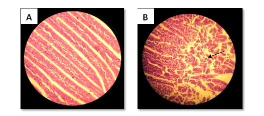
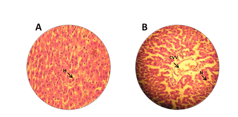
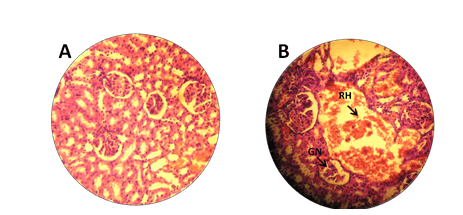
Histopathology enables the microscopic assessments of biological tissues to examine the form of diseased cells and tissues in very fine detail. The study has made an attempt to identify the adverse effects of S. dysentriae-07 and S. flexneri-277 administered intragstrically in rats. The rats that received these bacteria noticed injury to the intestinal tissue as confirmation by pathological alterations in the architecture of the intestine, namely, ulceration, epithelial desquamation and intense infiltration of PMNs (Figure 10A). Other vital organs like liver and kidney showed a normal structure; in the case of liver, the S. dysentriae-07 and S. flexneri-277 externally administered through intragastric route exhibited intense centrilobular necrosis, vacuolization and macro vesicular fatty change (CVV) as shown in Figure 10B; in the case of kidney, histological examination revealed that only S. dysentriae-07 and S. flexneri-277-induced rats showed intense damage to kidney tissue, including Renal Haemorrhage (RH) necrosis of Glomerular Cells (GN), Bowman capsule and proximal tubular in group as compared to control (Figure 10C).
Figure 10A: a) Histological Studies of treated rats in control and experimental groups infected with S. dysentriae-07 and S. flexneri-277 where, A: Section of intestine from a control rat showing normal architecture; b) Intense neutrophil infiltration, muscularis propria in epithelial lineage to severe hyperplasia in tissue treated with S. dysentriae-07 and S. flexneri-277induced rats.
Discussion
Shigellais a universal infection immoral for propagating fast in settings where there is overcrowding, inadequate cleanliness and lacking of clean water [22]. The variety of symptoms range from moderate watery diarrhea to fulminate bacillary dysentery, categorized by bloody stools, high fever, prostration, cramps, tenesmus and an array of severe intestinal and extraintestinal complications [21-22]. S. dysenteriaeand S. flexnerii are unique among theShigellaspp. in its ability to produce Shiga toxin and consequently cause the most serious forms of disease. Extensive use of a safe and effectiveShigellavaccine have been considered an attractive approach for fighting this naturally occurring illness and developing such a vaccine or alternative therapy is a high public health priority. The feature ofShigellato cause diarrheal disease is limited to human and nonhuman primate hosts. The lack of small animal model that fully replicates human bacillary dysentery is a major barrier to the advancement of successfulShigellavaccine or therapy [21-22]. The present study, estimated intragastric confront model of S. dysentriae-07 and S. flexneri-277 in rats for pre-clinical assessment of Shigella therapy. Even if many studies in rats challenged with S. dysentriae-07 and S. flexneri-277 have been reported earlier, a topical appraisal of this model was required before vaccine or for an alternative therapy evaluation. In humans, the infective dose for causing dysenteric diarrhea is only 10–100 bacteria [23]. In rats, diarrhea has been induced with 9 × 10⁸ organisms, while true dysentery was obtained with 12×108 S. dysentriae-07 and S. flexneri-277. The findings of our study indicate that direct intragastric inoculation of virulent S. dysentriae-07 and S. flexneri-277 in rats will result in successful clinical infection without the need for starvation and pre-treatment of animals. Within 24 hours of inoculation, the animals developed characteristic signs of Shigella species comparable to those noticed in humans [24]. In our study, symptoms included passage of frequent loose stool with blood and mucus, weight loss, and rise in body temperature (Figure 1). The knack of Shigella to cause diarrheal infection is limited to human and non-human primate hosts. Recently, a number of studies has been achieved using the rat model; even though not an intestinal model, the rat model was helpful only for testing the efficiency of vaccine or therapy candidates and associating the usefulness of immunological responses [16]. In another study, rats had been infected with Shigella dysentriae within 24 hours after infection; in addition, animals those orally immunized are conferred homologous defensive immunity against following challenges with the live strains. Even though these rat and rabbit ileal loop models [19] are helpful to study the pathogenesis and inflammatory responses, the simulated nature of these systems capacity hinder their ability to advance vaccine development in humans. In our current study, the detailed correlation of histological, biochemical studies and haematological analyses (Figure 8–10) in rats with shigellosis are comparable to those noticed in humans with shigellosis.
Conclusion
In conclusion, our results validate that the rat model directly imitates the diseases, immune response and inflammatory responses similar to responses are observed in humans. From the current study, we have received the affirmation that rats are the suitable species for use as an undertaking version with wild-kind Shigella pathogens; hence, we consider that this rat version or likely adjustments of it is going to be beneficial for the improvement of vaccine or an opportunity remedy and evaluation. Though there are similarities in clinical signs, flaking of the organisms, immune response, haematological and histopathological results among rats and humans infected with S.dysentriae-07 and S. flexneri-277 makes this animal model paramount at present for studying pathogenesis and infection-derived immunity.
Conflict of Interest Statement
We declare that no conflict of interest.
Authors Contribution
First and second author is responsible for carrying out the research work, data analysis and optimization of experimental work. Third author assisted with experiments and contributed in arranging tables, illustrations and preparing the manuscript. The corresponding author was responsible for research planning and execution as well as provision of other valuable input.
Acknowledgement
All the authors are profusely thankful to Department of Biotechnology (DBT), Govt. of INDIA, New Delhi. And Department of Biotechnology, Gulbarga University, Gulbarga for providing laboratory facilities to this work.
Ethical Approval
This research was conducted in compliance with the Institutional Animal Ethics Committee and other federal statute and regulations relating to animals (Lcp/PQ.col/IAEC/P/86/2016).
References
- Niyogi SK (2005) Shigellosis. J Microbiol 43:133-143
- Rambaud JC (2000) Traité de gastroentérologie. Flamm Méd Sci.
- Passwell JH, Harlev E, Ashkenazi S, Chu C, Miron D, et al. (2001) Safety and immunogenicity of improved Shigella O-specific polysaccharide-protein conjugate vaccines in adults in Israel. Infect Immun 69:1351-1357.
- Kouitcheu LB, Tamesse JL, Kouam J (2013) The anti-shigellosis activity of the methanol extract of Picralima nitida on Shigella dysenteriae type I induced diarrhoea in rats. BMC Complement Altern Med 13:1.
- Dutta S, Ghosh A, Ghosh K, Dutta D, Bhattacharya SK, et al. (2003) Newly emerged multiple-antibiotic-resistant Shigella dysenteriae type 1 strains in and around Kolkata, India, are clonal. J Clinic Microbiol 41:5833-5834.
- Sivapalasingam S, Nelson JM, Joyce K, Hoekstra M, Angulo FJ, et al. (2006) High prevalence of antimicrobial resistance among Shigella isolates in the United States tested by the National Antimicrobial Resistance Monitoring System from 1999 to 2002. Antimicrob Agents Chemother 50:49-54.
- kumar Oli A, Ashajyothi C, Chandrakanth RK (2015) Prevalence and antibiotic susceptibility pattern of fluoroquinolone resistant Shigella species isolated from infants stool in Gulbarga district, Karnataka, India. Asian Pac J Trop Dis 5:116-120.
- Clark AM (1996) Natural products as a resource for new drugs. Pharma Res 13:1133-1141.
- Cordell GA (2000) Biodiversity and drug discovery a symbiotic relationship. Phytochem 55:463-480.
- Rabbani GH, Albert MJ, Rahman H, Islam M, Mahalanabis D, et al. (1995) Development of an improved animal model of shigellosis in the adult rabbit by colonic infection with Shigella flexneri 2a. Infect Immun 63:4350-4357.
- Ahmed ZU, Sarker MR, Sack DA (1990) Protection of adult rabbits and monkeys from lethal shigellosis by oral immunization with a thymine-requiring and temperature-sensitive mutant of Shigella flexneri Y. Vaccine 8:153-158.
- Arm HG, Floyd TM, Faber JE, Hayes JR (1965) Use of ligated segments of rabbit small intestine in experimental shigellosis. J Bacteriol 89:803-809.
- Dutta NK, Habbu MK (1955) Experimental cholera in infant rabbits: a method for chemotherapeutic investigation. Br J Pharma Chemother 10: 153.
- Formal SB, Dammin GJ, Schneider H, LaBrec EH (1959) Experimental shigella infections characteristics of a fatal enteric infection in guinea pigs following the subcutaneous inoculation of carbon tetrachloride. J Bacteriol 78:800-804.
- Freter R (1956) Experimental enteric Shigella and Vibrio infections in mice and guinea pigs. J Experimen Med 104:411.
- René K, Vidal PK, Christine FM, Véronique PN, Magloire BS, et al. (2005) Shigella dysenteriae type 1-induced diarrhea in rats. Japanese J Infect Dis 58:335.
- Oyetayo VO, Adetuyi FC, Akinyosoye FA (2003) Safety and protective effect of Lactobacillus acidophilus and Lactobacillus casei used as probiotic agent in vivo. African J Biotechnol 2:448-442.
- Layton A, McKay L, Williams D, Garrett V, Gentry R, et al. (2006) Development of Bacteroides 16S rRNA gene TaqMan-based real-time PCR assays for estimation of total, human, and bovine fecal pollution in water. Appl Environ Microbiol 72:4214-4224.
- Moorthy G, Murali MR, Devaraj SN (2007) Protective role of lactobacilli in Shigella dysenteriae 1–induced diarrhea in rats. Nutrition 23:424-433.
- Bharucha C, Meyer H, Moody A, Carman RH (1970) Handbook of medical laboratory technology. Christian Medical College, vellore.
- Kweon MN (2008) Shigellosis: the current status of vaccine development. Curr Opin Infect Dis 21:313-318.
- Levine MM, Kotloff KL, Barry EM, Pasetti MF, Sztein MB, et al. (2007) Clinical trials of Shigella vaccines: two steps forward and one step back on a long, hard road. Nature Rev Microbiol 5:540-553.
- Sur D, Ramamurthy T, Deen J, Bhattacharya SK (2004) Shigellosis: challenges and management issues. Ind J Med Res 120:454.
- Islam D, Veress B, Bardhan PK, Lindberg AA, Christensson B, et al. (1997) Quantitative assessment of IgG and IgA subclass producing cells in rectal mucosa during shigellosis. J Clin Pathol 50: 513-520.
 Spanish
  Chinese
  Russian
  German
  French
  Japanese
  Portuguese
  Hindi
Hindi 