Clinical Image, J Clin Image Case Rep Vol: 3 Issue: 1
Gliomatosis Cerebri as a Rare Cause of Headache
Bindi Patel* and Ankit Agrawal
Rutgers Robert Wood Johnson Medical School - Saint Peter’s University Hospital, New Brunswick, New Jersey USA
*Corresponding Author: Bindi Patel
Rutgers Robert Wood Johnson Medical School - Saint Peter’s University Hospital, New Brunswick, New Jersey USA
Tel: +447447113330
E-mail: bp480@rwjms.rutgers.edu
Received: August 23, 2018 Accepted: September 1, 2018 Published: September 4, 2018
Citation: Sargsyan N (2019) Smoking on Long Term Oxygen Therapy. J Clin Image Case Rep 3:1.
Abstract
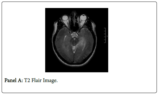
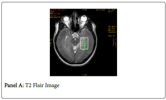
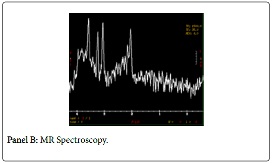
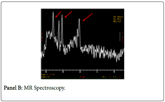
healthy 34 year-old male presented to the Emergency Department with a one month history of headaches with pain behind the left eye and associated with aura of scintillating scotomas, photophobia, and vomiting. Neurological examination was pertinent only for diffuse hyperreflexia. Complete blood count revealed leukocytosis of 14,300 per cubic millimeter (reference range 4,000-11,000). The rest of the biochemical parameters were within normal limits. MRI brain (Panel A) and MR Spectroscopy (Panel B) were consistent with gliomatosis cerebri. Patient was started on Dexamethasone, which was continued upon discharge [1]. At four month follow-up, patient is clinically asymptomatic and has deferred recommended biopsy of brain. Gliomatosis cerebri is a type of astrocytoma characterized by rapid and difficult to localize expansion infiltrating into multiple areas of the brain simultaneously. Fewer than 100 cases are diagnosed in the United States every year, and patients have a very poor prognosis [2].
Keywords: scotomas, photophobia,
A healthy 34 year-old male presented to the Emergency Department with a one month history of headaches with pain behind the left eye and associated with aura of scintillating scotomas, photophobia, and vomiting. Neurological examination was pertinent only for diffuse hyperreflexia. Complete blood count revealed leukocytosis of 14,300 per cubic millimeter (reference range 4,000-11,000). The rest of the biochemical parameters were within normal limits. MRI brain (Panel A) and MR Spectroscopy (Panel B) were consistent with gliomatosis cerebri. Patient was started on Dexamethasone, which was continued upon discharge [1]. At four month follow-up, patient is clinically asymptomatic and has deferred recommended biopsy of brain. Gliomatosis cerebri is a type of astrocytoma characterized by rapid and difficult to localize expansion infiltrating into multiple areas of the brain simultaneously. Fewer than 100 cases are diagnosed in the United States every year, and patients have a very poor prognosis [2].
 Spanish
  Chinese
  Russian
  German
  French
  Japanese
  Portuguese
  Hindi
Hindi 


