Case Report, Int J Ophthalmic Pathol Vol: 12 Issue: 2
Hybrid Cyst of the Eyelid: An Amalgamation of Epidermoid and Apocrine Differentiation
Rasheeda Mohamedali1*, RPS Punia1, Laxmi Tuli2 and Uma Handa1
1Department of Pathology, Government Medical College and Hospital, Chandigarh, India
2Department of Ophthalmology, Government Medical College and Hospital, Chandigarh, India
*Corresponding Author: Rasheeda Mohamedali
Department of Pathology,
Government Medical College and Hospital, Chandigarh, India
Tel:+917529888111
E-mail: rashi.reviews@gmail.com
Received date: 19 March, 2023, Manuscript No. IOPJ-23-92243;
Editor assigned date: 22 March, 2023, PreQC No. IOPJ-23-92243 (PQ);
Reviewed date: 05 April, 2023, QC No. IOPJ-23-92243;
Revised date: 12 April, 2023, Manuscript No. IOPJ-23-92243 (R);
Published date: 19 April, 2023, DOI: 10.4172/2324-8599.12.2.010
Citation: Mohamedali R, Punia RPS, Tuli L, Handa U (2023) Hybrid Cyst of the Eyelid: An Amalgamation of Epidermoid and Apocrine Differentiation. Int J Ophthalmic Pathol 12:2.
Abstract
Hybrid cysts are lesions that arise from different portions of the hair follicle. We describe a follicular hybrid cyst of the eyelid that contained features of epidermoid cyst and apocrine hidrocystoma in an elderly male. The unusual location, histopathologic and immunohistochemical features are discussed.
Keywords: Eyelid; Hybrid cyst; Apocrine hidrocystoma; Epidermoid cyst
Introduction
Hybrid cysts of the eyelid are a rare type of cystic lesion that displays features of two or more constituents of the pilosebaceous unit. They were first described in the literature by McGavran and Binnington in 1966 as a cutaneous cystic tumor with a dual, infundibular and trichilemmal lining, and since then, there have been several reported cases in the literature [1]. However, these cysts are rare in the eyelid and are often underdiagnosed due to the predominance of one of the lining epithelium or their resemblance to other types of cysts found in the eyelid. Here, we present a rare case of hybrid cyst of the eyelid which demonstrated both epidermoid and apocrine differentiation in an elderly. We discuss the clinical, histopathological and immunohistochemical features of this lesion.
Case Presentation
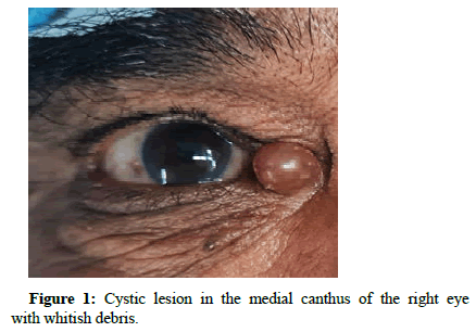
A 60-year-old male developed a cystic lesion in the right upper eyelid in a 5-year interval, which was increasing in size for 5 months. On examination, the lesion was circumscribed, non-tender, immobile, cystic lesion of 5 mm size and was filled with clear fluid which transilluminated (Figure 1).
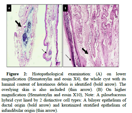
This lesion was not fixed to skin or underlying structures. There was no discharge or inflammation in the surrounding area. The rest of the ocular examination was unremarkable. The excision biopsy revealed a cyst in the dermis with an undulating lining epithelium and a lumen filled with keratinous material (Figure 2A). At higher magnification, the cystic wall was lined partly by stratified squamous epithelium which had a keratohyaline layer. This was admixed with a double-layered epithelium comprising of cuboidal and myoepithelial layer resembling the ductal epithelium (Figure 2B).
Figure 2: Histopathological examination: (A) on lower magnification (Hematoxylin and eosin X4), the whole cyst with its luminal content of keratinous debris is identified (bold arrow). The overlying skin is also included (thin arrow). (B) On higher magnification (Hematoxylin and eosin X10), Note: A pilosebaceous hybrid cyst lined by 2 distinctive cell types: A bilayer epithelium of ductal origin (bold arrow) and keratinized stratified epithelium of infundibular origin (thin arrow).
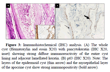
The transition between the two epitheliums was abrupt. The wall of the cyst showed few eccrine glands and pilosebaceous units. The overlying epidermis was unremarkable. Pancytokeratin immunostains these epidermis, hair structures, and the thin walls of apocrine hidrocystomas as well as the layers of the pilar portion of the cyst (Figure 3A). A deeper portion of an apocrine hidrocystoma manifests a continuous layer of p63 positive basal myoepithelial cells (Figure 3B). A diagnosis of hybrid cyst was rendered where the components of epidermoid cyst and apocrine hidrocystoma was identified in the same cyst.
Figure 3: Immunohistochemical (IHC) analysis. (A) The whole cyst (Hematoxylin and eosin X20) with pancytokeratin (IHC X20, inset) showing strong diffuse immunoreactivity of the entire cyst lining and adjacent lamellated keratin. (B) p63 (IHC X20). Note: The layers of the epidermoid cyst (thin arrow) and the myoepithelial layer of the apocrine cyst show strong immunopositivity (bold arrow).
Results and Discussion
A unique case of hybrid cyst exhibiting dual differentiation in the form of epidermoid cyst and apocrine hidrocystoma has been described in this case study. There are only three cases of hybrid epidermoid and apocrine cyst reported in the eyelid. Most of the studies have described cutaneous hybrid cyst as those occurring in overlapping anatomical segments of hair follicle [2-8].
Hybrid cysts of the eyelid clinically present as a painless, mobile, subcutaneous mass on the upper or lower eyelid. They can range in size from a few millimeters to several centimeters in diameter. On clinical examination, they can appear similar to other types of cystic lesions, such as chalazia or epidermoid cysts. However, some clinical features may suggest a diagnosis of a hybrid cyst, such as a translucent appearance, a bluish or yellowish hue, or a pedunculated appearance.
Hybrid cyst is simply the combination of epithelia in the pilosebaceous unit and sweat gland. The pathogenesis of these hybrid cysts has not been completely deciphered. These cysts are thought to form at the junction of keratinizing squamous epithelium and glandular portion. Other theories include, squamous metaplasia of apocrine hydrocystoma or collision tumour [9]. Apocrine glands are found predominantly in the anogenital and axillary regions, but also located in the eyelid (Moll’s gland), the areola. Hence, the distribution of apocrine cyst follows its anatomical distribution of its gland.
The epidermoid cyst is a common entity which is found in the head and neck regions, trunk and perineal areas. The cyst is line by stratified squamous epithelium with a well-defined and an intact kerato-hyaline layer. This cyst can be normal, atrophic or hyperplastic in nature. The apocrine hidrocystoma is an uncommon entity, usually arising in the head and neck region. Histologically, this cystic lesion is lined by a double layer of epithelial cells, the outer layer of flattened myoepithelial cells and the inner layer of cuboidal cells, with round or oval nuclei and eosinophilic cytoplasm, usually exhibiting snoutings at their apical ends with decapitation secretion [2,3].
The difficulty arises when the apocrine hidrocystoma is distended leading to flattening and atrophy of the epithelium. Hence, it becomes difficult to distinguish it from an eccrine hidrocystoma. Some authors have proposed the alternative term of “ductal hidrocystoma”, to reflect the possible dual occurrence. The closest hint to find out the possible ductal type in an eyelid cyst is the location. Apocrine glands are located only at the eyelid margins where there are no eccrine glands, and eccrine glands are located in the pretarsal and preseptal skin [10]. Eccrine duct cyst as a part of hybrid cyst could occur as an invagination into the cyst, as it is embryologically developed separately from the pilosebaceous unit. Hence, eccrine component in hybrid cyst is yet to be encountered [8].
Immunohistochemical analysis of apocrine hidrocystomas have been found to show immunoreactivity towards low molecular weight cytokeratin and carcinoembryonic antigen and are negative for high molecular weight cytokeratin [5]. In addition, p63 marker has shown to highlight the nucleus of basal myoepithelial cells. The stratified squamous epithelium shows immunoreactivity towards high molecular weight cytokeratin whereas these are negative for low molecular weight cytokeratins.
Conclusion
Hybrid cysts of the eyelid are a rare type of cystic lesion that can be difficult to diagnose clinically and warrants a histopathological examination. Surgical excision is the treatment of choice, with careful attention paid to the location of the lesion. Awareness of the clinical features and histopathological characteristics of hybrid cysts of the eyelid is essential for accurate diagnosis as these interesting lesions can easily be missed.
References
- McGavran MH, Binnington B. (1966) Keratinous cysts of the skin. Identification and differentiation of pilar cysts from epidermal cysts. Arch Dermatol 94:499–508.
[Crossref] [Google scholar] [Pubmed]
- Ma L, Jakobiec FA, Wolkow N, Dryja TP, Borodic GE. (2018) Multiple eyelid cysts (apocrine and eccrine hidrocystomas, trichilemmal cyst, and hybrid cyst) in a patient with a prolactinoma. Ophthalmic Plast Reconstr Surg 34:e83-e85.
[Crossref] [Google scholar] [Pubmed]
- Milman T, Iacob C, McCormick SA. (2008) Hybrid cysts of the eyelid with follicular and apocrine differentiation: An under-recognized entity? Ophthalmic Plast Reconstr Surg 24:122-125.
[Crossref] [Google scholar] [Pubmed]
- Requena L, Yus SE. (1991) Follicular hybrid cysts. An expanded spectrum. Am J Dermatopathol 13:228-233.
[Crossref] [Google scholar] [Pubmed]
- Seo HM, Jang JW, Park SK, Oh SU, Park HK, et al. (2023) A case of hybrid epidermoid and apocrine cysts of scalp. Ann Dermatol 35:84-85.
[Crossref] [Google scholar] [Pubmed]
- Kim MS, Lee JH, Son SJ. (2012) Hybrid cysts: A clinicopathological study of seven cases. Australas J Dermatol 53:49-51.
[Crossref] [Google scholar] [Pubmed]
- Serra F, Kaya G. (2021) A new case of hybrid epidermoid and apocrine cyst. Dermatopathology (Basel) 8:442-445.
[Crossref] [Google scholar] [Pubmed]
- Takeda H, Miura A, Katagata Y, Mitsuhashi Y, Kondo S. (2003) Hybrid cyst: Case reports and review of 15 cases in Japan. J Eur Acad Dermatol Venereol 17:83-86.
[Crossref] [Google scholar] [Pubmed]
- Andersen WK, Rao BK, Bhawan J. (1996) The hybrid epidermoid and apocrine cyst. A combination of apocrine hidrocystoma and epidermal inclusion cyst. Am J Dermatopathol 18:364-366.
[Crossref] [Google scholar] [Pubmed]
- Jakobiec FA, Zakka FR. (2011) A reappraisal of eyelid eccrine and apocrine hidrocystomas: microanatomic and immunohistochemical studies of 40 lesions. Am J Ophthalmol 151:358-74.e2.
[Crossref] [Google scholar] [Pubmed]
 Spanish
  Chinese
  Russian
  German
  French
  Japanese
  Portuguese
  Hindi
Hindi 


