Case Report, J Spine Neurosurg Vol: 8 Issue: 1
Luxation of the First Costovertebral Joint after Trauma of the Thorax
Van Groningen NJ1 and Wijffels MME2*
1Erasmusm Medical Center, Rotterdam, The Netherlands
2Department of Trauma Surgery, Erasmus Medical Center, Rotterdam, The Netherlands
*Corresponding Author : Wijffels MME
MD PhD, Erasmus MC, University Medical Center Rotterdam, Trauma Research Unit, Department of Surgery, P.O. Box 2040, 3000 CA Rotterdam, The Netherlands
Tel: +31107031050
E-mail: m.wijffels@erasmusmc.nl
Received: September 10, 2018 Accepted: October 6, 2018 Published: October 15, 2018
Citation: Van Groningen NJ, Wijffels MME (2019) Luxation of the First Costovertebral Joint after Trauma of the Thorax. J Spine Neurosurg 8:1. doi: 10.4172/2325-9701.1000309
Abstract
Thoracic trauma is common and may vary from simple contusions to lethal injuries. Uncommon thoracic injury is a costovertebral luxation of the first rib. Up till now only one other case report has been written in 1980. Therefore a case with this injury is presented.
Keywords: First rib; luxation; Thorax; Costovertebral; Shoulder
Introduction
A 38 year old male was presented to the Emergency Department after a prolonged suppression of the thorax. On arrival at the scene the patient was hemodynamically stable with a maximal Glasgow Coma Scale, but was intubated due to severe pain and a clinical flail chest [1].
Initially at the Emergency Department the patient remained hemodynamically stable. During secondary survey blood pressure decreased but patient proved to be a responder. Furthermore right sided radial and brachial artery pulsations were absent on palpation and with Doppler test.
Case Report
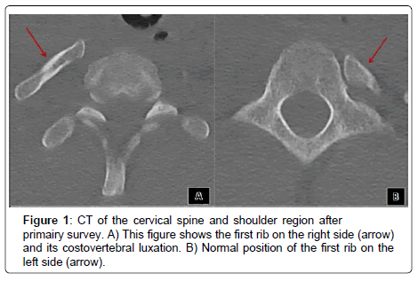
The Computed Tomography scan (CT-scan) of the thorax showed, among others, a transection of the right subclavian artery, a dissection of the right common carotid artery, a right-sided hemothorax, multiple high rib, scapulae and clavicular fractures on both sides and a costovertebral luxation of the first rib on the right side.
In acute setting a stent was placed using a rendez-vous procedure in the subclavian artery resulting in good peripheral vascularization. After this procedure patient was admitted to the intensive care unit (ICU).
Some of the fractures were treated operatively during ICU admission. After cessation of sedation, patient proved injury of the brachial plexus on the right side due to either primary pressure during in the injury or secondary to the fractured clavicular bone.
The costovertebral luxation of the first rib was treated nonoperatively because there were no radiological signs of compression of the brachial plexus or approximate vessels (Figure 1).
After a 10-day ICU stay, the patient showed recovery and regained strength of the right upper extremity reflecting brachial plexus recovery. The patient was discharged from the hospital after 37 days for outpatient clinical rehabilitation. The patient suffered at discharge from limited strength, motion and sensibility of the right upper extremity. During rehabilitation, the patient showed further progress within the aforementioned limitations of the right hand. Patient indicated pain during follow-up, deep in his neck on the right side.
At last follow-up; 13 months after trauma, discomfort in his neck remained constant, increasing with movement of the neck. Abduction of the right thumb and index finger remained diminished.
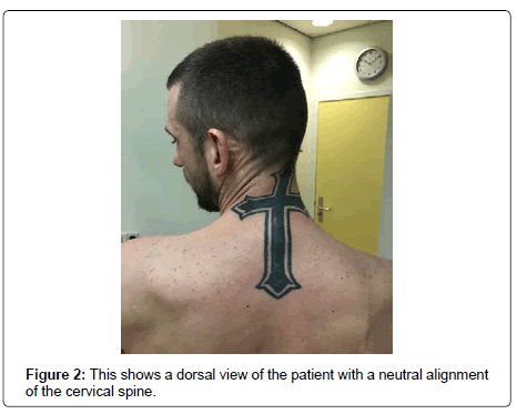
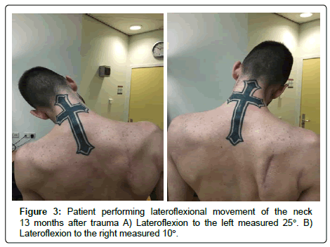
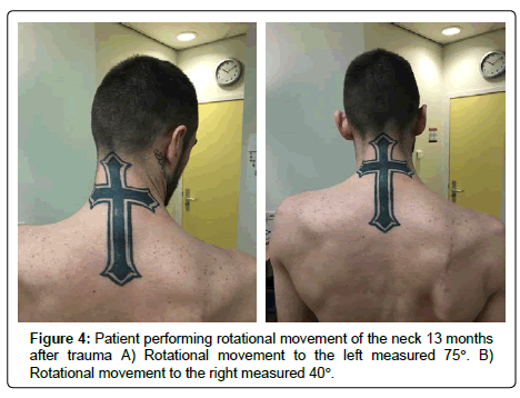
No residual respiratory complaints are indicated by the patient. Function of the neck during physical examination the cervical spine showed a neutral alignment (Figure 2). Lateroflexion to the right measured 10°, compared with 25° to the left (Figure 3). Rotation to the right measured 40° and to the left 75 (Figure 4); Ventral and dorsal flexion of the neck showed no limitations.
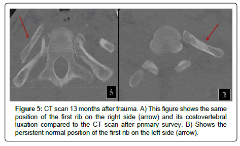
A CT-san of the neck during follow-up showed no changes in the luxated position of the first rib (Figure 5).
Discussion
In this case report, a posttraumatic costovertebral luxation of the first rib on the right side was presented. Nonoperative treatment showed remaining discomfort.
Chest wall movements are enabled by joints between ribs and spine. These joints consist of two parts, the costocentral joint (articulatio capitis costae) and the costotransverse joint (articulatio costotransversaria). The costocentral joint is an arthrodial joint connecting the caput of the rib to the lateral part of the corpus vertebrae. In all ribs, except for the first, there is a juncture of the ribs and two adjacent vertebrae. The first rib articulates with only one vertebra [2].
The costotransversal joint connects the tuberculum costae (dorsal side of the neck of the rib) and the processus transversus of the vertebrae. These joints are stabilized by three ligaments; the ligamentum costotransversarium, lateral costotransversarium and superior costotransversarium [1,3,4].
Despite the ligamentous stabilization, these joints are flexible but expansive force, could lead to fractures, luxation, and dislocation. Rib fractures within the spinal joints are seen in more than 10% of trauma patients suggesting, the fact that costovertebral luxations are less common, the strength of the joint stabilizing ligaments [2,5].
A luxation of the costovertebral joint can cause complications. One of the complications is pressure on the brachial plexus. However, it remains unknown if the brachial plexus lesion results from a first rib luxation or directly from the trauma these injuries are often seen together after high energy thoracic trauma [6]. If any suggestion of pressure on the brachial plexus by the luxated first rib; a releasing intervention may be mandatory. In this case the CT scan showed no compression on the brachial plexus, in presence of other explanations for nervous dysfunction. Therefore nonoperative treatment was chosen.
Another complication due to first rib luxation might be compression of vascular structures. Compression can be present on the subclavian or carotic arteries or its side branches. No literature is available on this subject. Compression of the subclavian vene might, theoretically, result in obstructed venous return and consequently thrombosis of the upper extremity. Depending on symptoms, in presence of vascular compression, a decompressive intervention should be considered. In this case no signs of arterial or venous compression were found clinically or on the CT-scan, justifying nonoperative treatment.
The presented patient showed discomfort and loss of motion of the neck, most likely due to the costovertabral luxation of the first rib. Many other injuries were found in this area, but the consolidated ribs and clavicular bone suggest that their influence on neck function is limited.
Only one study was found in literature concerning first costovertebral luxations [1]. Three cases with various traumas were presented. This study focused mostly on the mechanism of injury and treatment was not further specified, as is outcome. This indicates the need for future publishing cases and experience on this subject, to increase knowledge on costovertebral luxations of the first rib.
Conclusion
A luxation of the first costovertebral joint on the right side, treated nonoperatively was presented. This seems justifiable in absence of neurovascular compression. Long-term complaints of discomfort and limited loss of motion of the neck might be the result.
References
- Christensen EE, Dietz GW (1980) Injuries of the first costovertebral articulation. Radiology 134: 41-43.
- Ziegler DW, Agarwal NN (1994) The morbidity and mortality of rib fractures. J Trauma 37: 975-979.
- Platzer W (2009) Part one: Musculoskeletal system. Atlas of the anatomy. Stuttgart, Germany.
- Saker E, Graham RA, Nicholas R, D”Antoni AV, Loukas M, et al. (2016) Ligaments of the costovertebral joints including biomechanics, innervations, and clinical applications: A comprehensive review with application to approaches to the thoracic spine. Cureus 8: e874.
- Marasco S, Lee G, Summerhayes R, Fitzgerald M, Bailey M (2015) Quality of life after major trauma with multiple rib fractures. Injury 46: 61-65.
- Abu-Judeh HH, Contractor S, Bahramipour P, Sanches O, Biswal R, et al. (1999) Subclavian artery disruption following first rib dislocation. Emerg Radiol 6: 364-366.
 Spanish
  Chinese
  Russian
  German
  French
  Japanese
  Portuguese
  Hindi
Hindi 