Spanish
  Chinese
  Russian
  German
  French
  Japanese
  Portuguese
  Hindi
Hindi Research Article, J Liver Disease Transplant Vol: 6 Issue: 4
Metalloproteinase Inhibitor-1 Closely Correlates With the Severity of Liver Disease in Egyptian Patients
Khaled Metwally1*, Tamer Fouad1, Nashwa Shible1, Hassan Zaghla1, Eman Abdel sameea1, Mona M.Aref2 and Fatma A. Khalaf2
1Hepatology and gastroenterology Unit, National Liver Institute, Menoufiya University, Egypt
2Clinical Biochemistry Department, National Liver Institute, Menoufiya University, Egypt
*Corresponding Author : Dr. Khaled Metwally
National Liver Institute, Egypt
Tel: 00201000486019
Fax: + 20 48 2222740
E-mail: kh_m55555@yahoo.com
Received: December 08, 2017 Accepted: December 22, 2017 Published: December 29, 2017
Citation: Metwally K, Fouad T, Shible N, Zaghla H, Sameea EA, et al. (2017) Metalloproteinase Inhibitor-1 Closely Correlates With the Severity of Liver Disease in Egyptian Patients. J Liver Disease Transplant 6:4. doi: 10.4172/2325-9612.1000157
Abstract
Background: Liver disease prevalence is high in Egypt and finding a reliable seromarker is needed for diagnosis and prognosis of fibrosis. Tissue inhibitor of metalloproteinase -1 (TIMP-1) previously showed a good relation with fibrosis but few data are available from Egypt.
Objective: To test the correlation of TIMP-1 to the degree of liver fibrosis in Egyptian patients with and without cirrhosis, and to test its values in different degrees of liver dysfunction.
Methods: Forty Six adult patients (31 males and 15 females) with chronic liver disease were included in the current study, with age ranged from 42 –63 years were recruited from outpatient clinics at the National Liver Institute, Menoufiya University from February to July 2016, where sixteen patients had liver cirrhosis. Evaluation
of the degree of fibrosis and liver dysfunction was done by clinical examinations, laboratory tests, and imaging tests (Ultrasound and Fibroscan) with/ without histological examinations. TIMP-1 was determined in plasma samples using the MAC15 TIMP-1 ELISA.
Results: Median age was 51 years (42-63 years) and 67% were men. The primary cause of liver disease was hepatitis C, 89.1%. TIMP-1 values showed significant relation to the degree of fibrosis or cirrhosis assessed by liver biopsy or Fibroscan (p<0.001). Its level increases progressively with the development of encephalopathy and ascites and with the worsening of Child class (P=0.025, 0.018 and 0.039; respectively).
Conclusion: TIMP-1 increases significantly with liver cirrhosis and its degree of increase correlates with the degree of liver dysfunction.
Keywords: Fibrosis; Liver cirrhosis; TIMP-1; Liver dysfunction; ELISA
Introduction
Liver fibrosis and its end stage sequelae cirrhosis represent a major worldwide health problem. Liver fibrosis represents the final common pathological outcome for the majority of chronic liver insults (e.g. alcohol, autoimmune, or viral injury) [1]. Chronic liver injury, irrespective of the underlying disease, triggers a wound healing response that leads to fibrogenesis and can ultimately result in the development of liver cirrhosis. Patients with liver cirrhosis are at risk for mortality by complications caused by portal hypertension and liver failure. Moreover, underlying liver cirrhosis represents a major predisposition for the development of hepatocellular carcinoma (HCC) [2]. Liver fibrosis is a dynamic process. Extracellular matrix (ECM) deposition, mainly by hepatic stellate cells (HSCs), occurs secondary to chronic liver injury causing fibrosis. This process is opposed via degradation and remodelling of ECM by metalloproteinase enzymes (MMPs) which may lead to healing of fibrosis. Tissue inhibitors of matrix metalloproteinases (TIMPs) interfere with MMPs function and inhibit ECM degeneration [3].
By definition progressive fibrosis occurs when the rate of matrix synthesis exceeds matrix degradation. The central mediator of liver fibrosis is the hepatic stellate cell (HSC). During liver injury these cells proliferate and undergo a phenotypic transformation to myofibroblast-like cells, a process termed activation. The activated hepatic stellate cell (HSC) is the major source of collagens I and III and the tissue inhibitors of metalloproteinase-1 (TIMP-1), so the hypothesis that matrix degradation is inhibited during progressive fibrosis [4]. During spontaneous recovery from liver fibrosis, there is a decrease of TIMP expression, an increase in collagenase activity, and increased apoptosis of HSC, highlighting a potential role for TIMP-1 in HSC survival Recent work suggests that the HSCs are also a source of matrix degrading metalloproteinase (MMPs), indicating that, together with other cells, hepatic stellate cells (HSC) could participate in matrix remodelling [5].
Regardless of the etiologic factors, gross remodelling of ECM in the fibrotic liver is regulated by a balance of synthesis and enzymatic degradation of ECM. Matrix degradation is catalysed by the activity of matrix metalloproteinases (MMPs), the activities of MMPs are inhibited by tissue inhibitors of metalloproteinases (TIMPs) because of formation of a non-covalent complex with the MMPs. Matrix metalloproteinases (MMPs) are a family of over 24 zinc-dependent endopeptidases capable of degrading virtually any component of the ECM. MMPs have been categorized into five major groups according to their ECM substrate specificity: collagenases, gelatinases, membranetype, stromelysins and matrilysins. In addition to their recognized roles in ECM protein degradation an drearrangement, MMPs also act on non-ECM substrates, such as cytokines and chemokines, and have regulatory functions in inflammation and immunity. The regulation of MMP activity is a tightly controlled process and it takes place at transcriptional.
Tissue inhibitors of metalloproteinases (TIMPs) are a family of at least four identified physiological inhibitors (TIMP 1–4) capable of regulating proteolytic activities of post-transcriptional, and at protein levels. Dysregulation of MMP activity often results into tissue damage and functional alterations [6].
MMPs in tissues. TIMPs are secreted molecules that bind reversibly to MMPs in a 1:1 stoichiometric ratio. Alterations in MMP–TIMP balances have been linked to pathologies that require disruption of basement membranes, such as tumor invasion, angiogenesis, and wound healing Individual members of this family of proteins display selective affinities for different members of the MMP family of enzymes. TIMP-1 controls most MMP, in particular, MMP-1 [7].
In regulating MMPs, TIMP1 plays a crucial role in extracellular matrix (ECM) composition, wound healing, and pregnancy. The dysregulated activity of TIMP1 has been implicated in cancer. Transcription of TIMP 1 gene is highly inducible in response to many cytokines and hormones. Increased expression of TIMP1 has been found to be associated with worse prognosis of various tumors, such as laryngeal carcinoma or melanoma [8]. TIMP-1 previous studies showed that its level increase with hepatic fibrosis [5], but few or scarce data are available from Egypt. Our study will test TIMP- 1levelsin hepatic patients and its correlation with the degree or severity of liver disease.
Although, liver biopsy is the gold standard to assess hepatic tissue, yet it has many limitations. For instance it doesn’t reflect the changes in the entire liver, different fibrotic stages could appear from different biopsy sites, a chance of serious complications could happen beside the patient refusal to do it in most of the time. That is why the need for seromarker is rising especially when we need to reassess the hepatic tissue to evaluate the treatment success [9].
The Aim of The Study
The present study was aimed to test the correlation of TIMP-1 to the degree of liver fibrosis in Egyptian patients with and without cirrhosis, and to test its values in different degrees of liver dysfunction.
Subjects and methods
Forty Six adult patients (31 males and 15 females) with chronic liver disease were included in the current study, with age ranged from 42 –63 years. They were collected from the outpatient Clinic of Hepatology Department, National Liver Institute (NLI) -Menoufiya University, between February and July 2016. Patients included two groups:
I. Cirrhotic Group: 16 patients with liver cirrhosis on regular follow up visits.
II. Non Cirrhotic Group: 30 patients suffered chronic HCV. They were newly discovered hepatitis C (HCV) and were coming for assessment and treatment.
Evaluation of the liver dysfunction and the degree of fibrosis was done by: Clinical features, laboratory tests and imaging tests (Ultrasound and Fibroscan), with/ without biopsy histological examinations.
Noninvasive approaches to assess histology in CHC patients include clinical symptoms and signs, routine lab-oratory tests, serum markers of fibrosis and inflammation, quantitative assays of liver function, and radiologic ima
AST/Platelets ratio (APRI score) was calculated [10].
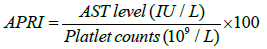
2 cut-off points were chosen to predict the absence (APRI<0.50) or presence (APRI <1.50) of significant fibrosis. Similarly, 2 cut-off points were chosen to predict the absence (APRI <1.00) or presence (APRI _ 2.00) of cirrhosis.
Child – Pugh scoring was done for the cirrhotic patients, used to assess the prognosis of chronic liver disease, mainly cirrhosis [11]. The score employs five clinical measures of liver disease (Table 1). Each measure is scored 1–3, with 3 indicating most severe derangement. Chronic liver disease is classified into Child–Pugh class A to C, employing the added score from above [11].
| Measure | 1 point | 2 points | 3 points |
|---|---|---|---|
| Total bilirubin (mg/dL) | (<2 | 2–3 | >3 |
| Serum albumin, g/dL | >3.5 | 2.8–3.5 | <2.8 |
| Prothrombin time | <4.0 | 4.0–6.0 | > 6.0 |
| Ascites | None | Mild (or suppressed with medication) | Moderate to severe (or refractory) |
| Hepatic encephalopathy | None | Grade I–II | Grade III–IV |
Table 1: Score employs five clinical measures of liver disease.
- Class A: 5-6 Points
- Class B: 7-9 Points
- Class C: 10-15 Points
Exclusion criteria
The following were excluded from the study:
a. Patients diagnosed with malignancy to avoid any effect on the TIMP-1 reading [12]
b. Patients with any degree of renal impairment to avoid any result misinterpretation
The research protocol was approved by the local Ethics Committee of the National Liver Institute, Menoufiya University and a written informed consent was obtained from all subjects participated in the study.
The following was done for the patients:
a. Full history and clinical examination: (for symptoms and signs of ascites and encephalopathy)
b. Abdominal Ultrasonography
c. Fibroscan
The following laboratory investigations were done:
Peripheral venous blood samples were drawn from all patients and the following laboratory investigations were done:
a. Liver enzymes: {alanine aminotransferase (ALT), Aspartate aminotransferase (AST), Bilirubin and Albumin} were measured on COBAS 6000 (Roche Diagnostics)
b. Complete Blood Count (WBC, Platelets) performed on 3 parts. Differential automated cell counter Sysmex KX- 21N (Sysmex, Kobe, Japan)
c. Prothrombin Time (INR)
Determination of plasma TIMP-1
Using the MAC15 TIMP-1 ELISA, this has a high sensitivity and specificity [13].
Blood collections and plasma separation
• Peripheral blood was drawn with minimal stasis into citrate, EDTA, or heparin collection tubes mixed by 5 times inversion, and immediately chilled on ice.
• As soon as possible (no later than 1.5 h after collection) the plasma was separated from blood cells by centrifugation at 4°C at 1200 g for 30 min, and stored frozen at –80°C prior to assay.
• When analysed, the samples were thawed quickly in a water bath at 37°C and placed on ice until the 1:100 plasma dilutions were made.
TIMP-1 ELISA
• A sensitive and specific sandwich ELISA was developed, using TIMP-1 antibodies
• A sheep polyclonal anti-TIMP-1 antibody was used for catching, a murine monoclonal anti-TIMP-1 IgG1 (MAC- 15) for detection of the antigen, and a rabbit anti-mouse immunoglobulins–alkaline Phosphatase conjugate enabled kinetic rate assay.
• The latter conjugate was supplied preabsorbed against human IgG, thus avoiding cross-reaction with IgG in the plasma samples.
• As the monoclonal detection antibody MAC-15 recognizes both free TIMP-1 and TIMP-1 in complex with MMPs ,the total TIMP-1 content of the measured sample captured by the sheep polyclonal anti-TIMP-1 antibody was determined by the ELISA.
• Immunoassay plates (96-well) (Maxisorp, Nunc, Roskilde, Denmark) were coated for 1 h at 37°C with 100 μl per well of polyclonal sheep anti-TIMP-1 (4 mg l–1) in 0.1 mol l–1 carbonate buffer, pH 9.5.
• Then the assay wells were rinsed twice with 200 μl per well of SuperBlockTM solution (Pierce Chemicals, Rockford, IL, USA) diluted 1:1 with phosphate-buffered saline (PBS).
• The immunoassay plates were stored for up to 14 days at –20°C.
• On the day of use the plates were thawed at room temperature and washed five times in PBS containing 1 g l–1 Tween-20.
• Wells were then treated for 1 h at 30°C with 100 μl per well of triplicate 1:100 dilutions of plasma made in a sample buffer consisting of 50 m mol l–1 phosphate, pH 7.2, 0.1 mol l–1 sodium chloride (NaCl), 10 g l–1 bovine serum albumin (Fraction V, BoehringerMannheim, Penzberg, Germany) and 1 g l–1 Tween-20.
• On every assay plate the first three columns of wells (each column consisting of eight wells) were incubated with a series of standards, consisting of seven serial dilutions in triplicate of purified recombinant human TIMP-1, starting at 10 μg l–1 then 5, 2.5, 1.25, 0.625, 0.313 and 0.156 μg l–1.
• Also included on each plate were triplicate blank wells containing only sample dilution buffer, and two sets of triplicate wells of a 1:100 dilution of a control citrate plasma pool; the first set of triplicate plasma pool was added as the first sample to the assay plate and the second set of triplicate plasma pool was added as the last.
• After TIMP-1 binding, the wells were washed five times, then treated for 1 h at 30°C with 100 μl per well of the purified murine monoclonal anti-TIMP MAC-15 (0.5 mg l-1) in sample dilution buffer.
• After another five washes, the wells were incubated for 1 h at 30°C with 100 μl per well of rabbit anti-mouse immunoglobulins–alkaline phosphatase conjugate diluted 1:2000 in sample dilution buffer.
• Following five washes with washing solution and three washes with pure water, 100 μl of freshly made p-nitrophenyl phosphate (Sigma, St Louis, MO, USA) substrate solution (1.7 g l–1 in 0.1 mol l–1 Tris-HCl, pH 9.5, 0.1 mol l–1 NaCl, 5 mmol l–1 magnesium chloride) was added to each well and the plate was placed in a Ceres 900TM plate reader (Bio-Tek Instruments, Winooski, VT, USA).
• The yellow colour development at 23°C was monitored automatically, with readings taken at 405 nm against an air blank every 10 min for 60 min.
• KinetiCalc II software was used to manage the data, calculate the rate of colour change for each well (linear regression analysis) and compute from the rates for the TIMP-1 standards a 4-parameter fitted standard curve, from which the TIMP-1 concentration of each plasma sample was calculated.
Statistical Methods
The data were collected and statistically analysed using SPSS computer program version 21. The data were expressed as mean ± SD and differences between 2 groups were analysed by student t test for parametric variable distribution or Mann-Whitney test for nonparametric. One way analysis of variance (ANOVA) F-test was used for comparison between more than 2 groups. Pearson’s correlation coefficient was used to test the relationship between various variables. P value is considered significant if <0.05.
Results
Forty six patients (31 males and 15 females) with chronic liver disease were included in the current study, with age ranged from 42 –63 years. Thirty patients suffered chronic HCV and Sixteen patients had liver cirrhosis. The primary cause of liver disease was hepatitis C (HCV) 89.1% (n=41), NASH (n=3) and unknown cause (n=2).
Table 2 shows the characteristics of patients in the two patient groups. TIMP-1 values showed significant relation to the degree of fibrosis or cirrhosis assessed by liver biopsy or Fibroscan (p<0.001).
| Non-cirrhotic (N = 30) Mean +/- SD | Cirrhotic (N = 16) Mean +/- SD | P value | |
|---|---|---|---|
| AST | 41 +/- 25 | 72 +/- 97 | |
| ALT | 47 +/- 33 | 50 +/- 59 | |
| Albumin | 4.4 +/- 0.3 | 2.6 +/- 0.5 | |
| Platelets | 206 +/- 62 | 85 +/- 24 | |
| Bilirubin | 0.7 +/- 0.5 | 1.9 +/- 1.3 | |
| WBC | 6.2 +/- 1.9 | 8.6 +/- 4.7 | |
| TIMP-1 | 156 +/- 93 | 269 +/- 103 | P <0.001 ** |
| APRI | 0.5 +/- 0.3 | 2.5 +/- 3.6 | |
| Ascites | N = 10 (tense 3) | ||
| Encephalopathy | N = 7 |
Table 2: Patient’s characteristics.
Table 3 shows the different TIMP-1 mean values in relation to grades of fibrosis. When we simplify it into two groups, cirrhotic or non-cirrhotic, it was also highly significant (p=0.001).
| Grades of fibrosis | Patients number | TIMP-1 means | |
|---|---|---|---|
| No fibrosis | 2 | 51 +/- 29 | P < 0.001 |
| Mild | 22 | 142 +/- 60 | |
| Moderate | 6 | 243 +/- 143 | |
| Cirrhosis | 16 | 269 +/- 103 |
Table 3: TIMP-1 means in different grades of fibrosis.
Interesting significant results appeared when we tested different patients’ liver parameters against TIMP-1, but when we tested these significant parameters in their patient groups (i.e. cirrhotic or noncirrhotic) we had insignificant results for most of them, which means the significance was related to liver dysfunction itself not these parameters. Only AST, ALT and APRI values were significant when we tested them in their patient group. Table 4 shows p values of these variables against TIMP-1 in different patient groups. Then we tested the cirrhotic group alone, using the Child – Pugh score, to see the effect of liver dysfunction degree on TIMP-1 levels, Cirrhotic patients were classified into Child A (n=4), Child B (n=6), Child C (n=6). Our results showed a significant correlation between the severity of liver dysfunction and TIMP-1 level. Again, when we tested its values against the presence or absence of hepatic encephalopathy or the patients’ grade of ascites, we had the same significant correlation. Table 5 below shows the different hepatic dysfunction parameters we used against TIMP-1.
| AST | ALT | APRI | |
|---|---|---|---|
| All patients (N = 46) | P = 0.001 | P = 0.002 | P = 0.002 |
| Non- cirrhotic (N = 30) | P = 0.003 | P = 0.005 | P < 0.001 |
| Cirrhotic (N = 16) | 0.013 | 0.025 | 0.019 |
Table 4: AST, ALT and APRI values tested against TIMP-1 in different patient groups.
| Patients number | TIMP-1 means | p values | ||
|---|---|---|---|---|
| Child – Pugh score | A | 1 | 150 | P = 0.025 |
| B | 8 | 229 +/- 55 | ||
| C | 7 | 368 +/- 120 | ||
| Hepatic encephalopathy | Absent | 9 | 217 +/- 53 | P = 0.018 |
| Present | 7 | 335 +/- 117 | ||
| Ascites | Absent | 6 | 219 +/- 47 | P = 0.039 |
| Mild | 7 | 247 +/- 83 | ||
| Moderate | 1 | 398 | ||
| Tense | 2 | 400 +/- 140 | ||
Table 5: Liver dysfunction in relation to TIMP-1.
Discussion
Finding a seromarker which could correlate well with the degree of fibrosis saves us the need for liver biopsy. Although we won’t probably be able to avoid completely the usage of biopsy, as it is the only test showing us what is really going on in the liver tissue, but we will be able to decrease its usage so much. Nowadays with the new drugs development for various liver diseases, including liver fibrosis, we need to reassess the degree of fibrosis more than one time but it is so difficult to convince your patient to have a second biopsy. Different types of markers showed good relation to fibrosis and TIMP-1 was one of those. It has been evaluated and validated in many studies and showed very good correlation to fibrosis [14]. That is becauseTIMP-1 is the important inhibitor of MMPs, and the MMP - TIMP balance determine the degree of fibrosis. It was agreed to be used in different fibrosis scoring systems like the Enhanced Liver Fibrosis (ELF) score [15]. What makes it a good marker is that, its level doesn’t change by sex, age, time of taking the sample or even the active alcohol consumption [16].
Because, there is a few or scarce published studies on TIMP-1 levels in Egyptian patients with chronic liver disease, we aimed to test its correlation with the degree of liver fibrosis in our patients with and without cirrhosis. Also we aimed to test it against the degree of liver dysfunction. We considered that Child class, presence or absence of encephalopathy or the patients’ grade of ascites, if present, are good parameters to evaluate hepatic dysfunction. We also tested it against APRI score which is one of the easiest and productive scores to use in evaluation of liver fibrosis. Our main findings are 1) TIMP-1 values significantly increase with the advancement of fibrosis and reach its highest levels with cirrhosis, 2) TIMP-1 also significantly increases with AST and ALT levels both in cirrhotic and noncirrhotic groups, 3) It also correlate well with the APRI score in both patient groups, 4) TIMP-1 showed good correlation with the degree of liver dysfunction and portal hypertension as its level increases progressively with the development of encephalopathy and with the worsening of Child class or ascites. Our results go with [17,18] who found TIMP-1 to be much higher in cirrhotic than non-cirrhotic. Again Busk et al. [13] showed the same results, besides their finding of its correlation to liver cirrhosis severity and portal hypertension. Why does the presence of encephalopathy or the advancement of ascites grade in cirrhotic patients showed significant increase in TIMP-1 levels, while other parameters failed to do so? This may be explained by that these two conditions could represent an advanced stage of decompensation. As shown in Table 5, we tested TIMP-1 in a cirrhotic patient group with about half of them had a sort of marked decompensation (Child C and encephalopathy in 43% for each and Ascites in 62.5% of patients) and this makes our results more reliable when we conclude that there is significant increase in TIMP-1 with the advancement of liver decompensation and its level could be used as an indirect measurement of the hepatic function state. We still need more studies with larger population to assess and examine a cut off value for cirrhosis and another one for hepatic decompensation, if possible.
Acknowledgments
We are indebted to all subjects for agreeing to participate in this study.
Declaration of Interest
There is no conflict of interest that could be perceived as prejudicing the impartiality of the research reported.
References
- Iredale JP (1997) Tissue inhibitors of metalloproteinases in liver fibrosis. Int. J Biochem. Cell Biol 29: 43-54.
- Thiele ND, Wirth JW, Steins D, Koop AC, Ittrich H, et al. (2017) IMP-1 is upregulated, but not essential in hepatic fibrogenesis and carcinogenesis in mice. Sci Rep 7: 714.
- Murphy FR., Issa R., Zhou X, Ratnarajah S, Nagase H, et al. (2002) Inhibition of Apoptosis of Activated Hepatic Stellate Cells by Tissue Inhibitor of Metalloproteinase-1 Is Mediated via Effects on Matrix Metalloproteinase Inhibition: implications for reversibility of liver fibrosis. J Biol Chem 277: 1069-1100.
- Benyon RC, Iredale JP, Goddard S, Winwood PJ, Arthur MJ (1996) Expression of tissue inhibitor of metalloproteinases 1 and 2 is increased in fibrotic human liver. Gastroenterology 110: 821-831.
- Boeker KH, Haberkorn IC, Michels D, Flemming P, Manns MP, et al. (2002) Diagnostic potential of circulating TIMP-1 and MMP-2 as markers of liver fibrosis in patients with chronic hepatitis C. Clin. Chim. Acta 316: 71-81.
- Yoshiji H, Kuriyama S, Yoshii J, Ikenaka Y, Noguchi R, et al. (2002) Tissue Inhibitor of Metalloproteinases-1 Attenuates Spontaneous Liver Fibrosis Resolution in the Transgenic Mouse. Hepatology 36: 850-860.
- Duarte S, Baber J, Fujii T, Coito AJ (2015) Matrix metalloproteinases in liver injury, repair and fibrosis. Matrix Biol 44-46: 147-156.
- Brew K, Nagase H (2010) The tissue inhibitors of metalloproteinases (TIMPs): an ancient family with structural and functional diversity. Biochim Biophys Acta 1803: 55-71.
- Borsoi VMS, Takei K, Collarile YDC, Guz B, Strauss E (2009) Use of AST platelet ratio index (APRI Score) as an alternative to liver biopsy for treatment indication in chronic hepatitis C. Ann Hepatol 8: 26-31.
- Wai CT, Greenson JK, Fontana RJ, Kalbfleisch JD, Marrero JA, et al. (2003) A simple noninvasive index can predict both significant fibrosis and cirrhosis in patients with chronic hepatitis. Hepatology 38: 518-526.
- Cholongitas E, Papatheodoridis GV, Vangeli M, Terreni N, Patch D, et al. (2005) Systematic review: The model for end-stage liver disease-should it replace Child-Pugh's classification for assessing prognosis in cirrhosis? Aliment Pharmacol Ther 22: 1079-89.
- Lai R, Rassidakis GZ, Medeiros LJ, Ramdas L, Goy AH, et al. (2004) Signal transducer and activator of transcription-3 activation contributes to high tissue inhibitor of metalloproteinase-1 expression in anaplastic lymphoma kinase-positive anaplastic large cell lymphoma. Am. J. Pathol 164: 2251-2258.
- Holten-Andersen MN, Murphy G, Nielsen HJ, Pedersen AN, Christensen IJ, et al. (1999) Quantitation of TIMP-1 in plasma of healthy blood donors and patients with advanced cancer. Br. J. Cancer 80: 495-503.
- Rosenberg WM, Voelker M, Thiel R, Becka M, Burt A, et al. (2004) Serum markers detect the presence of liver fibrosis: a cohort study. Gastroenterology 127: 1704-1713.
- Lichtinghagen R, Pietsch D, Bantel H, Manns MP, Brand K, et al. (2013) The Enhanced Liver Fibrosis (ELF) score: normal values, influence factors and proposed cut-off values. J. Hepatol 59: 236-242.
- Ponomarenko Y, Leo MA, Kroll W, Lieber CS (2002) Effects of alcohol consumption on eight circulating markers of liver fibrosis. Alcohol Alcohol 37: 252-255.
- Fontana RJ, Goodman ZD, Dienstag JL, Bonkovsky HL, Naishadham D, et al.(2008) Relationship of serum fibrosis markers with liver fibrosis stage and collagen content in patients with advanced chronic hepatitis C. Hepatology 47: 789-798.
- Busk TM, Bendtsen F, Nielsen HJ, Jensen V, Brünner N, et al. (2014) TIMP-1 in patients with cirrhosis: relation to liver dysfunction, portal hypertension, and hemodynamic changes. Scand. J. Gastroenterol 49: 1103-1110.