Spanish
  Chinese
  Russian
  German
  French
  Japanese
  Portuguese
  Hindi
Hindi Research Article, J Clin Exp Oncol Vol: 15 Issue: 1
Multitargets and Interactions of Wortmannin with MSC-Mitogenic Proteins Validate the Enhanced Antiproliferative Effect of MSCSecretome on Breast Cancer Michigan Cancer Foundation-7 Cells
Doha F. Ismail1, Saad M Elgendy2, Mai M El-Keey1 and Mohamed Hessien1*
1Department of Biochemistry, Tanta University, Tanta, Egypt
2Department of Cancer Biology, National Cancer Institute, Cairo University, Cairo, Egypt
*Corresponding Author: Mohamed Hessien
Department of Biochemistry, Tanta University, Tanta, Egypt
E-mail: Mohamed.Hussien1@science.tanta.edu.eg
Received date: 28 November, 2024, Manuscript No. JCEOG-24-153516; Editor assigned date: 03 December, 2024, PreQC No. JCEOG-24-153516 (PQ); Reviewed date: 17 December, 2024, QC No. JCEOG-24-153516; Revised date: 03 April, 2025, Manuscript No. JCEOG-24-153516 (R); Published date: 10 April, 2025, DOI: 10.4172/2324-9110.1000346
Citation:Ismail DF, Elgendy SM, El-Keey MM, Hessien M (202) Multitargets and Interactions of Wortmannin with MSC-Mitogenic Proteins Validate the Enhanced Antiproliferative Effect of MSC-Secretome on Breast Cancer Michigan Cancer Foundation-7 Cells. J Clin Exp Oncol 14:2.
Abstract
Keywords: MSC; PI3K/Akt/mTOR; Wortmannin; Breast cancer; Target prediction; Chemo-regenerative therapy
Introduction
Mesenchymal Stromal Cells (MSCs) are distinguished by renewal, clonality and differentiation into multilineage cells. Since their emergence about five decades ago, MSCs have been commonly presented as a promising cellular regenerative therapy. However, numerous concerns have been raised about the complications associated with MSC transplantation due to the adverse conditions they challenge after transplantation, such as loss of the Extracellular Matrix (ECM), hypoxia and host-related inflammatory responses. For these reasons, the cell-free approach was strongly suggested as a novel therapeutic approach, which depends on the MSC-secretome and the Extracellular Vesicles (EVs) that contain considerable amounts of biologically active growth factors and cytokines. Surprisingly, these regulatory molecules include a mix of mitogenic (proliferative), antiproliferative and anti-inflammatory factors. Mitogenic growth factors stimulate cell proliferation and trigger differentiation-related events including cell cycle progression, DNA replication and mitosis. In this context, previous studies demonstrated the role of MSCs in tumor development via the pro-survival or anti-apoptotic factors including VEGF, FGF, PDGF, TGF-β, SDF-1α and HGF they secret. Also, VEGF and FGF can boost the expression of the antiapoptotic gene Bcl-2, whereas PDGF and TGF-β enhance VEGF and FGF expression to support tumor angiogenesis. Other studies reported that SDF-1α, can protect leukemia cells against spontaneous apoptosis. Similarly, the crosstalk between MSC-secretome and cancer cells has been found to regulate Epithelial-Mesenchymal Transition (EMT), metastasis and the development of cell resistance to chemotherapy and immunotherapy. These scenarios depict that the co-existence of MSCs in the tumor microenvironment offers cancer cells proliferative advantages [1].
In another context Wortmannin (Wortmn), a steroidal metabolite originally isolated from Penicillium wortamanni, is known as a potent inhibitor of Phosphatidylinositol 3-Kinases (PI3Ks) and Phosphatidylinositol 3-Kinase-related Kinases (PIKKs). Wortmn anticancer effect is established via inhibiting the P13K/Akt-mTOR signaling that manages cell growth, proliferation, survival and motility. Abnormalities in P13k/Akt pathways are often associated with the overexpression of PI3K catalytic subunits (p85 and p110) that derive cancer development, progression, metastasis and drug resistance. However, Wortmn has no clinical applications due to the associated severe hepatotoxicity and pharmacological challenges. To ease its clinical use, several approaches have been suggested to minimize its hepatoxicity and enhance its water solubility via structural modifications, generating structural derivatives, conjugation with water-soluble compounds or nanoformulation. Also, an alternative administration approach was invested to improve its efficacy and reduce the associated complications. Accordingly, it is suggested that combining MSC-CM with drugs that inhibit PI3K/AktmTOR pathway, may reduce drug-associated complications and enhance the therapeutic effect of MSC-CM. At this point, it is important to define Wortmn cellular targets in cancer cells and know how far it can interact with MSC-mitogenic proteins. So, this research was designed to explore the interaction of Wort with MSC-derived mitogenic proteins in silico and evaluate the cytotoxic effect of MSCCM combined with Wortmn on breast cancer cells [2].
Materials and Methods
Chemicals and reagents
1-(alpha, 11 alpha)-11-Acetyloxy-1-methoxymethyl-2- oxaandrosta-5,8-dieno (6,5,4-bc) furan-3,7,17-trione (Wortmannin) was delivered by Toronto Research Chemicals, Canada (Cat. no. W499400). MTT from Sigma, Acridine Orange (AO), ethidium bromide and DAPI were from InvitrogenTM. Tissue culture media (Minimum Essential Medium-alpha (MEM-α) with nucleosides, Lglutamine- containing Dolbecco’s Minimum Essential Medium (DMEM) and cell culture supplements including antibioticantimycotic (Pen/Strep), Fetal Bovine Serum (FBS) and Trypsin- EDTA were from Lonza PharmaBiotech [3].
BM-MSCs isolation and preparation of conditioned media
Initially, MSCs were isolated from a male rat (Lewis, 200 g body weight) bone marrow as previously reported following the ethical regulation. The work design was approved by the ethical committee, Faculty of Science, Tanta University (IACUC: SCI-TU-0214). The cavities of the femoral and tibia bones were flushed in MEM-α, cells were incubated overnight at 37°C and 5% CO2. Next, MSCs were propagated to the fourth passage using standard procedure. To authenticate MSC cells, their mesenchymal surface markers were evaluated by flow cytometry to determine the expression pattern of CD105 and CD45. Briefly, cells were incubated for 30 min with CD marker-specific monoclonal antibodies and then analyzed by a flow cytometer. To prepare conditioned media, MSCs were maintained in 0.1% FBS media for 48 h, after which media were recovered [4].
Cancer cells and treatment
Michigan Cancer Foundation-7 (MCF-7) breast cancer cells were delivered by VACSERA, Cairo. MCF-7 cells were cultured in Lglutamine- containing DMEM complete media (supplemented with 10% heat-inactivated FBS, 1% penicillin/streptomycin), incubated in humidified conditions at 5% CO2 and 37°C and then treated with 100 nM Wortmn or MSC-CM containing 100 nM Wortmn [5].
Assessment of cell viability
The viability of cells was evaluated by MTT assay, where they were cultured (at 1 × 10⁴ cells/well), incubated overnight in a 96-well plate at 37°C and 5% CO2 and treated with Wortmn in DMEM or Wortmncontaining conditioned media (MSC-CM). Next, cells were labeled with MTT and incubated at 5% CO2 and 37°C for 4 h. After medium decantation, acidified isopropanol was added and the OD was measured at 630 nm. The percentage of viable cells was normalized against the corresponding drug-free cells [6].
Apoptosis assessment by fluorescence microscopy
To assess apoptosis-mediated cell death, morphology and membrane permeability of treated MCF-7 cells, AO/EB staining was used. Briefly, cells (105 cells/well) were incubated in Wortmn-free MSC-CM or Wortmn-containing MSC-CM for 48 h. After old media removal, cells were washed with PBS, dually stained with AO/EB working solution for 5 min and visualized under a fluorescence microscope. To monitor apoptosis and nuclear changes, DAPI staining was carried out on cells (2 × 10⁴ cells/well) in 96-well plates. After overnight incubations, cells were treated as shown above for 48 h. Next, cells were stained with DAPI for 10 min, washed with PBS and fixed with formaldehyde. Finally, cells were observed under a fluorescence microscope [7].
Target fishing
The cellular targets of Wortmn were predicted using the Similarity Ensemble Approach (SEA) (https://sea.bkslab.org/), TargetNet (http:// targetnet.scbdd.com/), Polypharmacology Browser-2 (PPB2) (https:// ppb2.gdb.tool), Swiss target prediction (http://www.swiss targetprediction.ch/), and SuperPred (https://prediction.charite.de/). The Comparative Toxicogenomics Database, (www.CTD.org), Metascape (https://metascape.org) and GeneMania (https://genemania.org) were used to define target-related cellular activities, gene ontology, and other analyses.
Docking analysis
Ligands structures (Wortmn and paclitaxel) were downloaded from PubChem site. The Three-Dimensional (3D) structures of MSCderived mitogenic proteins were retrieved from the Protein Database (PDB). Molecular operating environment software was utilized for docking analysis. The 3D structure of the ligand binding active site was performed using BIOVIA Discovery Studio Visualizer. All structure minimizations were conducted until RMSD gradient of 0.05 kcalâ??mol-1Å-1 with MMFF94x force field and partial charges were automatically calculated for EGFR (PDB:1M17), G-CSF (PDB: 1CD9) and TGF-β3 (PDB: 1TGK). Initially, the analysis procedure was validated by self-docking of the co-crystallized ligand near the binding sites of targets. Then, the authenticated docking protocol was used to study the ligand-protein interactions at the binding site for the reported factors to predict their binding mode and affinity. The inhibitory activity of Wortmn was compared to paclitaxel through computational analysis-based investigations [8].
Statistical analysis
The results of cell viability were expressed as means of at least 3 data sets (± standard deviation). The significance of differences between means was analyzed by ANOVA and post hoc Tukey’s honestly significant difference tests. Differences were considered statistically significant at p<0.05.
Results
Antiproliferative effect of MSC-CM/Wortmn combination MSCs were isolated from the bone marrow cells of a rat (Figure 1A) and then maintained to the fourth passage. Cells were morphologically characterized, where they demonstrated spindle fibroblast-like shape under an inverted (phase contrast) microscope (Figures 1B and C). Also, cells were highly and minimally expressing CD105 and CD45, respectively indicating their mesenchymal characteristics (Figures 1D and E). Treatment of MCF-7 cells with MSC-CM or Wortmn-containing MSC-CM for 48 h led to cell death. Morphologically, cells demonstrated apoptosis features such as cell rounding, shrinkage, and floating plate detachment (Figures 2A-C). Also, the MTT assay demonstrated a significant decrease in MCF-7 viability to 70.2% when they were incubated with MSC-CM and a further reduction (P<0.001) in cell viability (49.25%) in cells incubated in Wortmn-containing MSC-CM (Figure 2D). Moreover, AO/EB dual staining and fluorescent microscopy demonstrated the development of apoptosis where cell nuclei demonstrated dark orange to red fluorescent because of the interaction of EB indicating the disintegration of the cell membrane and a massive infusion of EB that interacted with nuclear DNA (Figure 2E-H). Also, nuclear staining with DAPI demonstrated the progressive dye influx into the cells with chromatin condensation and fragmentation (Figures 3A-D) [9].
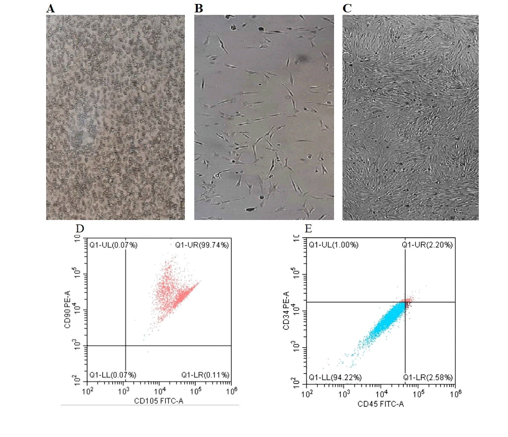
Figure 1: Morphological and phonotypical characterization of BMMSCs utilized to obtain MSC-CM. A-C are representative phasecontrast micrographs of cells aspirated brown marrow cells (A), BMMSCs at day 4 (B), and confluent 4th passage cells (C). “D” and “E” are scatter plots of the expression of the mesenchymal surface marker (CD105), and the hematopoietic surface marker (CD45).
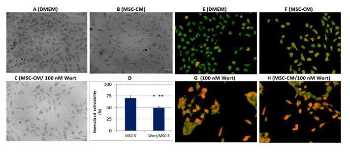
Figure 2: Cell-death effect of MSC-CM and Wort-containing MSC-CM on MCF-7. A-C are representative photomicrographs of untreated cells (A), cells treated with MSC-CM (B), and cells treated with 100 nM wortmn-containing MSC-CM (C). (D) is a bar graph of normalized cell viability of cells incubated for 48 h in MSC-CM without or with 100 nM Wortmn. (E) to (H) are micrographs demonstrating the nuclear morphology using dual staining with acridine orange and ethidium bromide (AO/EB). Note: ***indicates P<0.001.
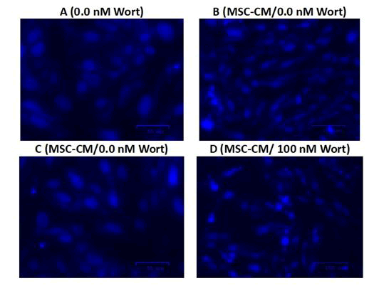
Figure 3: Fluorescent microscopy for apoptosis detection by nuclear staining of treated cells with DAPI.
Prediction of wortmannin cellular targets
In silico studies predicted that Wortmn specifically inhibits PI3Krelated proteins in cancer cells including the integral P110-alpha and P85-alpha catalytic subunits as three (out of five databases) nominated PI3K subunits including p110-delta, p110-beta and delta. Furthermore, the top 10 targets included other kinases such as Myosin light chain kinase, Ser/Thr-protein kinase mTOR, Ser/thr-protein kinase PLK1 and Serine/threonine-protein kinase PLK3. In parallel, other target prediction databases (TargerNet, http://targetnet.scbdd.com) nominated additional candidate genes including aromatase, arachidonate 15-lipoxygenase (ALOX15B), heat shock protein HSP-90 (HSP90AA1), Receptor- Interacting Serine/Threonine-Protein Kinase 2 (RIPK2), M-phase inducer phosphatase-2, acetylcholine receptor M4 (CDC25B), perilipin-5 (Plin5) and Hepatocyte Nuclear Factor 4-alpha (HNF 4A) with high probability (>0.9). As Wortman is metabolized to the hydroxylated derivative (17-hydroxy Wortmn), which is more potent than Wortman, the PPB2 database predicted 65%, 30% and 5% of Wortmn targets are enzymes, membrane receptors, or transcription factors, respectively. The hydroxylated derivative, however, targets more transcription factors (10%) and other unclassified proteins. According to Swiss target prediction, 66.7% of Wortmn targets are cellular kinases, whereas 33.3% are other enzymes (Figures 4A-C). To explore the widespread genome and gene-gene interactions of Wortmn the Comparative Toxicogenomics Database (CTD) was employed. The direct effect of Wortmn on the genome of the treated cells is restricted to intercellular adhesion molecule 1 (ICAM) and cycle D2 (CCDN2). However, its widespread effect on cellular proteins nominated 485 targets in human cells. In human breast cancer, the CTD database nominated 33 genes involved by protein kinase, 30 genes involved in ERK1 and ERK2 pathways, 38 genes involved in cell migration (metastasis) and 33 genes involved in EGF are negatively affected by Wortmn. Gene lists and the corresponding gene-gene interactions are shown in Figures 4 D-G.
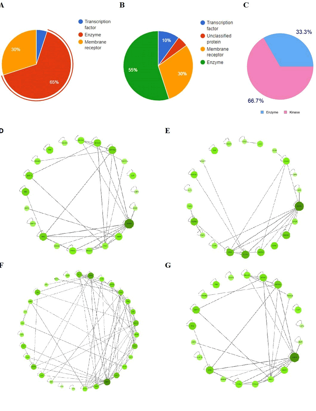
Figure 4: Classification of wortmannin cellular targets and genegene interaction cytoscape graphs in human breast cancer. A and B demonstrate cellular targets of wortmannin (A) and its metabolite (17- hydroxy wortmannin) (B), as predicted by PPB2 and Swiss prediction databases (C). D-G are gene-gene interaction pathways of genes modulated in human breast cancer by Wortmn and involved in Akt signaling (D), ERK1 and ERK2 pathways (E), cell migration (metastasis) (F), and EGF regulation (G). Gene network interactions are represented and retrieved by CTD database.
Wortmannin interaction with MSCs secretome
Next, the interaction forms between Wortmn and some mitogenic proteins commonly secreted by MSCs were analyzed in silico and compared with paclitaxel as a reference drug commonly utilized in many cancers including breast cancer. The molecular interactions analysis revealed that Wortmn docking scores ranged from -6.643 to -4.4185, as it interacts with TGF-β3 (Docking energy -4.4492 kcal/ mol), G-CSF (Docking energy -4.412 kcal/mol), and EGFR (Docking energy -6.53 kcal/mol) (Figures 5A-C). The close similarity between Wortmn and PTX is supported by the high number of signaling events (371) commonly modulated by both drugs (Figure 6).
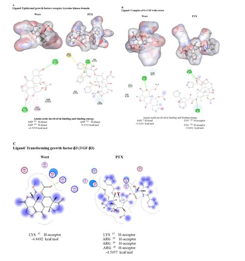
Figure 5: Molecular interactions of wortmannin with the crystal structures of mitogenic growth factors released by MSCs including the Epidermal Growth Factor Receptor (EGFR) (A), Granulocyte-Colony Stimulating Factor (G-CSF) (B), and Transforming Growth Factorbeta3 (TGF-β3). The docking energy (in kcal/mol), structures and amino acids involved in binding are shown next to each factor.
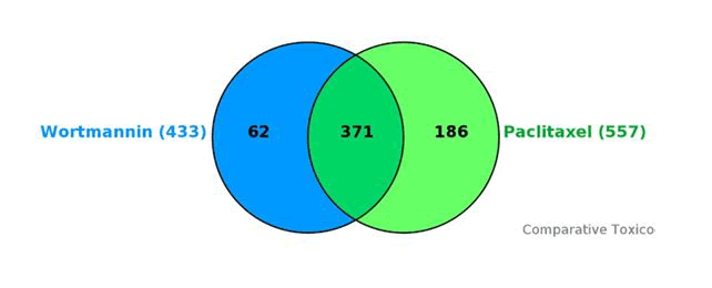
Figure 6: Enriched pathways commonly modulated by wortmannin and paclitaxel generated by CTD Vinn Viewer. The diagram refers to cellular signaling events (371) commonly regulated by wortmannin and paclitaxel.
Discussion
This work demonstrated the additive anticancer effect of Wortmn with MSC-conditioned media, where they showed antiproliferative effects against breast cancer cells. The mechanism seems to involve the direct and combined apoptotic effect of Wortmn in addition to its role in targeting PI3K catalytic subunits and other cellular proteins. Moreover, Wortmn revealed a reasonable molecular interaction with growth factors commonly released by MSCs and mitogenic receptors commonly utilized in cell growth and differentiation. This interaction may abolish the proliferative effect of mitogenic components, thus synergistically enhancing the anticancer effect of MSC-CM. These observations demonstrate that the regenerative potential of MSCs relies on their paracrine effect via the growth factors, cytokines, chemokines, interleukins and growth factors they contain. Previously,we and other investigators reported the anti-inflammatory, antioxidant and antiproliferative role of MSCs and their secretomes. The anticancer effect of secretome derived from umbilical cord or adipose tissue MSCs, for example, was repeatedly reported in several types of cancers including breast cancer. Conversely, the localization of MSCs in the tumor microenvironment promoted tumor growth and enhanced breast cancer metastasis. In a similar context in regenerative-based wound healing, MSCs enhanced skin fibroblast proliferation. To eliminate such mitogenic effect of MSCs we combined Wortmn with MSC-CM to promote their antiproliferative effect against breast cancer cells. Similar studies utilized other anticancer drugs such as Paclitaxel (PTX), that demonstrated noticeable inhibition on the survival and metastasis of the highly invasive Triple-Negative Breast Cancer (TNBC) cells. In agreement with previous studies, Wortmn alone or loaded on MSC-CM, induced apoptosis-mediated cell death, as it inhibits PI3K classes (IA, IB, II and III), leading to the inhibition of PI3K/Akt-mTOR cascade. Although the drug has a superior anticancer effect in vitro and in vivo, it is encountered with hepatotoxicity, water insolubility, pharmacological and delivery challenges, resulting in its unsuccessful utilization in clinical applications. To solve this problem, it is important not only to enhance the antiproliferative effect but also to bypass the post-transplantation complications associated with MSC engraftment. The in silico analysis we conducted raised two important issues. The first is concerned with the widespread effect of Wortmn on cellular proteins, where targets PI3K catalytic subunits (α, β, δ and γ). Additionally, Wortmn targets other cellular proteins indicating its promising anticancer potential. Moreover, the analysis predicted that Wort interacts with many mitogenic factors commonly released in MSCCM with binding energies close to paclitaxel, commonly used as anticancer chemotherapy in breast cancers (Commercially known as Taxol, Taxan, or Unitaxel). These interactions may tilt the proliferation/antiproliferation potential of MSC-CM towards the anticancer effect. Further, we precisely explored the phenotypes associated with Wortmn interactions in human breast cancer using CTD database, where gene-gene interaction suggested different sets of genes negatively modulated by Wortmn including those involved in the regulation of protein kinase B signaling, ERK1/2 pathways, cell migration and EGF-related events.
Moreover, two target fishing tools nominated other targets including PRKDC, MYLK, mTOR, PLK1/3, aromatase, HSP90, CDC25B, CHRM4 and HNF4A.
This gene list was utilized to analyze the signaling pathways, their interaction and colocalization. Although PI3K-mTOR pathway is the classical signaling pathway targeted by Wortmn, other signaling pathways were identified indicating the PI3K-independent cytotoxic effect. Wortmn affects major cellular processes including protein phosphorylation, cell response to osmotic stress and regulation of protein transport. Cell metabolic process dominates the role of Wortmn target genes.
Conclusion
In conclusion, the present study suggests that loading MSCs-CM with a pan PI3K/Akt inhibitor, such as Wortmn, is a promising strategy in cancer treatment. The antiproliferative effect of Wortmnimpregnated MSC-CM was mediated by apoptosis-mediated cell death via Wortmn-dependent PI3K/Akt-mTOR inhibition. This may be explained to the direct impact of Wortmn on cell survival and via the interaction of Wortmn with several mitogenic proteins released by MSCs. Merging traditional anticancer drugs with MSCs secretome or extracellular Merging traditional anticancer drugs with MSCs secretome or extracellular vesicle may present a new therapeutic strategy having several advantages compared to chemotherapy or cellbased transplantation chemotherapy or MSC transplantation. First, the anticancer drug is diluted to less immunogenic conditions. Second: The synergistic anticancer effect may permit the utilization of lower doses of chemotherapy, thus minimizing the associated hepatotoxicity. Further studies should be undertaken to explore the direct impact of Wortmn and similar compounds, on the immunomodulatory proteins of MSCs secretome and their therapeutic efficacy.
Authors’ Contributions
Conceptualization: MH; Methodology and investigations: DI, SSA, SelG and MelK; Software and data analysis: MH, SSA; Draft preparation: MH and DI; Review and editing: MH, SelG. All the authors have read and approved the manuscript.
Funding
Science and Technology Research Fund (STDF), Postgraduate Support Grant (PGSG) program (Grant number 48609), and the Academy of Scientific Research and Technology (ASRT), Egypt (grant ID: RESPECT21-9999).
Data Availability
The data supporting the findings of this study are available from the corresponding author HM, upon reasonable request.
Competing Interest
All authors declare that they have no competing interests.
Acknowledgment
The authors would like to thank The Academy of Scientific Research and Technology (ASRT), and the Science and Technology Development Fund (Postgraduate Support Grant program), for their financial support.
References
- Salem HK, Thiemermann C (2010) Mesenchymal stromal cells: Current understanding and clinical status. Stem Cells 28: 585-596.
[Crossref] [Google Scholar] [PubMed]
- Baldari S, di Rocco G, Piccoli M, Pozzobon M, Muraca M, et al. (2017) Challenges and strategies for improving the regenerative effects of mesenchymal stromal cell-based therapies. Int J Mol Sci 18: 2087.
[Crossref] [Google Scholar] [PubMed]
- Vakhshiteh F, Atyabi F, Ostad SN (2019) Mesenchymal stem cell exosomes: A two-edged sword in cancer therapy. Int J Nanomedicine 14: 2847-2859.
[Crossref] [Google Scholar] [PubMed]
- Hung SC, Pochampally RR, Chen SC, Hsu SC, Prockop DJ (2007) Angiogenic effects of human multipotent stromal cell conditioned medium activate the PI3K-Akt pathway in hypoxic endothelial cells to inhibit apoptosis, increase survival, and stimulate angiogenesis. Stem Cells 25: 2363-2370.
[Crossref] [Google Scholar] [PubMed]
- Dias S, Choy M, Alitalo K, Rafii S (2002) Vascular endothelial growth factor (VEGF)-C signaling through FLT-4 (VEGFR-3) mediates leukemic cell proliferation, survival and resistance to chemotherapy. Blood 99: 2179-2184.
[Crossref] [Google Scholar] [PubMed]
- Konig A, Menzel T, Lynen S, Wrazel L, Rosen A, et al. (1997) Basic fibroblast growth factor (bFGF) upregulates the expression of bcl-2 in B cell chronic lymphocytic leukemia cell lines resulting in delaying apoptosis. Leukemia 11: 258-265.
[Crossref] [Google Scholar] [PubMed]
- Brogi E, Wu T, Namiki A, Isner JM (1994) Indirect angiogenic cytokines upregulate VEGF and bFGF gene expression in vascular smooth muscle cells, whereas hypoxia upregulates VEGF expression only. Circulation 90: 649-652.
[Crossref] [Google Scholar] [PubMed]
- Burger JA, Tsukada N, Burger M, Zvaifler NJ, Dell'Aquila M, et al. (2000) Blood-derived nurse-like cells protect chronic lymphocytic leukemia B cells from spontaneous apoptosis through stromal cell-derived factor-1. Blood 96: 2655-2663.
[Google Scholar] [PubMed]
- Ridge SM, Sullivan FJ, Glynn SA (2017) Mesenchymal stem cells: Key players in cancer progression. Mol Cancer 16: 31.
[Crossref] [Google Scholar] [PubMed]