Spanish
  Chinese
  Russian
  German
  French
  Japanese
  Portuguese
  Hindi
Hindi Research Article, J Ergon Res Vol: 1 Issue: 1
Neck-Shoulder Main Musculature is the Major Cervical Compression Producers during Single-Hand Lifting
1Department of Biomedical Engineering, Construction Engineering, University of Nebraska-Lincoln, Lincoln, NE, USA
2Department of Industrial and Management Systems Engineering, University of Nebraska-Lincoln, Lincoln, NE, USA
*Corresponding Author : Mohamed R. Amar
Department of Biomedical Engineering, Construction Engineering, University of Nebraska-Lincoln, Lincoln, NE, USA
Tel: +1-402-326-0805
E-mail: m.ammar.82@huskers.unl.edu
Received: December 21, 2017 Accepted: December 29, 2017 Published: March 07, 2018
Citation: Amar MR, Cochran D (2018) Neck-Shoulder Main Musculature is the Major Cervical Compression Producers during Single-Hand Lifting. J Ergon Res 1:1.
Abstract
Objective: Cervical spine injuries have been associated with work tasks requiring heavy arm and shoulder exertions. Current biomechanical models established to estimate compressive forces acting on the cervical spine do not include the contributions of the muscles surrounding the neck in supporting the shoulder and arm during these exertions. This paper presents a biomechanical analysis of compressive force as a result of single-hand lifting. Method: It was hypothesized that contraction of the three main shared muscles are the major producers of the cervical compression. To test this hypothesis, bilateral Electromyography data of major shared musculature (upper trapezius, sternocleidomastoid, and levator scapula) and posture data were collected from twenty subjects preforming lifting of five different weights from twenty different locations produced by the interaction of varying heights, reach distance, and angles simulating the work done by assembly line workers. All lifting tasks were done by the right hand. Analysis of the moment produced by these muscles forces and the head’s mass at the C4/C5 cervical spine intervertebral disc were performed to investigate moment balance. A coordinate system was set on segment at the C4-C5 disc center and the moment was calculated around two axes; the X, which divides the segment into front and back, and around Y, which divides the segment into right and left. Results: A t-test of the summation of moments around each axis was performed and results showed that the moment was balanced. Conclusion: A balance in moment indicates that the shared muscles considered are the major producers of cervical compression during single hand lifting. These findings also demonstrate that any attempt to develop a biomechanical modeling to estimate the cervical compression must include the contribution of hand lifting activities
Keywords: Normalized electromyography; Maximum voluntary contraction; Shared musculature; Cervical spine; Musculoskeletal disorders; Moment balance; Compressive forces.
Introduction
Current biomechanical estimation of the cervical spine compression does not account for arm work contribution. The existing biomechanical models of the cervical spine have adopted a cutting-plane approach and have only dealt with muscle forces generated in response to external neck moments [1-3]. The existence of an external moment on worker’s head while performing hand work activities, like work in assembly lines, appears to be not realistic; therefore, compressive forces acting on the cervical spine could only be the result of the shared muscles contracting to support the shoulder during hand activities and the mass of the head. Our previous work has demonstrated that the main shared musculature the upper trapezius, sternocleidomastoid, and levator scapula muscles on both sides of the neck were active during single hand lifting activities and the muscle activities are scientifically affected by work layout factors [4]. This paper presents a test hypothesis that most of the forces on cervical spine are generated by the contraction of the major shared muscles between the neck and the shoulder and the mass of the head. If true, future attempts to estimate the compression of the cervical spine must account for hand lifting activities. This paper utilizes data and findings presented in our previous paper summarized in the method section [4].
Literature Review
Neck region is a common site of work-related musculoskeletal injuries and disorders [5]. The frequent sites of neck injury are C5 level (74%), C4 level (16%), and C6 level (10%) [6]. The use of upper extremities in working activities has been linked to neck musculoskeletal injuries. In work related activities neck pain has been reported to be as high as 37% for food packers, 31% for cash register operators, 27% for office workers and 63% for welders [7-9]. It was concluded from epidemiologic studies that there is an ‘evidence’ connecting forceful exertion of the arm and the occurrence of musculoskeletal injuries in the neck [5,10]. Hand lifting is one of the causes of the cervical-disc complex disorders [11]. A variety of occupations and/or work activities have been studied experimentally to understand the factors associated with neck musculoskeletal disorders. Surface EMG (EMGs) of the neck muscles is often used to understand the mechanism of neck MSD. Lannersten and Harms Ringdahl, for example, studied the effect different cash registers had on the EMGs activity levels of the infraspinatus, trapezius, and erector spinae muscles for cash register operators [12]. Based on the pattern of EMG activities of the muscles studied, the authors concluded that keyboard and pen reader registers generated lower EMGs values than scanners in which the cashier needs to lift the product and scan it. In a similar study, Takala and Viikari-Juntura found that reducing the height of the service counter by 25 cm reduces the EMG amplitudes for the right upper trapezius of female bank cashiers [13]. Dennerlein and Johnson studied the effect of different positions of computer mouse within computer workstations to evaluate biomechanical risk factors across different mouse positions [14]. The high mouse position resulted in the highest level of muscle activity. Anton, Rosecrance, Gerr, Merlino, and Cook studied construction workers in a laboratory setting to evaluate the effect of two different types of concrete blocks [15]. Their results showed that the activity in the upper trapezius muscle was not affected by the block weight, but increased as the height of the wall increased. Lindberg, Frisk-Kempe, Linderhed, and Eklund studied upper trapezius muscle activities for manual vs. automated fabric-seaming tasks [16]. Analysis EMG amplitude revealed a higher risk of MSD for the manual seaming than the automated seaming tasks. In a similar study, Pitts, Aghazadeh, and Harvey evaluated ten dentists using EMG [17]. He found that EMG of the upper trapezius muscle revealed signs of fatigue at the end of 8 hours shift. Nimbarte also measured EMG in the sternocleidomastoid and the upper trapezius muscles during several different types of two handed lifting tasks under 25%, 50%, and 75% exertion of their maximum strength [18]. It was found that EMG magnitude of the upper trapezius as % MVC is higher than the % MVC observed in the sternocleidomastoid. The author concluded that both load lifted and the vertical position significantly affect the activation of these muscles with female contract their sternocleidomastoid harder. With such evidence of active shared muscles during hand-lifting activities, current biomechanical estimation of the cervical spine compression does not account for arm work contribution. The existing biomechanical models have only dealt with muscle forces generated in response to external neck moments [1-3]. The existence of an external moment on worker’s head while performing hand work activities, like work in assembly lines, appears to be not realistic; therefore, compressive forces acting on the cervical spine could only be the result of the shared muscles contracting to support the shoulder during hand activities and the mass of the head.
Materials and Methods
Subjects
Ten subjects, five males and five females, participated and gave their informed consent to the procedure, which was approved by the University of Nebraska-Lincoln Institutional Review Board. The mean height of the subjects was 170.8 (SD 6.01) cm, body weight 69.68 (SD 10.57) kg and age 29 (SD 4.96) years. All subjects were right handed and were screened for health history and were accepted only if they were without a history of back, neck, shoulder, arm, wrist, or hand pathology.
Experiment
This study used a 5 × 2 × 2 × 5 design with the 5 weights, 2 heights, 2 reach distances, and 5 angles (distribution). Each subject lifted five different weights (1, 1.5, 2, 2.5, and 3) kg from 20 different locations at varies heights, reach distances, and angles. Subjects were asked to sit in a chair and adjust the chair height until their elbows were parallel to the table surface. A foot rest was provided when needed. These loads were randomly distributed on the table in front of the subject and they were lifted using just the right hand. The subjects were instructed to lift each weight slightly (about 2 inches) off the surface, maintain that for 2-3 seconds, and then place it back on the surface. Each subject performed a total of 100 trials in random order. As a result, each muscle of the six muscles studied had a total of 100 responses. While performing these lifting tasks, the EMGs and the posture data were recorded using the EMGs system and Motion capture system respectively. A rest period of one to two minutes was provided between each trial with additional time upon request. After all trials had been successfully performed, the series of maximum voluntary muscle contractions were repeated. More details of the experimental layout, equipment, and data processing can be found on our previous paper [3].
EMGs normalization
For each trial, EMG values were normalized based on a maximal voluntary contraction (MVC) for each of the six muscles in the study. A normalized EMG value was calculated as:
NEMG = EMGTask /MVC
Where, EMGTask was the measured filtered and integrated EMG value for a particular experimental trial, and MVC was the highest value based on a series of MVC trials for each muscle. The MVC normalization procedures were conducted consistent with a previous work [2,12,14,16,18,19].
Analysis
NEMG -Force relation
As isometric force increases, the magnitude of EMG signal also increases [20-26]. Muscle force has been found to have a nonlinear relationship to the normalized EMG [23]. This non-linear relationship was experimentally quantified in isometric contractions by [27-29], given as:
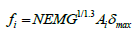
Where fi is the ith muscle force in Newton, Ai is the muscle cross-sectional area in m2,δmax is the maximum muscle force per unit of cross-sectional area that is equal to (3.5×10⁵ N.m2), NEMG is normalized EMG 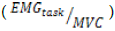 [3].
[3].
Moment calculations
Three bilateral pairs of neck muscles were included in the moment analysis. The muscles originating or inserting at the shoulder level, crossing the C4-C5 level and running parallel to the cervical spine, were included in the model. The muscles that were included are; Right and Left Sternocleidomastoid. Right and Left Levator scapula. Right and Left upper trapezius. An origin of the coordinate system was set at the C4-C5 disc center, with the positive X axis along the right (lateral) direction, and the positive Y axis along the anterior direction, with the positive Z axis acting upward (Figure 1). Moment at C4-C5 level created by six shared muscles is calculated using muscle forces, acquired by NEMG-Force relation, their line of action and anatomical characteristics. The information of muscle locations, line of action, and area are adapted from Moroney, S.P and presented in Table 1 [2]. Using the muscle forces of the shared muscles and their anatomical location and orientation, the moment was calculated around the X-axis, which divides the segment into front and back, and around Y-axis, which divides the segment into right and left. In addition, the moment analysis was done such that it was portioned into moment produced by the right-side muscles, the left side muscles, the front side muscles, and the back side muscles of the segment taken at the C4/C5 level (Figure 1). Moments around both axes were produced by vertical component (Z-component) and the head’s mass.
| Muscle | Area | Centroid | Line of Direction | |||
|---|---|---|---|---|---|---|
| X | Y | X-α | Y-β | Z-λ | ||
| Sternocleidomastoid | 0.0301 | 0.396 | 0.088 | 75 | 58 | 37 |
| Levator scapula | 0.0228 | 0.323 | 0.147 | 110 | 90 | 20 |
| Upper trapezius | 0.0144 | 0.188 | 0.373 | 120 | 90 | 30 |
Table 1: Shared Muscle geometry. Areas are expressed in ratio to the product of the neck width (mediolateral) and depth (anterior-posterior) and centroids are expressed in ratio to the neck width and depth.

Figure 1: A cross-sessional cut at C4-C5 level indicating the three muscles included in the biomechanical model. 1. Sternocleidomastoid (S), 2. Levator scapula (L), 3. Upper trapezius (T) adapted from (Morony et al. 1988).
Moment balance Testing
Balance of moments created on each axes (Y-Axis and X-Axis) in both directions (counter clockwise and clockwise) and the head’s mass was tested. Posture data was used to determine the direction of the moment created by the head’s mass for each lift. A t-test with an (α = 0.05) was used to determine if there would be a statistical difference in the moments, in opposing directions, on each axis.
Results and Discussion
T-test showed that the moment around the Y-axis is balanced. There was no significant difference in the total moment produced by the muscles the left side and muscles on the right side of the segment (P-value = 0.711). Similarly-test results also showed that the moment around the X-axis is balanced. There was no significant difference in the total moment produced by the back side and front. Side of the segment (P-value=0.15). It was noticed that subjects manipulated their heads toward the left side (away from the lifting arm) and forward when lifting. As the hand loads increases, posture data showed that subjects increase their neck lateral flexion (Figure 2). This appears to be an attempt to use the head mass to create a moment that stabilizes the head by assisting in countering the moment created by the active muscles in the right side which is relatively higher than the moment created by their co-contracting muscles on the left side. As Figure 2 shows, the head moment increases as an increase in loads. It was hypothesized in this research that the main shared musculature between the neck and shoulder are the major producers of the compressive forces on the cervical spine during hand lifting activities. Analysis of moment balance on a coordinate system that was set on a neck segment at the C4-C5 disc center was conducted. The vertical components of the muscle forces create pressure on the cervical spine and also create a moment around the two other axes. The hypothesis in this paper was tested such that whether the moments produced around the neck in the opposite directions are not significantly different. Results of the t-test showed that moments produced by the muscles located in the left side of the neck segment are not significantly different from the moments produced by the muscles located in right side of the neck segment. Similarly, t-test results showed that the moments produced the muscles located in the front side of the neck segment are not significantly different the muscles located in the back side of the segment. Portion of the moment created by the head balances the total moment. This was a significant observation during the experiment. It was observed that participants move their head to left side while using only the right hand to perform the lifting activity. The moment created by the head balanced the moment around the axes by assisting in countering the moment created by the active muscles in the right side which is relatively higher than the moment created by their co-contracting muscles on the left side [4]. Therefore, it can be concluded that active shared muscles and their co-contracting counterparts are the main generators of the compressive forces on the cervical spine when the hand is used extensively in carrying loads. It can also be comprehended that the effect of the other muscles in the cervical spine might be considered negligible under which criteria this research was conducted.

Figure 2: The effect of Load and Neck later flexion interaction on head moment.
Conclusion
While results of this experiment show significant muscular activity associated with the single-handed lifting, it should be realized that the subjects participating in this experiment were university students, and differences may be found if experienced workers were used. This limitation has the potential to influence the external validity and occupational applicability of this research. Additionally, muscle information used for calculation was adapted from the literature which might slightly differ from the actual muscle information if measured by muscle imaging systems. The dynamic task considered for this experiment was simplified to a single (static) point in time at the initiation of the lift. Analysis of the complete trajectory or consideration of different discrete points in the lift may have resulted in other conclusions. It is difficult to generalize these results to a dynamic task or for a repetitive lifting task. Additionally, our aim was to examine the hypothesis that main shared musculature is the major producer of the cervical compression and only three pairs were included. Other shared muscles might be active during hand lifting activities. Our analysis and results could further be used to build a comprehensive biomechanical model that is capable of taking into account the forces generated on the cervical spine as a result of hand usage.
References
- Kumar S, Scaife W (1979) A precision task, posture, and strain, J Safety Res 11: 28-36.
- Moroney P, Schultz B, Miller A (1988) Analysis and measurement of neck loads, J Orthop Res 6: 713-720.
- Choi H, Vanderby R (1999) Comparison of biomechanical human neck models Muscle forces and spinal loads at C4/5 level, J App Biomech 15: 120-138.
- Mohamed Amar, Jeffery Woldstad, David Cochran (2017) The effect of work layout factors on the activation level of the active neck-shoulder shared musculature during single-handed lifting tasks and how they active musculature activation influences its co-contracting counterpart, J Int Biomech 4: 1-8.
- National Institute for Occupational Safety and Health (1997) Musculoskeletal disorders and workplace factors: A critical review of epidemiological evidence for work-related musculoskeletal disorders of the neck, upper extremity, and low back, 97-141.
- Torg S, Pavlov H, O’neill J, Nichols E, Sennett B (1991) The axial load teardrop fracture: a biomechanical, clinclinical,and roentgenographic analysis, Am J Sports Med 19: 355-364.
- Ohara H, Aoyama H, Itani T (1976) Health hazard among cash register operators and effects of improved working conditions, J Hum Ergol 5:31-40.
- Luopajarvi T, Kuorinka I, Virolainen M, Holmberg M (1979) Prevalence of tenosynovitis and other injuries of the upper extremities in repetitive work. Scan, J Work Environ Health 3: 48-55.
- Toner M, Zetterberg C, Anden U, Hansson T, Lindell V (1991) Workload and musculoskeletal problems: A comparison between welders and office clerks, Ergon 34: 1179-1196.
- Aaras A, Ro O (1997) Electromyography (EMG).Methodology and application in occupational health, J Indus Ergon 20: 207-214.
- Borenstein D, Wiesel S, Boden S (1998) Neck pain: Medical diagnosis and comprehensive management, Ame J Phy Med Rehab 77: 58.
- Lannersten L, Harms-Ringdahl K (1990) Neck and shoulder muscle activity during work with different cash register systems, Ergon 33: 49-65.
- Takala P, Viikari-Juntura E (1991) Muscular activity in simulated light work among subjects with frequent neck-shoulder pain, Int J Indus Ergon 8: 157-164.
- Dennerlein T, Johnson W (2006) Different computer tasks affect the exposure of the upper extremity to biomechanical risk factors, Ergon 49(1): 45-61.
- Anton D, Rosecrance C, Gerr F, Merlino A, Cook M (2005) Effect of concrete block weight and wall height on electromyographic activity and heart rate of masons, Ergon 48: 1314-1330.
- Lindberg M, Frisk-Kempe K, Linderhed H, Eklund J (1993) Musculoskeletal disorders, posture and emg temporal pattern in fabric-seaming tasks, Int J Indus Ergon 11: 267-276.
- Pitts M, Aghazadeh F, Harvey C (2005) Musculoskeletal disorders in dentistry: An epidemiological study XVIII, Las Vegas, Nevada.
- Nimbarte D (2014) Risk of neck musculoskeletal disorders among males and females in lifting exertions. Int J Indus Ergon, 44: 253-259.
- DeLuca C (1997) The use of surface Electromyography in Biomechanics, J App Biomech 135-163.
- Dolan P, Adams M (1993) The relationship between EMG activity and extensor moment generation in the erector spinae muscles during bending and lifting activities, J Biomech 26: 513-522.
- Clancy E, Hogan N (1997) Relating agonist-antagonist electromyograms to joint torque during isometric, quasi-isotonic, non-fatiguing contractions, IEEE Trans Biomed Eng 44: 1024-1028.
- Dolan P, Kingman I, DeLooze M (2001) An EMG technique for measuring spinal loading during asymmetric lifting, Clin Biomech 16: S17-S24.
- Stokes I, Moffroid M, Rush S, Haugh L (1989) EMG to torque relationship in rectus abdominis muscle: results with repeated testing, Spine 14: 857-861.
- Vogt R, Nix W, Pfeifer B (1990) Relationship between electrical and mechanical properties of motor units, J Neurol Neurosurg Psychiatry, 53: 331-334.
- Herzog, W. (1998). EMG-force relation in dynamically contracting cat plantaris muscle, J Electromyogr Kinesiol 8: 147-155.
- Lloyd D, Besier T (2003) An EMG-driven musculoskeletal model to estimate muscle forces and knee joint moments in vivo, J Biomech 765-776
- Cholewicki J, McGill S, Norman R (1995) Comparison of muscle forces and joint load from an optimization and EMG assisted lumbar spine model: Towards development of hybrid approach, J Biomech 28: 321-331.
- Lawrence S (1987) The epidemiology of degenerative joint disease: Occupational and ergonomic aspects, in HJ Helminen, I Kiviranta, M Tammi, A-M Saamanen, K Paukonen, and J Jurvelin (eds.), Joint Loading: Biology and Health of Articular Structures, Bristol, UK: Wright, 1987.
- Nygren A, Berglund A, von Koch M (1995) Neck-and-shoulder pain, an increasing problem. Strategies for using insurance material to follow trends, Scand J Rehabil Med Suppl 32:107-112.