Research Article, J Womens Health Vol: 12 Issue: 6
Neural Correlates of Memory Function and Emotional Processing During Late Pregnancy: Evidence from Event Related Potentials
Sivan Raz1,2* and Ora Fiterman1,3
1Department of Behavioral Sciences, The Max Stern Yezreel Valley College, Nazareth, Israel
2Department of Psychology, Tel Hai College, Israel
3Department of Psychology, University of Haifa, Nazareth, Israel
- *Corresponding Author:
- Sivan Raz
Department of Behavioral Sciences,
The Max Stern Yezreel Valley College,
Nazareth,
Israel,
Tel: +972546634301;
E-mail: sivanr@yvc.ac.il
Received date: 25 May, 2023, Manuscript No. JWHIC-23-99946;
Editor assigned date: 29 May, 2023, PreQC No. JWHIC-23-99946 (PQ);
Reviewed date: 12 June, 2023, QC No. JWHIC-23-99946;
Revised date: 05 March, 2024, Manuscript No. JWHIC-23-99946 (R);
Published date: 12 March, 2024, DOI: 10.4172/2325-9795.1000488
Citation: Raz S, Fiterman O (2024) Neural Correlates of Memory Function and Emotional Processing During Late Pregnancy: Evidence from Event Related Potentials. J Womens Health 13:2.
Abstract
Studies on cognitive and emotional function during pregnancy draw a complex picture and do not entirely support the frequently self-reported cognitive decline among pregnant women. Research concerning neural changes accompanying pregnancy-related cognitive changes is scarce. We investigated behavioral and neural correlates of cognitive-affective processing in pregnant women (third trimester) compared with non-pregnant controls. Electrophysiological brain activity was recorded using a 64-channel EEG-ERP system while participants completed an emotional word recognition task. This task included an initial presentation of a continuous sequence of emotional and neutral words and a subsequent recognition memory test in which participants had to indicate for each word whether it was 'new' or 'old'. Contrary to the prevalent subjective perception, results indicated that recognition ability was not compromised during late pregnancy, since no group differences were found in error rates. However, pregnant women had slower reaction times than controls. Electrophysiological results indicated that pregnant women exhibited more pronounced amplitudes of the N1, P2 and N400 ERP components. Augmentation of these ERPs may reflect the recruitment of additional brain resources for perceptual processing. Pregnancy status interacted with emotional content of stimuli so that pregnant women had more pronounced N1 and N400 to negative words, but not to positive and neutral words. Pregnant women also had more pronounced N1 to 'new' words but not to 'old' words. These results suggest that during late pregnancy, women show increased sensitivity and responsiveness to new/unfamiliar stimuli in their environment, and particularly to negative stimuli that may indicate potential threat or danger. This may lead to more cautious behavioral style, which may be advantageous in optimizing fetal growth and development.
Keywords: Pregnancy; Electrophysiological; Neuroanatomical; Brain; Socioeconomic
Introduction
A growing body of literature provides evidence for cognitiveaffective changes during pregnancy. Studies in which pregnant women subjectively rate their own cognitive abilities (e.g., attention, concentration, and memory) show that many perceive their cognitive functioning as being adversely affected during pregnancy and postpartum. Some empirical studies, but not all, support the claim that cognitive functions are adversely affected during pregnancy, especially in tasks testing visual memory, explicit memory, and implicit memory, working memory, attention functions and visual motor speed processing. This decline was attributed to multiple potential pregnancy-related factors such as mood changes, anxiety, changes in the structure and quality of sleep, energy trade-offs, and hormonal changes [1,2]. There is also evidence that the trimester of pregnancy may be an important factor, although data is not consistent: Some studies showed that cognitive deficits are particularly marked in the second trimester of pregnancy, few studies suggested that pregnant women have poorer memory performance across all three trimesters and others found that memory impairments are particularly apparent during the third trimester. In a recent meta-analysis regarding pregnancy and cognitive function, authors concluded that cognitive function among pregnant women is significantly poorer as compared to control group, mainly during the third trimester [3]. However, they noted that the effect sizes were small to moderate, and the performance of both groups remained within the normal ranges of general memory and cognitive functioning.
Pregnancy and memory functions
To date, empirical studies in the field of memory function during pregnancy have yielded inconsistent results. As noted above, in selfreport questionnaires, pregnant women usually report a memory decline. This self-reported memory decline has been supported by some studies but not all several studies found no evidence of any pregnancy-related memory impairment and some even reported an improved memory performance during pregnancy and suggested specific pregnancy-related cognitive advantages. For instance, animal studies link pregnancy and motherhood with improved spatial learning and memory functions [4].
There is considerable evidence that pregnancy is associated with a significant decline when memory is tested using free recall. However, results from studies using recognition tasks are inconsistent: While some studies found no differences between pregnant women and controls. Others suggested that recognition memory is possibly enhanced by pregnancy [5,6]. These discrepancies between studies may be due to methodological differences, e.g., using tasks that examine different aspects of memory. The self-reported memory complaints during pregnancy are generally confined to declarative memory. The declarative memory system involves memories for facts and events that can be consciously recalled or recognized and the most common tests in this field include free recall, cued recall and recognition. In a review of the impact of pregnancy on memory functions, Henry and Rendell covered a total of 14 studies published between 1991 and 2007. All studies included a sample of pregnant and/or postpartum women, in addition to a non-pregnant control group. Each study also included at least one measure of either the storage or executive component of working memory or LTM long term memory [7]. The authors reported that pregnant women are impaired on some, but not all, measures of memory. Specifically, pregnant women are impaired on measures of free recall and on tasks that place relatively high demands on executive functioning related to cognitive control.
The present study focused on emotional recognition memory during late pregnancy. Recognition memory is a subcategory of declarative memory. In order to assess recognition memory, participants are usually presented with previously studied items that are combined with new items and are required to categorize these items as 'old' (previously studied) or 'new'. Recognition memory performance, a judgement of prior event, can be based on two different processes, often referred to as familiarity and recollection [8]. While familiarity-based recognition is fast, automatic and reflects the assessments of continuous memory strength without retrieving contextual information, recollection reflects the retrieval of qualitative information about a prior studied event and results in slow and more effortful judgments.
Pregnancy and affective changes
Relatively little is known about pregnant women's emotion processing style. Empirical studies suggest that pregnancy-related gender hormones influence emotion-processing systems and increase sensitivity to emotional content. Levels of gender hormones such as estrogen and progesterone rise from early to late pregnancy. Pearson, et al., examined women during early pregnancy (before 14th gestational week) and again in late pregnancy (after 34th gestational week) using an emotional recognition task and a clinical interview. They found that during late pregnancy women had a better ability to encode emotional faces. Moreover, anxiety symptoms were associated with greater accuracy to encode faces indicating threat (fearful and angry faces). During late pregnancy, compared with early pregnancy, women were more accurate when they asked to encode emotional expressions indicating threat or harm (fearful, angry, and disgusted faces) [9].
Neural activity during pregnancy
Despite the public and scientific interest in cognitive-affective function among pregnant women, and the stigmas surrounding this issue, not much is known about brain neurofunctional and neuroanatomical changes accompanying human pregnancy. Findings regarding pregnancy-related brain changes come from basic research in laboratory animals or some non-invasive imaging studies in humans.
Event-Related Potentials (ERPs) may be an ideal method to study pregnancy, as it is a non-invasive safe technique that does not involve any harmful, stressing, or painful procedures. In addition, ERP offers excellent temporal precision and resolution in measuring brain dynamics underlying sensory, cognitive, and emotional processing. Despite these advantages, ERP studies in pregnancy are surprisingly scarce. Examination of the literature from 1980 to 2019 in Google Scholar and PubMed using the terms “pregnancy”, “pregnant-women” and “event-related potentials” revealed only six studies. However, only three of these studies focused on cognitive functioning during pregnancy. and there are no previous pregnancy studies examining ERPs during a memory recognition task [10].
+Raz, assessed ERPs to emotional and non-emotional stimuli using an oddball task. Results showed that among pregnant women, compared to non-pregnant controls, behavioral and ERP alterations were significantly more pronounced when processing emotional rather than non-emotional stimuli. Differences in ERP components were revealed in a reduced P300 and heightened N170 to emotional targets, suggesting that women in late pregnancy may have altered attentional capacity allocation when exposed to emotional content [11]. Fiterman, et al., examined brain ERPs and response inhibition function in pregnant women during a stop-signal task. Pregnant women had longer response times to target stimuli but similar error rates as nonpregnant controls. Furthermore, compared to controls, pregnant women demonstrated superior response inhibition abilities. ERP alterations were reflected in greater amplitudes of P2 in response to target stimuli and lesser amplitudes of P1 in response to stop-signals. It has been suggested that women in late pregnancy have more cautious and controlled, less impulsive, response patterns.
Taken together, there are few studies looking at the neural bases of cognitive function in pregnancy. Although scarce, findings do suggest neural changes during late pregnancy and point to alternations in ERPs related to cognitive and emotional processes. These findings highlight the need for more research on the neural bases underling cognitive and emotional function during pregnancy.
The current study
This study aimed at investigating the behavioral (accuracy, Reaction Time (RT)) and neural (ERPs) correlates of cognitive-affective processing in late pregnancy. More specifically, we sought to answer two questions at both behavioral and neural levels.
• Do pregnant women, compared to matched non-pregnant controls,
react differently to emotional (negative/positive) and neutral words?
• Do pregnant women, compared to a control group, react differently
to ‘old’ (previously presented) ‘new’ words? Cognitive function
(recognition memory) and neural activity were evaluated using
scalp-recorded ERPs during an emotional word recognition task.
This task included an initial presentation of a continuous sequence
of emotional and neutral words and a subsequent recognition
memory test in which participants had to indicate for each word
whether it was 'new' or 'old'. To our knowledge, there are no
previous pregnancy studies examining both behavioral and neural
indices of recognition memory and related processing of verbal
emotional content using ERP.
The most intensively studied language-related ERP component is the N400. It is a negative-going voltage deflection (tends to be largest over centro-posterior sites) starting around 250 ms and peaking around 400 ms after the onset presentation of a word, sentence, or other potentially meaningful stimuli. N400 amplitude is sensitive to a variety of stimulus and context manipulations, including word frequency, repetition, sentence and discourse congruity, lexical association, concreteness and semantic richness, semantic processing load and semantic priming. N400 amplitude is considered an index of the difficulty of accessing and retrieving stored conceptual knowledge associated with a word. Given its sensitivity to various semantic manipulations such as word repetition, it has been suggested that the N400 may serve as an effective dependent variable for studying semantic memory and recognition memory.
Besides N400, ERP studies identified the visual N1 and P2 components as relevant to early lexical processing, semantic access, and word recognition. The N1 is a negative-going polarity peaking between 100 and 200 msec over posterior scalp locations, that is considered an index of selective attention and thought to reflect visual discrimination processes. The N1 attention effect seems to be more prominent when subjects are required to discriminate between stimuli than when they must merely detect the presence of a stimulus. Selective attention mechanisms regulate behavioral responses through enhancement of the processing of relevant information while suppressing the processing of irrelevant information. Selectivity prevents reflexive reactions to stimuli in the environment and allows for behavioral flexibility. The P2 is a positive-going component occurring approximately 200-300 ms post-stimulus onset. It originates in the inferior occipital (extrastriate) cortex and has been identified in many different cognitive tasks, including stimulus classification/ discrimination, response inhibition, selective attention, and short-term memory. It was also reported that P2 can be modulated by the valence of stimuli in the context of affective tasks, being most pronounced in response to unpleasant/negative visual stimuli. In lexical tasks, both N1 and P2 have shown sensitivity to word frequency and predictability and to word emotional valence.
Hypotheses
At the behavioral level, based on prior studies supporting the claim that visual motor speed processing is adversely affected during pregnancy, we expected slower RTs among pregnant women compared with controls. Based on Henry, et al., and Sharp, et al., we did not expect a main effect of group in accuracy (percent of errors).
At the electrophysiological level, given the nature of the task (visual recognition with emotional and neutral words as stimuli), and based on the literature we focused on the N1, P2 and the N400 ERP components at anterior and at posterior-parietal scalp locations. We expect pregnant women, compared with non-pregnant women, to demonstrate differences in mean amplitudes of the selected components. We also expect ERPs to interact with the type of word stimuli such that pregnant women will have more pronounced neural responses to emotional words, especially those with negative emotional content.
Materials and Methods
Participants
Participants included 22 pregnant women in the third trimester of pregnancy and 25 non-pregnant controls matched to the pregnant group for age, number of children, ethnicity, mother tongue and educational level as shown in Table 1. The sample consisted of undergraduate and graduate students as well as college administrative and academic staff with no history of neurological or psychiatric conditions. All of them had normal or corrected-to-normal visual acuity. Inclusion criteria for the pregnancy group were: ≥ 18 years of age, gestational age ≥ 26 weeks, singleton pregnancy, having normal current pregnancy and without a history of adverse pregnancy-related conditions or terminations. In both groups, women were excluded from the study if they had children younger than 1 year of age [12]. Written informed consent was obtained from all participants. All subjects participated voluntarily. The study was approved by the Max Stern Yezreel valley college review board.
| Pregnant (n=22) | Non-pregnant (n=25) | P | |||||
|---|---|---|---|---|---|---|---|
| Mean | SEM | Range | Mean | SEM | Range | ||
| Age (years) | 33.36 | 0.97 | 23-42 | 31.88 | 1.22 | 22-44 | 0.21 |
| Education (years) | 17.59 | 0.62 | 12-21 | 17.2 | 0.47 | 13-21 | 0.47 |
| Number of children | 0.82 | 0.19 | 0-2 | 1 | 0.23 | 0-3 | 0.55 |
| Gestational age (week) | 32.95 | 0.82 | 26-31 | - | - | - | - |
Table 1: Sample characteristics.
Measures
Emotional word recognition task: We used a classic old/new word recognition task consisting of a test phase in which subjects are asked to judge whether visual stimuli were previously presented in an initial learning phase ('old') or not ('new'). Due to the length of this task, it was divided into two blocks of 90 items, with each block consisting of 30 words in the learning phase, followed by a recognition test consisting of 60 words (30 taken from the original study list and 30 new words). In each block, participants were first presented with 30 words, one word at a time, at the center of a computer monitor. A third of the words were emotionally neutral (e.g., the Hebrew version for: "Door”, “car”, “shoe”), a third were emotionally negative (e.g., the Hebrew version for: "Rape", “pain”,“murder”) and a third were emotionally positive (e.g., the Hebrew version for: "Happiness", “hug”, “success”).
Words ranged between three and six letters. Each condition (negative/positive/neutral) consisted of equal number of short (3 letters), medium (4-5 letters), and long (6 letters) words [13]. All trials consisted of a 1000 ms stimulus display followed by a blank screen for an inter-trial interval of 1500 ms. During this phase, participants were instructed to study the words without any active response. Subsequently, participants performed a recognition test in which they were presented with 60 words (half were taken from the previously studied list i.e., 'old', and half were not previously presented i.e., 'new') and were required to judge whether each word had appeared previously in the learning phase or not. Again, a third were neutral, a third positive and a third negative. Responses were made by pressing the left or right button of the computer mouse. Participants were allowed a short rest period between the two blocks and were informed that there is no connection between the two lists of words in each block. The two blocks as well as the words in each of them, were presented in random order [14].
Procedure
Upon arrival at the lab, participants completed a demographic and personal details questionnaire and a computerized emotional word recognition task with EEG-ERP recording. During the EEG-ERP session, the subjects were seated in an armchair, 80 cm away from a 19" computer screen [15]. During the emotional word recognition task, they were instructed to focus their gaze on the word stimuli to be presented in the center of the screen (learning phase) and then to report (test phase) whether or not each word had appeared previously in the learning phase by pressing the left or right button of the computer mouse. RTs and error rates were recorded.
EEG recording; data acquisition and target-evoked ERP components
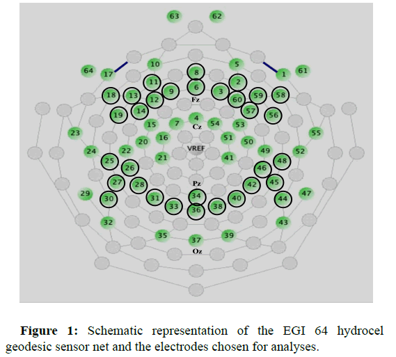
EEG was recorded continuously as participants engaged in an emotional word recognition task using a 64-channel hydrocel geodesic sensor net, net amps 300 amplifier, and net station, version 4.2, software (Electrical Geodesics Inc, Eugene, OR) at 250 Hz with 0.1 Hz high-pass and 100 Hz low-pass filtering. Electrode impedances were maintained below 50 kΩ. During acquisition, all channels were referenced to the vertex electrode. All stimulus presentations and behavioral response collections were controlled by a PC computer running E-prime 2.0 software (Psychology Software Tools Inc., PA). Data preprocessing was performed offline following acquisition using net station, (Waveform Tools Package), version 4.2, software (Electrical Geodesics Inc, Eugene, OR). Continuous EEG was filtered with a 1-30 Hz band-pass filter and segmented by condition into 900 ms stimulus-locked epochs (i.e., time-locked to stimulus onset) going from 100 ms pre-stimulus to 800 ms post-stimulus. Segments contaminated with vertical eye movements (eye blinks; ± 140 μV) and horizontal eye movement ( ± 55 μV) artifacts, as automatically identified by the built-in net station artifact detection tool, were eliminated. Following the manufacturer's recommendation and as is customary in other studies that used the same system recording segment was marked as 'bad' if it contained ten or more bad channels (15% of the total number of electrodes; bad channel: ± 200 μV for the entire segment). Individual bad channels were replaced on a segmentby- segment basis using the built-in net station bad channel replacement tool. Participants were excluded from further analysis if they had fewer than 85% good trials per experimental condition, which result in a minimum of 34 trials per condition [16]. Averaged ERP data was baseline corrected (100 ms pre-stimulus onset) and re-referenced into an average reference frame. N1 (100-180 ms post-stimulus), P2 (180-320 ms post-stimulus) and N400 (320-500 ms post-stimulus) components were chosen for analyses based on inspection of the grand average ERPs, scalp topography distributions of both the pregnant and non-pregnant groups, and the above-mentioned a-priori hypotheses. Mean amplitudes of N1, P2 and N400 were analyzed at two preselected scalp regions of interest: Anterior-frontal (average of channels 2,3,6,8,9,11,12,13,14,18,19,56,57,58,59,60), and posterior-parietal (average of channels 25,26,27,28,30,31,33,34,36,38,40,42,44,45,46,48). For the electrode array, as shown in Figure 1.
Polarity conventions at the field of ERP studies are highly inconsistent. ERP waveforms can be plotted with upward deflections indicating positive or negative potentials at the active electrode relative to the reference electrode. Both conventions are used in the literature and no consensus exists as to which is preferable. When comparing waveforms to those in the literature, it is essential to consider differences in recording system and reference method. In this study, all channels were referenced to the vertex sensor during acquisition and then re-referenced into an average reference frame. This resulted in negative going posterior N1 and N400 and positive going posterior P2 with a corresponding frontal/anterior polarity reversal mirroring posterior effects.
Data analysis
Data analysis was performed using SPSS 25.0.
Behavioral data analysis: To examine whether pregnant women, compared to control group, react differently to 'old' and 'new' negative, positive and neutral words, group differences and interaction effects in error rates and RTs were analyzed using a 2 × 2 × 3 repeated measures (mixed-design) ANOVA with group (pregnant/non-pregnant) as the between-subject factor, and condition (old/new) and emotional content (negative/positive/neutral) as the within-subject factors. Independent sample t-tests were used for post-hoc comparisons [17].
ERP analysis: To assess the relationship between pregnancy and brain activity, we performed separate 2 × 3 (group × emotional content) and 2×2 (group × condition) mixed-model ANOVAs to analyze mean amplitudes for the selected N1, P2 and N400 ERP components.
In both the behavioral and ERP analyses, significance levels were adjusted using the Huynh-Feldt correction when needed (the degrees of freedom indicated in the text are always those before the Huynh- Feldt correction, but the p-values are always those after the correction). Follow-up independent sample t-tests were used to break down interaction effects. The Bonferroni correction was applied and only results that remained significant after the correction are reported. A total of 42 subjects were included in behavioral and ERP analyses (pregnant=22, non-pregnant=20). Five participants from the nonpregnant group were excluded due to excessive artifacts in the ERP data (fewer than 85% good trials per experimental condition). Throughout the results section, numeric and graphical results are presented as mean ± SEM (the standard error of mean) [18].
Results
Behavioral results
A 2 × 2 × 3 repeated measures ANOVA for RTs revealed a significant group × condition × emotional content interaction effect (F(2,80)=4.27, p=0.017, ηp2=0.10). Subsumed under this interaction there was a main effect of condition (F(1,40)=86.39, p=0.0001, ηp2=0.68)-RTs to new words (840.92 ms ± 19.45) were greater than to old words (751.36 ms ± 18.37) and a main effect of emotional content (F(2,80)=4.70, p=0.012, ηp2=0.11)-RTs to negative (803.54 ms ± 20.40) and positive (804.65 ms ± 19.73) words were greater than to neutral words (780.39 ms ± 16.61). There was also a condition × emotion interaction effect (F(2,80)=21.29, p=0.0001, ηp2=0.35). More importantly, analysis revealed a main effect of group (F(1,40)=6.02, p=0.019, ηp2=0.13) such that pregnant women had significantly slower RTs (825.13 ms ± 20.08) than controls (767.26 ms ± 14.58). No group × condition, nor group × emotional content interaction effects were found.
No significant 2 × 2 × 3 repeated measures ANOVA was found for the measure of accuracy (percent of errors). Pregnant and nonpregnant women did not differ with respect to error rates, and there were no group × condition, nor group × emotional content interaction effects. Taken together, test performance results indicate that pregnant women were slower to react but as accurate as non-pregnant controls [19].
ERP results
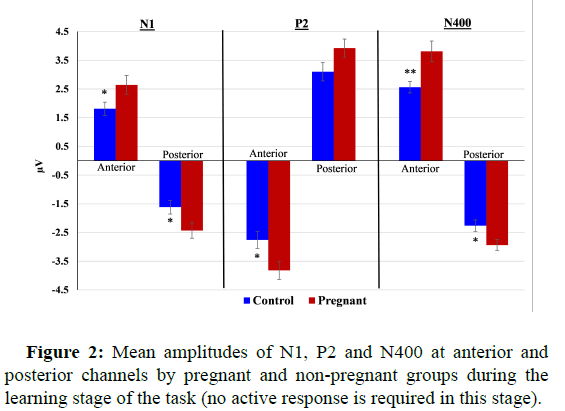
Learning stage: Analysis of ERPs during the passive learning stage of the task revealed that pregnant women had significantly more pronounced N1 (Anterior: F(1,40)=3.99, p=0.05, ηp2=0.09; posterior: F(1,40)=4.73, p=0.036, ηp2=0.11), P2 (Anterior: F(1,40)=5.68, p=0.022, ηp2=0.12; posterior: N.S), and N400 (Anterior: F(1,40)=8.18, p=0.007, ηp2=0.18; posterior: F(1,40)=5.62, p=0.023, ηp2=0.12) compared with non-pregnant controls (Figure 2). Group did not interact with emotional content.
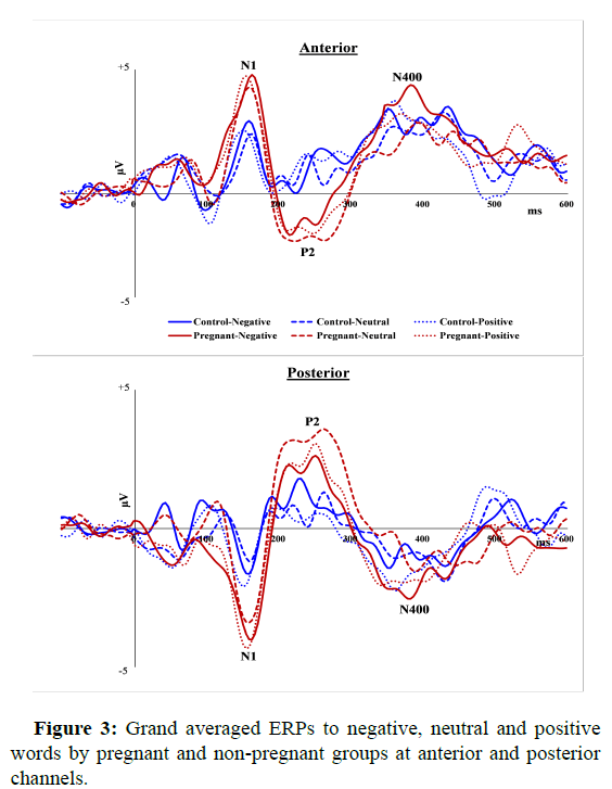
ERP results recognition test phase-emotional content (old/new combined): Figure 3 depicts the grand averaged ERPs to negative, neutral and positive words by pregnant and non-pregnant groups at anterior and posterior channels.
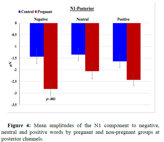
N1 (120-180 ms post-stimulus): Analysis for the anterior cluster revealed a significant group difference in N1 amplitude (F(1,40)=5.56, p=0.023, ηp2=0.12), such that pregnant women had more pronounced N1 (2.58 μV ± 0.33) compared to non-pregnant women (1.62 μV ± 0.28). No interaction was found between group and emotional content. At posterior channels, analysis revealed a main effect of emotional content (F(2,80)=4.71, p=0.012, ηp2=0.11)-N1 to negative and positive words was greater than to neutral words a main effect of group (F(1,40)=6.25, p=0.017, ηp2=0.14)-pregnant women had more pronounced N1 than controls and, a group × emotional content interaction (F(2,80)=3.14, p=0.049, ηp2=0.07). Follow-up comparisons showed that pregnant women had significantly more pronounced N1 to emotional negative words than non-pregnant women (t(40)=-3.18, p=0.003, Cohen’s d=0.99), while no such significant group difference was found for neutral (p=0.12) and positive (p=0.044) words (Figure 4).
P2 (180-320 ms post-stimulus): Analysis for both the anterior and posterior locations revealed a main effect of group (F(1,40)=5.97, p=0.019, ηp2=0.13; F(1,40)=4.35, p=0.043, ηp2=0.10, respectively) such that pregnant women had greater P2 amplitude (anterior: -2.35 μV ± 0.28; posterior: 2.72 μV ± 0.35) compared with non-pregnant controls (anterior: -1.56 μV ± 0.19; posterior: 1.91 μV ± 0.22). No interaction effects were found between group and emotional content.
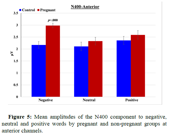
N400 (320-500 ms post-stimulus): Analysis for the anterior channels revealed a main effect of emotional content (F(2,80)=4.26, p=0.024, ηp2=0.10)- N400 was more pronounced to negative than to neutral words a main effect of group (F(1,40)=5.60, p=0.023, ηp2=0.12) pregnant women had more pronounced N400 than non-pregnant women and, a significant group × emotional content interaction (F(2,80)=3.67, p=0.039, ηp2=0.08) i.e., group difference in N400 amplitude was evident only for negative words t(40)=4.44, p=0.0001 Cohen’s d=1.38 and not for neutral (p=0.37) or positive (p=0.36) words. At posterior channels, there was a main effect of emotional content F(2,80)=4.10, p=0.02, ηp2=0.09- N400 to negative and neutral words was greater than to neutral words. There was also a main effect of group F(1,40)=6.89, p=0.012, ηp2=0.15: Pregnant women had greater (more negative) N400 (-2.10 μV ± 0.16) than non-pregnant women (-1.63 μV ± 0.15). The interaction between group and emotional content did not reach statistical significance (Figure 5).
ERP results recognition test phase-condition (negative/ positive/neutral combined)
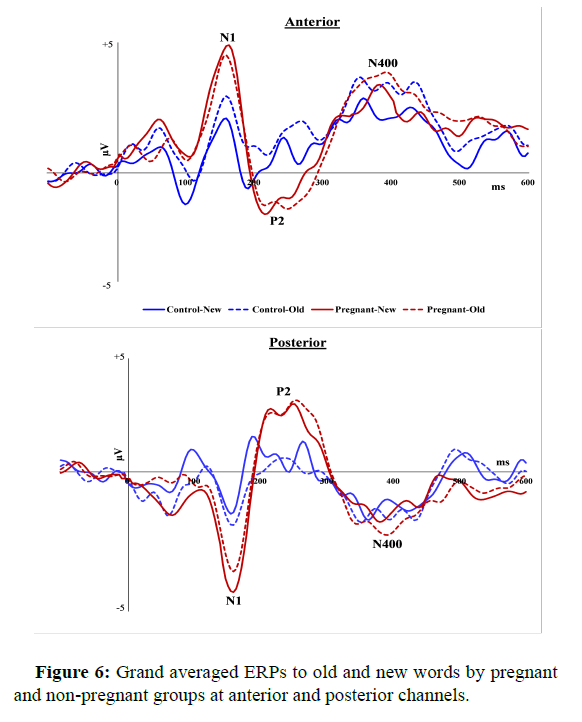
Figure 6 depicts the grand averaged ERPs to old and new words by pregnant and non-pregnant groups at anterior and posterior channels [20].
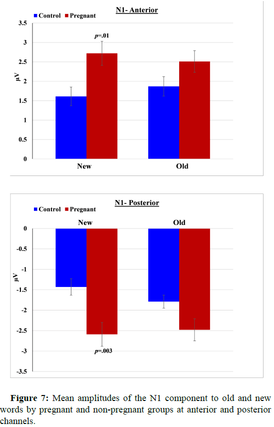
N1 (100-180 ms post-stimulus): Analysis for anterior channels showed a significant group difference in N1 amplitude (F(1,40)=5.26, p=0.027, ηp2=0.12), such that pregnant women had more pronounced N1 than controls as well as a group × condition interaction effect (F(1,40)=4.11, p=0.049, ηp2=0.09): The difference in N1 between pregnant and controls was significant only for new words (t(40)=2.70, p=0.01, Cohen’s d=0.84) but not for Old words (p=0.10). Similar results were found for posterior channels: Pregnant women had greater N1 than controls (F(1,40)=8.01, p=0.007, ηp2=0.17), and there was a significant interaction between group and condition (F(1,40)=4.43, p=0.042, ηp2=0.10). The difference in N1 between pregnant and nonpregnant women was evident especially in response to New words (t(40)=-3.21, p=0.003, Cohen’s d=1.00) compared to old words (p=0.04) (Figure 7).
P2 (180-320 ms post-stimulus): Analysis of both the anterior (F(1,40)=8.24, p=0.005, ηp2=0.18) and posterior (F(1,40)=6.27, p=0.016, ηp2=0.14) locations showed a significant effect of group, such that pregnant women had more pronounced P2 (anterior: -2.23 μV ± 0.27; posterior: -2.54 μV ± 0.26) compared with controls (anterior: 1.33 μV ± 0.15; posterior: 1.74 μV ± 0.19). There were no interactions between group and condition.
N400 (320-500 ms post-stimulus): Analysis of both the anterior (F(1,40)=5.76, p=0.021, ηp2=0.13) and posterior (F(1,40)=6.25, p=0.017, ηp2=0.14) locations showed a significant effect of group, such that pregnant women had more pronounced N400 (anterior: 2.43 μV ± 0.13; posterior: -1.89 μV ± 0.14) compared with non-pregnant women (anterior: 2.00 μV ± 0.15; posterior: -1.46 μV ± 0.12). No group × condition interactions were found.
Discussion
In the present study, we examined behavioral and brain function measures to assess cognitive-affective processing in pregnant women at third trimester of pregnancy as compared to non-pregnant comparison women. We examined the neurological correlates of pregnancy by recording ERPs during a visual emotional word recognition task. This task assesses measures of declarative memory and early processing of verbal emotional content. At the behavioral level, our hypothesis that pregnant women would perform as accurate, but slower, than non-pregnant controls, was supported. These results are in line with previous studies that reported women’s tendency to react more slowly during late pregnancy and with some recognition studies that found no differences in accuracy between pregnant women and controls. Contrary to the prevalent subjective perception among pregnant women, the current results suggest that recognition ability, in terms of achieved levels of accuracy, is not compromised during late pregnancy. However, measuring up to accuracy levels of non-pregnant women comes at the cost of significantly slower reaction times.
At the electrophysiological level, we hypothesized that pregnant women would differ from non-pregnant controls in their mean amplitudes of several task-related ERP components, with an emphasis on N1, P2 and N400. During the learning/study phase of the task, when participants were instructed to carefully observe and try to memorize the target words, pregnant women exhibited larger N1, P2 and N400 amplitudes at both anterior and posterior sites. These results may suggest that the pregnant group required additional processing in comparison with the non-pregnant group. In the recognition test phase (active discriminating response is required), when 'old' and 'new' stimuli were combined into one general variable of emotional content (negative/neutral/positive), our results again showed significant group differences in all pre-selected components at anterior and posterior channels. Pregnant women had more pronounced mean amplitudes of N1, P2 and N400 compared to non-pregnant women. Moreover, with respect to N1 and N400, significant interactions were found between pregnancy status and words’ emotional content. N1 (posterior) and N400 (anterior) were significantly greater in pregnant women only for the emotional negative words, while no such between-groups difference was found for emotional positive and neutral words (quite similar interactions were also evident for anterior N1 and posterior N400 but after the Bonferroni correction results did not reach the required level of statistical significance). The finding of such heightened neural responses to stimuli bearing negative content is in line with Pearson, et al., who reported that during late pregnancy, women were more accurate when they asked to encode emotional expressions indicating threat or harm (fearful, angry and disgusted faces). The researchers suggested that these findings might be explained by the influence of high levels of estrogen and other gender hormones on amygdala and/or serotonin functioning during late pregnancy. Future studies should further investigate the possible relations between pregnancy-related steroid hormones as well as serotonin function and behavioral and neural bias toward negative/ threat-related environmental stimuli.
When negative, positive and neutral stimuli were combined into one general variable of condition (old/new), our results again revealed that pregnant women had more pronounced mean amplitudes of N1, P2, and N400, compared to non-pregnant women at both anterior and posterior locations. Also, a significant interaction between pregnancy status and old/new condition was found for N1. N1 amplitude of pregnant women was significantly greater than that of controls only in response to the 'new' words, while no such difference was found for 'old' words. Taken together, the current results indicate general greater neural reactivity to word stimuli during the recognition task, and especially to emotionally negative words and to new/unfamiliar words in late pregnancy.
N1, P2 and N400 components are highly relevant to processing of visual verbal information. The visual N1 component is thought to reflect early perception, selective attention and the operation of a discrimination process within the focus of attention. It has been suggested that greater N1 amplitude may reflect generally greater sensory sensitivity or increased arousal. N1 amplitude is also affected by perceptual load, thus larger N1 is typically seen in tasks that require greater allocation of perceptual processing resources. Early ERP effects around 100 ms post stimuli, during a word recognition task, are proposed to index early attentional resource allocation to rapidly process potentially meaningful information. The P2 ERP component is thought to index the recruitment of early attentional resources that forms a basis for subsequent cognitive processing. It has been identified in tasks involving short term memory and stimulus classification found that pregnant women, compared to controls, had greater P2 amplitude in response to target stimuli during a stop signal task. N1 and P2 amplitudes are also sensitive to word stimuli emotional content larger N1 and P2 are evident in response to emotional relative to neutral words. The N400 component reflects a neural response to words in all modalities. It is thought to index the difficulty of retrieving stored conceptual knowledge associated with a word and is modulated in amplitude when prior exposure turns semantic processing to easier. There is an ongoing debate in the literature, in several respects, regarding the interpretation of the N400 effect in the context of semantic processing during recognition tasks. For instance, some researchers draw a distinction between two hypothesized processes that contribute to performance in tests of recognition memory- familiarity and recollection, and suggest that these processes are reflected in two distinct ERP effects: Frontally distributed FN400 reflecting familiarity-based recognition, and parietal N400 reflecting semantic processing. Other researchers doubted this discrimination and claimed that FN400 and N400 are actually electro-physiologically and functionally identical. Either way, it has been shown that N400 amplitude is modulated by a wide variety of stimulus and context factors (e.g., priming manipulations, semantic context, word frequency, lexical class in sentences as well as the specific method of EEG recording, referencing and ERP analysis). Going into a more thorough review of the N400 literature is beyond the scope of this paper and exceeds the immediate goals of the present study.
Augmentation of N1, P2 and N400 may reflect the recruitment of additional brain resources for perceptual processing of attended emotional stimuli. Such augmentation in ERP responses may also suggest that women in late pregnancy had to recruit additional brain processing resources in order to successfully perform the task and may partly explain their slower response times. The greater N1, P2 and N400 amplitudes found in pregnant women relative to controls may indicate that they are generally more alert, and specifically more sensitive and reactive to novel and emotional negative signals in their environment.
The present research has some limitations that deserve to be addressed. First, our pregnancy sample included both multigravid and primigravid women. Although they were carefully matched with multiparous and nulliparous controls, future studies may attempt to further control for reproductive history and parenthood status. Second, we investigated women during late pregnancy. It would be of interest to explore the development of ERP differences from early to late pregnancy as well as from late pregnancy to postpartum. Finally, being undergraduate/graduate students and college administrative and academic staff, participants in this study had a relatively high educational level which may affect their attitude, motivation, performance and compliance during the experimental session. To establish the generalizability of the current results this study should be replicated in more representative samples of varying education, socioeconomic status, and ethnicity.
Conclusion
In conclusion, our results shed new light on memory function and neural activity during late human pregnancy. Contrary to popular belief, pregnant women did not differ from non-pregnant women in their memory task performance (i.e., accuracy levels achieved). They did, however, differ from non-pregnant women in RTs and in their neural response patterns. To our knowledge, this study is the first to examine both behavioral and neural measures of recognition memory and related early processing of verbal emotional content, during late pregnancy, using event related potentials. The current results may suggest that at early stages of neural processing, women at late pregnancy are generally more hypervigilant to novel environmental stimuli and are particularly more sensitive to stimuli with negative content. Heightened precautionary behavior, reflected, among other things, in an increased sensitivity towards environmental novel stimuli and towards signals of threat and harm may be advantageous in optimizing fetal growth and development and preparing women for the unique demands of motherhood.
References
- Alderman BL, Olson RL, Bates ME, Selby EA, Buckman JF, et al. (2015) Rumination in major depressive disorder is associated with impaired neural activation during conflict monitoring. Front Hum Neurosci 9:269.
[Crossref] [Google Scholar] [PubMed]
- Anderson MV, Rutherford MD (2011) Recognition of novel faces after single exposure is enhanced during pregnancy. Evol Psychol 9:47-60.
[Google Scholar] [PubMed]
- Bai Y, Yao Z, Cong F, Zhang L (2015) Event-related potentials elicited by social commerce and electronic-commerce reviews. Cogn Neurodyn 9:639-648.
[Crossref] [Google Scholar] [PubMed]
- Barber HA, Kutas M (2007) Interplay between computational models and cognitive electrophysiology in visual word recognition. Brain Res Rev 53:98-123.
[Crossref] [Google Scholar] [PubMed]
- Barry RJ, Clarke AR, Johnstone SJ (2003) A review of electrophysiology in attention- deficit/hyperactivity disorder: II. Event-related potentials. Clin Neurophysiol 114:184-198.
[Crossref] [Google Scholar] [PubMed]
- Bayet L, Saville A, Balas B (2021) Sensitivity to face animacy and inversion in childhood: Evidence from EEG data. Neuropsychologia 156:107838.
[Crossref] [Google Scholar] [PubMed]
- Bornkessel-Schlesewsky I, Schlesewsky M (2019) Toward a neurobiologically plausible model of language-related, negative event-related potentials. Front Psychol 10:298.
[Crossref] [Google Scholar] [PubMed]
- Boksem MA, Meijman TF, Lorist MM (2005) Effects of mental fatigue on attention: An ERP study. Cogn Brain Res 25:107-116.
[Crossref] [Google Scholar] [PubMed]
- Brett M, Baxendale S (2001) Motherhood and memory: A review. Psychoneuroendocrinology 26:339-362.
[Crossref] [Google Scholar] [PubMed]
- Bridger EK, Bader R, Kriukova O, Unger K, Mecklinger A (2012) The FN400 is functionally distinct from the N400. Neuroimage 63:1334-1342.
[Crossref] [Google Scholar] [PubMed]
- Brindle PM, Brown MW, Brown J, Griffith HB, Turner GM (1991) Objective and subjective memory impairment in pregnancy. Psychol Med 21:647-653.
[Crossref] [Google Scholar] [PubMed]
- Buckwalter JG, Stanczyk FZ, McCleary CA, Bluestein BW, Buckwalter DK, et al. (1999) Pregnancy, the postpartum, and steroid hormones: Effects on cognition and mood. Psychoneuroendocrinology 24:69-84.
[Crossref] [Google Scholar] [PubMed]
- Burkhart MA, Thomas DG (1993) Event-related potential measures of attention in moderately depressed subjects. Electroencephalogr Clin Neurophysiol 88:42-50.
[Crossref] [Google Scholar] [PubMed]
- Cardenas EF, Kujawa A, Humphreys KL (2019) Neurobiological changes during the peripartum period: Implications for health and behavior. Soc Cogn Affect Neurosci 15:1097-1110.
[Crossref] [Google Scholar] [PubMed]
- Carreiras M, Vergara M, Barber H (2005) Early event-related potential effects of syllabic processing during visual word recognition. J Cogn Neurosci 17:1803-1817.
[Crossref] [Google Scholar] [PubMed]
- CarretiE L, Mercado F, Tapia M, Hinojosa JA (2001) Emotion, attention, and the ‘negativity bias’, studied through event-related potentials. Int J Psychophysiol 41:75-85.
[Crossref] [Google Scholar] [PubMed]
- Casey P (2000) A longitudinal study of cognitive performance during pregnancy and new motherhood. Arch Womens Ment Health 3:65-76.
- Casey P, Huntsdale C, Angus G, Janes C (1999) Memory in pregnancy. II: Implicit, incidental, explicit, semantic, short-term, working and prospective memory in primigravid, multigravid and postpartum women. J Psychosom Obstet Gynaecol 20:158-164.
[Crossref] [Google Scholar] [PubMed]
- Christensen H, Poyser C, Pollitt P, Cubis J (1999) Pregnancy may confer a selective cognitive advantage. J Reprod Infant Psychol 17:7-25.
- Cheyette SJ, Plaut DC (2017) Modeling the N400 ERP component as transient semantic over-activation within a neural network model of word comprehension. Cognition 162:153-166.
[Crossref] [Google Scholar] [PubMed]
