Research Article, Cell Biol Henderson Nv Vol: 6 Issue: 2
New Insights into the Tissue Homeostasis between TIMP and MMP in Dupuytren’s Disease
Annika Borgschulze, Benita Sahlender, Tim Lögters, Joachim Windolf and Vera Grotheer*
Department of Trauma and Hand Surgery, Medical Faculty, Heinrich-Heine- University, Düsseldorf, Germany
*Corresponding Author : Vera Grotheer
Department of Trauma and Hand Surgery, Medical Faculty, Heinrich-Heine-University, Moorenstr. 5, 40225 Düsseldorf, Germany
Tel: +49 211 302039-234
E-mail: vera.grotheer@med.uni-duesseldorf.de
Received: May 03, 2017 Accepted: May 17, 2017 Published: May 22, 2017
Citation: Borgschulze A, Sahlender B, Lögters T, Windolf J, Grotheer V (2017) New insights into the Tissue Homeostasis between TIMP and MMP in Dupuytren’s Disease. Cell Biol (Henderson, NV) 6:2.doi: 10.4172/2324-9293.1000132
Abstract
Dupuytren’s disease is a fibrotic disorder of the palmar fascia, leading to a functional restriction of the affected finger. The etiological causes of Dupuytren’s Disease are multifactorial, with a genetic disposition concerning predominantly men in the middle age. In, this study on the one hand gene expression was analysed in Dupuytren’s tissue directly after surgery to determine the balance between the tissue inhibitors of matrix metalloproteinases and the matrix metalloproteinases (TIMP/MMP). And on the other hand Dupuytren’s fibroblasts were preincubated with nterleukin-1β (Il-1β) and transforming growth factor-β (TGF-β) to analyze wound healing characteristics. Our data suggest that Dupuytren’s fibroblasts show an overshooting inflammatory response, which could, in turn, influence the TIMP/MMP axis. Furthermore the analysis of the TIMP/MMP ratio shows a dichotomous imbalance, which could additionally provide tissue fibrosis.
Keywords: Dupuytren’s disease; Il-1β; TIMP; MMP; TGF- β; TNF-α
Introduction
Dupuytren’s Disease (DD) is one of the most common genetic disorders of the connective tissue [1]. In most cases, the course of this benign fibromatosis begins with the formation of subcutaneous nodules followed by fibrotic cords [2]. A coincidence besides genetic disposition [1,3] is observed with regard to epidemiological data with nicotine [4,5] diabetes mellitus [6,7] liver damage, alcohol abuse [5,8], epilepsy [9] manual traumas [10] and changes of the immune system, especially increased appearance in connection with HIV [11].
The clinical symptoms of DD are described as an excessive proliferation of fibroblasts and their reinforced differentiation to myofibroblasts [12]. Myofibroblasts contain intracellular bundles of microfilaments [13]. Accordingly, a higher concentration of α-smooth muscle actin (α-SMA) protein is found in Dupuytren’s tissue [14] and serves as a parameter for their differentiation to myofibroblasts. It has been shown that transforming growth factor- β1 (TGF-β) upregulates myofibroblasts differentiation in Dupuytren’s fibroblasts, and it is assumed, that uncontrolled myofibroblasts proliferation induced by TGF- β1 leads to symptomatic nodule formation [15]. Furthermore, overexpression of TGF-β induces and supports disturbed balance of matrix metalloproteinases (MMP), diminishing the extracellular matrix (ECM) (extracellular matrix) and their inhibitors, the tissue inhibitor of metalloproteinases (TIMP). These enzymes, in turn, influence the synthesis and deposition of collagen, and therefore authoritatively strengthen the origin of fibrosis [16].
Additionally, the expression of IL-1β is also increased in Dupuytren’s tissue [17] and interestingly -as a master regulator of inflammation a [18] IL-1β induces the proliferation of fibroblasts followed by an elevated collagen synthesis. So an elevated expression of IL-1β seems to be associated with fibrotic diseases [19,20] and regulates inflammation related proteins as MMP and TIMP [21]. Furthermore IL-1β attracts immune cells, which again lead to the extensive alterations in protein expression levels of inflammatory and wound healing parameters [18].
So the present study gives a brief insight into Dupuytren’s Disease evaluating the ratio of TIMP to MMP. Furthermore, wound healing parameters in Dupuytren’s fibroblasts preincubated with TGF-β and IL-1β are analysed. We could demonstrate that under these parameters, Dupuytren’s fibroblasts show an overshooting cytokine expression and disturbed MMP/TMP homeostasis compared to control palmar fascia fibroblasts (NPF).
Material and Methods
Patients
Study approval was obtained from the Ethics Review Board of the Medical Faculty, HeinrichHeine-University Düsseldorf (Study No. 3634). The study included [22,23] patients (3 women and 20 men; average age 62 years) with DD, who underwent fasciectomy at the Department of Trauma and Hand Surgery, University Hospital, Düsseldorf, Germany. NPF from patients with carpal tunnel release served as control (n = 24; 16 women and 8 men; average age 52,5 years). Informed consent was obtained from all the patients. All patient-related data were anonymized for analysis.
Isolation and culture of the cells
Minced tissue was incubated at 37°C in collagenase-solution (0.1 M CaCl, 0.005 M Glucose, 0.1 M Hepes, 0.12 M NaCl, 0.05 M KCL + 1.5 % BSA and 0.2 % collagenase type 1) for 45 min, filtrated through a nylon strainer, centrifuged (1200 rpm for 5 min at 4°C) and pellet resuspended in NaCl-solution. After centrifugation the pellet was resuspended in medium (RPMI 1640 with 10 % FBS, 1 x NEAA, 1 % Penicillin/Streptomycin, 1 x So-Pyruvat) and incubated at 37°C, 5 % CO2.
Stimulation of the cells
Confluent cells were stimulated over 18 hours with 500 U/ml rh IL- 1β (R&DSystems) or over 5 days with 2 ng/ml TGF- β1 (ImmunoTools rh TGF- β1) for extracting RNA as described previously [22,23]. After stimulation for over 3 days and 5 days either with IL-1β or with TGF- β1, the cells were harvested for immunohistochemistry.
Immunohistochemistry
Sub confluent cells grown on cover slips were fixed with 4% Paraformaldehyde. Then, the cells were washed with PBS and permeabilized with 0.2 % Triton X-100. Unspecific bindings were saturated with 4 % Bovine Serum Albumin for 30 min at 37°C and 5 % CO2. The samples were washed and immunolabeled with anti-α– SMA antibody (Abcam) for 60 min at 37°C. The next steps were undertaken in accordance with the manufacturer’s instruction (Dako REAL Detection System Alkaline Phosphatase/RED, Rabbit/Mouse). The samples were drained by an ascending alcohol series (80%, 96 % and 100 % of EtOH) and rehydrated by Xylol. The cells were fixed on the glass cover slips with a drop of DePeX and light microscope images were taken with Zeiss Axioskop 40.
RNA extraction from tissue and cultured cells
RNA was extracted using TRI Reagent after and a sonification step of 10 - 20 times. Following an incubation time of 5 min at room temperature (RT), chloroform was added and the samples were vortexed. The phases were separated and centrifuged (11,200 rpm for 15 min at 4°C). The colourless phase was taken and mixed with Isopropanol. After incubation for 10 min and centrifugation (11,200 rpm for 10 min at 4°C), the pellet was resuspended in of 75 % EtOH. Then the samples were centrifuged (8,800 rpm for 7 min at 4°C), EtOH was removed and the cells were resuspended in RNAse free water. RNA was purified with DNAfree KIT (Ambion) according to the manufacturer’s instructions.
Western blot analysis
α-SMA protein expression was measured with Western Blot Analysis. Protein concentration was determined with Pierce BCA Protein Assay Kit, following the manufacturer’s instructions. 10 μg of protein were mixed with Laemmli buffer (4 x Tris-glycin-SDS sample buffer, 252 mmol TrisHCL pH 6.8; 40 % Glycerin; 8 % SDS; 0.01 % bromphenol blue + 20 % mercaptoethanol), denaturized for 5 min at 95°C and separated on a 12 % sodium dodecyl sulphatepolyacrylamide gel (SDS-PAGE)). Separated proteins were transferred to a nitrocellulose membrane. Membrane was saturated with 5 % BSA in TBST (Tris-buffered saline, 0.1% Tween 20) for 1 h (RT) and immunolabeled with anti-α–SMA (Abcam) (1 : 1000 in 3 % BSA in TBST) over night at 4°C or (Glyceraldehyde 3-phosphate dehydrogenase) antibody (Dako) (1 : 6000 in 3 % BSA in TBST) for 1 h. After washing with TBST, the membrane was incubated with horseradish peroxidase (HRP)-conjugated secondary antibody (Dako) for 1 h. Bound antibodies were detected using Clarity TM Western ECL Substrate and analysed with the Quantity One 1-D Analysis Software Version 4.6.5 (BioRad).
Polymerase Chain Reaction (PCR) of IL-1, IL-6, IL-8, TGF-β2, TNFα, MMP-2, -3, -9, TIMP1,-2,-3,-4
500 ng RNA were reverse transcribed to cDNA using RT-Puffer, d-NTPs, Oligo dT, RNaseInhibitor, Omniskript RTase and RNasefree water.
Gene-specific primer in pairs was applied
IL-1 forward: AGGGCCAATCCCCAGCCCTTT; IL-1 reverse:
GCCGTGGTTTCTGTCAGGCGG; IL-6 forward: TGTAGCCGCCCCACACAGACA; IL-6
reverse: CTGCCAGTGCCTCTTTGCTGC; IL-8 forward:
GAGTGGACCACACTGCGCCA; IL-8 reverse: TCCACAACCCTCTGCACCCAGT; TGF-
β2 forward: CGAGAGGAGCGACGAAGAGTA; TGF-β2 reverse:
CACTGAGCCAGAGGGTGTTGT; TNFα forward: CGCTCTTCTGCCTGCTGCACT;
TNFα reverse: GCCTGGGCCAGAGGGCTGATT; MMP 2 forward:
AACCAGCTGGCCTAGTGATGATGT; MMP 2 reverse:
GGCAGCCATAGAAGGTGTTCAGGT; MMP 3 forward:
TTTTGGCCCATGCCTATGCCCC; MMP3 reverse: ACCCAGGGAGTGGCCAATTTCAT;
MMP 9 forward: TCAGTGCCGGAGGCGCTCAT; MMP 9 reverse:
GGTGGTGGTTGGAGGCCGTG; TIMP-1 forward: GACCTACACTGTTGGCTGTGA;
TIMP-1 reverse: GTCCGTCCACAAGCAATGAG; TIMP-2 forward:
AGGAAGTGGACTCTGGAAACG; TIMP-2 reverse: CCTTCTTTCCTCCAACGTCCA;
TIMP-3 forward: CTCTGTGGCCTTAAGCTGGA; TIMP-3
reverse:ACCGATAGTTCAGCCCCTTG; TIMP-4 forward:
TGTGGTGTGAAACTAGAAGCCA; TIMP-4 reverse:
GCTCGATGTAGTTGCACAGATG; GAPDH forward: CCCGCTTCGCTCTCTGCTCCT;
reverse: TGACCAGGCGCCCAATACGAC).
Relative gene expression was measured with ABI Prism 7300 according to the manufacture’s protocol with the following cycling conditions: 95°C, 10 min (1 cycle); 95°C, 15 s, 60°C, 60 s (40 cycles); 4°C hold with SYBR Green (Applied Biosystems). All the samples were run in triplicates. Expression of each target gene was normalized to the housekeeping gene Glycerinaldehyd-3-Phosphat-Dehydrogenase (GAPDH).
Statistical analysis
Statistical analyses were performed by two-tailed t-test or Mann- Whitney-U test. Data were expressed as mean value and standard deviation (SD). The level of significance was considered to be p ≤ 0.05 and was designated with asterisks.
Results
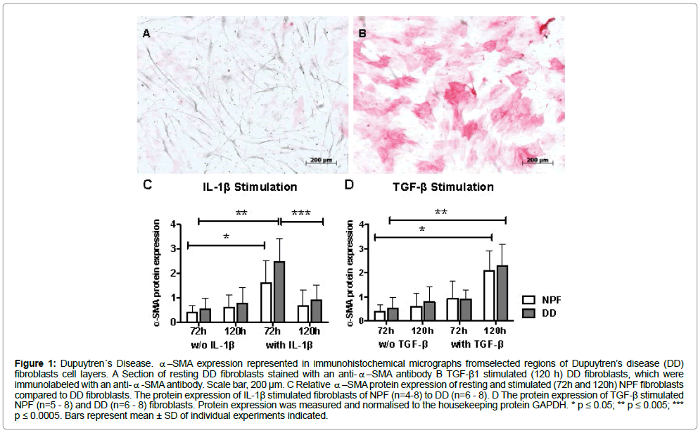
Relative α-SMA Protein expression of IL-1β or TGF-β1 stimulated fibroblasts stimulation with TGF-β1 resulted in a significantly enhanced expression of α-SMA in DD and NPF fibroblasts after 120 h (Figure1A and B). IL-1β stimulation of NPF led to a significant increase in α-SMA expression with a peak after 72 h. In DD fibroblasts treated with IL-1β demonstrated after 72 h a higher α-SMA protein expression, which was significantly reduced after 120 h compared to 72 h measurements (Figure 1C). Stimulation with TGF-β1 resulted in a significantly enhanced expression of α-SMA in DD and NPF after 120 h (Figure 1D).
Figure 1: Dupuytren´s Disease. α–SMA expression represented in immunohistochemical micrographs fromselected regions of Dupuytren’s disease (DD) fibroblasts cell layers. A Section of resting DD fibroblasts stained with an anti-α–SMA antibody B TGF-β1 stimulated (120 h) DD fibroblasts, which were immunolabeled with an anti-α-SMA antibody. Scale bar, 200 μm. C Relative α–SMA protein expression of resting and stimulated (72h and 120h) NPF fibroblasts compared to DD fibroblasts. The protein expression of IL-1β stimulated fibroblasts of NPF (n=4-8) to DD (n=6 - 8). D The protein expression of TGF-β stimulated NPF (n=5 - 8) and DD (n=6 - 8) fibroblasts. Protein expression was measured and normalised to the housekeeping protein GAPDH. * p ≤ 0.05; ** p ≤ 0.005; *** p ≤ 0.0005. Bars represent mean ± SD of individual experiments indicated.
Relative gene expression of TGF-β2 and TIMP3 (Tissue)
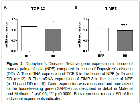
TGF- β2 and TIMP3 expressions were significantly decreased in tissue of DD patients in comparison to the tissue of NPF (Figure 2A and 2B).
Figure 2: Dupuytren´s Disease. Relative gene expression in tissue of normal palmar fascia (NPF) compared to tissue of Dupuytren’s disease (DD). A The mRNA expression of TGF-β in the tissue of NPF (n=9) and DD (n=12). B The mRNA expression of TIMP-3 in the tissue of NPF (n=11) and DD (n=16). Gene expression was measured and normalized to the housekeeping gene (GAPDH) as described in detail in Material and Methods. * p<0.05; *** p<0.0005. Bars represent mean ± SD of the individual experiments indicated.
The balance of TIMP/MMP
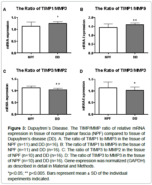
The balance of TIMP1 to MMP3 and MMP9 gene expressions were significantly increased in tissue of DD patients in comparison to tissue of NPF (Figure 3A and 3B). The ratio of TIMP3 to MMP2 and MMP3 were significantly downregulated (Figure 3C and 3D).
Figure 3: Dupuytren´s Disease. The TIMP/MMP ratio of relative mRNA expression in tissue of normal palmar fascia (NPF) compared to tissue of Dupuytren’s disease (DD). A: The ratio of TIMP1 to MMP3 in the tissue of NPF (n=11) and DD (n=16). B: The ratio of TIMP1 to MMP9 in the tissue of NPF (n=11) and DD (n=16). C: The ratio of TIMP3 to MMP2 in the tissue of NPF (n=10) and DD (n=16). D: The ratio of TIMP3 to MMP3 in the tissue of NPF (n=10) and DD (n=16). Gene expression was normalized (GAPDH) as described in detail in Material and Methods.
Relative gene expression of IL-1β stimulated fibroblasts
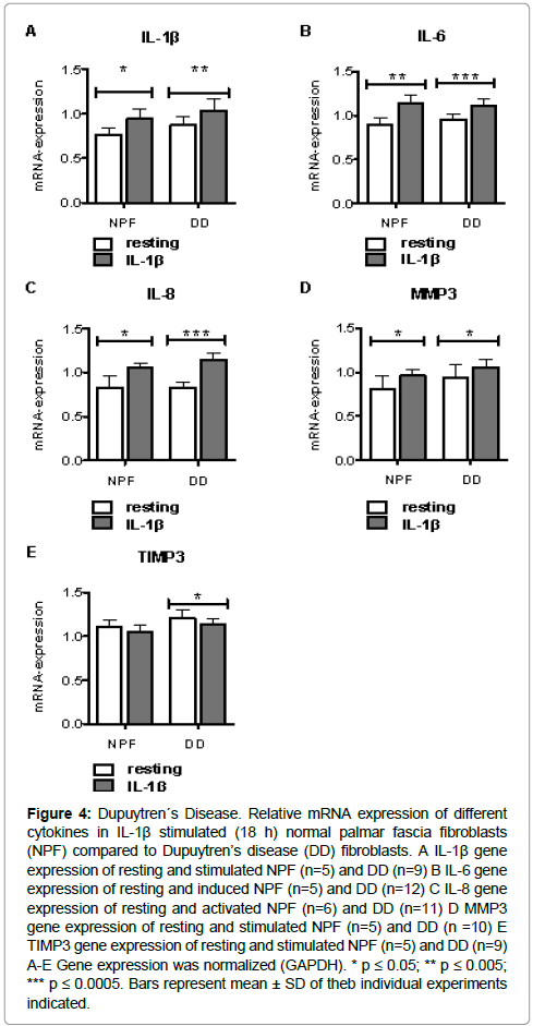
The expression of IL-1β, IL-6, IL-8 and MMP3 was significantly higher after IL-1β stimulation in DD fibroblasts related to NPF compared to the respective controls (Figure 4A, 4B, 4C, 4D). On the contrary, stimulation caused significant reduction in TIMP3 expression of DD fibroblasts (Figure 4E).
Figure 4: Dupuytren´s Disease. Relative mRNA expression of different cytokines in IL-1β stimulated (18 h) normal palmar fascia fibroblasts (NPF) compared to Dupuytren’s disease (DD) fibroblasts. A IL-1β gene expression of resting and stimulated NPF (n=5) and DD (n=9) B IL-6 gene expression of resting and induced NPF (n=5) and DD (n=12) C IL-8 gene expression of resting and activated NPF (n=6) and DD (n=11) D MMP3 gene expression of resting and stimulated NPF (n=5) and DD (n =10) E TIMP3 gene expression of resting and stimulated NPF (n=5) and DD (n=9) A-E Gene expression was normalized (GAPDH). * p ≤ 0.05; ** p ≤ 0.005; *** p ≤ 0.0005. Bars represent mean ± SD of theb individual experiments indicated.
Relative gene expression of TGF-β1 stimulated fibroblasts
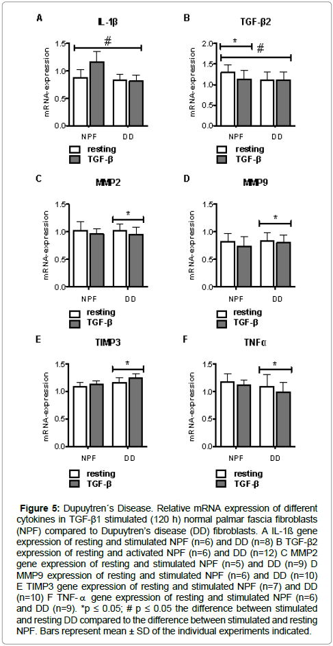
NPF responded significantly different to TGF-β1 stimulation compared to DD fibroblasts (Figure 5A, 5B). The gene expression of TGF-β2 in NPF was significantly down regulated after TGF-β1 stimulation (Figure 5B). DD fibroblasts showed significant relief in MMP2, MMP9 and TNFα expression after stimulation (Figure 5C, 5D, 5F), while TIMP3 expression was significantly raised (Figure 5E).
Figure 5: Dupuytren´s Disease. Relative mRNA expression of different cytokines in TGF-β1 stimulated (120 h) normal palmar fascia fibroblasts (NPF) compared to Dupuytren’s disease (DD) fibroblasts. A IL-1ß gene expression of resting and stimulated NPF (n=6) and DD (n=8) B TGF-β2 expression of resting and activated NPF (n=6) and DD (n=12) C MMP2 gene expression of resting and stimulated NPF (n=5) and DD (n=9) D MMP9 expression of resting and stimulated NPF (n=6) and DD (n=10) E TIMP3 gene expression of resting and stimulated NPF (n=7) and DD (n=10) F TNF-α gene expression of resting and stimulated NPF (n=6) and DD (n=9). *p ≤ 0.05; # p ≤ 0.05 the difference between stimulated and resting DD compared to the difference between stimulated and resting NPF. Bars represent mean ± SD of the individual experiments indicated.
Conclusions
Apart from the genetic disposition DD is also associated with environmental factors such as alcohol and nicotine abuse, epilepsy, diabetes, androgens, but also immunological factors as repeated manual traumas, and HIV infection [15], etc. seem to be involved in the progression of this disease. Till date, it has not been possible to simulate these multiple factors supporting the symptoms of this disease in cell culture conditions, so this study inter alia evaluates gene expression in Dupuytren’s tissue directly after surgical extraction. So in the present work in the directly examined tissue a significantly decreased TGF-β2 concentration could be observed (Figure 2A), and according to the results shown in scleroderma, this could support fibrosis formation [24]. Furthermore, a decreasing level of TIMP3 could be determined (Figure 2b). Our findings were confirmed from Kassiri and Fan [25,26] demonstrating, that the loss or deficiency of TIMP3 enhances fibrosis. In addition, the decreased expression of TIMP3 could be the cause for the inadequate capability of Dupuytren’s fibroblasts to undergo apoptosis [27] because TIMP3 also mediates apoptosis in human colon carcinoma cells [28].
In general, TIMPs regulate the degradation or synthesis of the ECM and their function is dependent on the specific interaction with particular MMP within a given tissue. It is generally accepted that the TIMP/MMP axis determines the ECM turnover, and disturbance of this balance may lead to diseases of fibrotic natures [29]. For this reason we compared the TIMP/MMP ratio in DD to NPF. In detail the ratio between TIMP1/MMP3 and TIMP1/MMP9 was analysed, because TIMP1 is a strong inhibitor of multiple MMPs and it was observed that TIMP1 concentration is elevated in fibroproliferative disorders [30]. And so in the present study it could be demonstrated, that the expression of TIMP1 is significantly increased in relation to MMP3 and MMP9 in DD compared to NPF. MMP9 and MMP3 degrade laminin, fibronectin and several collagens. And MMP9 cleaves collagens more efficiently than other MMPs [31] and it activates further MMP, thereby also providing the symptoms of this disease. Furthermore, an inhibition of MMP3 by TIMP1 in the Dupuytren’s tissue could be a possible explanation for disease progression, because MMP3 especially degrades collagen III and it is known that the collagen III content in Dupuytren’s tissue is propagated and associated with diseased cord formation [32]. Additionally, microarray gene analysis also provides our findings, since a diminished level of MMP3 could also be determined in Dupuytren’s fibroblasts by the working group of Sprung [33].
Interestingly, if the ratio of TIMP3 and MMP2, respectively MMP3 (Figure3C, D) was calculated, the ratio is changed for the benefit of MMP2 and MMP3 in Dupuytren’s tissue compared to NPF. It was shown from Kassiri et al., that an imbalance in favor of MMP released sequestered TGF-β1 [34] within the ECM, which in turn leads to ECM deposition and fibrosis. And as mentioned above, a lower level of TIMP3 supports the formation of DD, revealing that loss of TIMP3 potentially induces renal fibrosis [25] and myocardial fibrosis through regulating inflammation and TGF-β1 activation as seen in injured TIMP3-/- mice [35].
Interestingly, MMP2 and MMP3 cleave IL-1β, whereas a long time incubation of IL-1β with MMP3 leads to a degradation of IL-1β [36]. This could be a potential explanation why our data could not provide an elevated level of IL-1β in Dupuytren’s tissue (Figure 5A) as described by other working groups [37]. Our results confirm, that the MMP/TIMP axis in DD is disturbed both in favor of an increasing level of TIMP1 in comparison with MMP3 and MMP9 and to a decreasing level of TIMP3 in relation to MMP2 and MMP3.
Moreover, cytokines such as IL-1β are overexpressed in the proliferative DD [37]. In order to analyse the special effect of this pleiotropic cytokine in Dupuytren’s Disease, in this study Dupuytren’s fibroblasts and NPF were analysed after a preincubation with IL1β (Figure 4). In relation to their resting controls, Dupuytren’s fibroblasts stimulated with IL-1β express more α-SMA (Figure 1B) and significantly more IL-1β, IL-6, IL-8 and less TIMP3 (Figure 4). And interestingly, the difference between the resting controls to their preincubated counterparts is bigger in Dupuytren’s fibroblasts than in NPF. So we could determine, that the expressions of these proinflammatory cytokines are increased in strength in Dupuytren’s fibroblasts. This could be relevant and important, especially as it affects the pyrogens IL-1β, IL-6 and IL-8.
Furthermore, IL-1β is a highly active cytokine, which can induce a systemic or local inflammation, playing an important role in fibrosis in vivo [38]. Also IL-6 and IL-8 play key roles in disturbed wound healing and are also involved in the pathogenesis of fibrosis [39,40].
If NPF are preincubated with TGF-β1 (Figure 5) the expression of IL-1β increases, whereas the expressions in Dupuytren’s fibroblasts remain unchanged. Additionally, the expression of TGF-β2 was diminished in NPF activated with TGF-β1, whereas, the TGF-β2 expression does not change in preincubated Dupuytren’s fibroblasts. This is consistent with our findings made in directly analysed tissue (Figure 2A). These results may indicate, that the gene expression in Dupuytren’s fibroblasts is also regulated in a dichotomous way, depending on which wound healing parameter is prevailing.
So as shown here Dupuytren’s fibroblasts seem to have an overshooting, partially changed cytokine expression in consequence of a treatment with inflammatory key mediators.
Furthermore we could determine, that in Dupuytren’s Disease the tissue homeostasis between MMP and TIMP is disturbed. But in what specific manner individual TIMP interact with particular MMP has to be elucidated in future work, because the role of TIMP regulating MMP is dichotomous and is dependent on the tissue origin [34]. But in conclusion, we have discovered additional clues that the TIMP/ MMP axis is a well-balanced homeostatic system, and if its balance lost, fibrotic diseases as DD could be intensified.
Acknowledgements
We thank Jutta Schneider, Christa-Maria Wilkens and Samira Seghrouchni for technical assistance. Data are provided from the doctoral thesis submitted from Annika Borgschulze manufactured in the Department of Trauma and Hand Surgery in the Faculty of Medicine of the Heinrich-Heine-University Düsseldorf, Germany.
Declaration
All experiments conformed to the respective national guidelines.
Sources of funding
This work was supported by the Faculty of Medicine of the Heinrich-Heine- University, Düsseldorf.
Conflict of Interest
No competing financial interests exist.
References
- Becker K, Tinschert S, Lienert A, Hennies HC (2015) The importance of genetic susceptibility in Dupuytren's disease. Clinical genetics 87: 483-487.
- Mikkelsen OA (1976) Dupuytren's disease--a study of the pattern of distribution and stage of contracture in the hand. The Hand 8: 265-271.
- Millesi H (1965) On the pathogenesis and therapy of Dupuytren's contracture. (A study based on more than 500 cases). Ergebnisse der Chirurgie und Orthopadie 47:51-101.
- An HS, Southworth SR, Jackson WT, Russ B (1988) Cigarette smoking and Dupuytren's contracture of the hand. J Hand Surg Am 13: 872-874.
- Burge P, Hoy G, Regan P, Milne R (1997) Smoking, alcohol and the risk of Dupuytren's contracture. The Journal of bone and joint surgery British volume 79: 206-210.
- Heathcote JG, Cohen H, Noble J (1981) Dupuytren's disease and diabetes mellitus. Lancet (London, England) 8235: 14-20.
- Noble J, Heathcote JG, Cohen H (1984) Diabetes mellitus in the aetiology of Dupuytren's disease. J Bone Joint Surg Br 66: 322-325.
- Noble J, Arafa M, Royle SG, McGeorge G (1992) The association between alcohol, hepatic pathology and Dupuytren's disease. Journal of hand surgery 17: 71-74.
- Pojer J, Radivojevic M, Williams TF (1972) Dupuytren's disease. Its association with abnormal liver function in alcoholism and epilepsy. Arch Intern Med 129: 561-566.
- Mikkelsen OA (1978) Dupuytren's disease--the influence of occupation and previous hand injuries. The Hand 10: 1-8.
- Bower M, Nelson M, Gazzard BG (1990) Dupuytren's contractures in patients infected with HIV. BMJ 300: 164-165.
- Shih B, Bayat A (2010) Scientific understanding and clinical management of Dupuytren disease. Nature reviews Rheumatology 6: 715-726.
- Gokel JM, Hubner G (1977) Occurrence of myofibroblasts in the different phases of morbus Dupuytren (Dupuytren's contracture). Beitrage zur Pathologie 161: 16675.
- Hindman HB, Marty-Roix R, Tang JB, Jupiter JB, Simmons BP, et al. (2003) Regulation of expression of alpha-smooth muscle actin in cells of Dupuytren's contracture. J Bone Joint Surg Br 85: 448-455.
- Picardo NE, Khan WS (2012) Advances in the understanding of the aetiology of Dupuytren's disease. The surgeon : journal of the Royal Colleges of Surgeons of Edinburgh and Ireland 10: 151-158.
- Bisson MA, Beckett KS, McGrouther DA, et al. (2009) Transforming growth factor-beta1 stimulation enhances Dupuytren's fibroblast contraction in response to uniaxial mechanical load within a 3-dimensional collagen gel. The Journal of hand surgery 34: 1102-1110.
- Rayan GM (2007) Dupuytren disease: Anatomy, pathology, presentation, and treatment. The Journal of bone and joint surgery 89: 189-198.
- Dinarello CA (2011) A clinical perspective of IL-1beta as the gatekeeper of inflammation. European journal of immunology 41:1203-1217.
- Borthwick LA (2016) The IL-1 cytokine family and its role in inflammation and fibrosis in the lung. Seminars in immunopathology 38: 517-534.
- Prabhu SD, Frangogiannis NG (2016) The Biological Basis for Cardiac Repair After Myocardial Infarction: From Inflammation to Fibrosis. Circulation research 119: 91-112.
- Liu W, Ding I, Chen K, Olschowka J, Xu J, et al. (2006) Interleukin 1beta (IL1B) signaling is a critical component of radiation-induced skin fibrosis. Radiat Res 165: 181-191.
- Satish L, Gallo PH, Baratz ME, Sandeep K (2011) Reversal of TGF-beta1 stimulation of alphasmooth muscle actin and extracellular matrix components by cyclic AMP inDupuytren's-derived fibroblasts. BMC musculoskeletal disorders 12: 113-357.
- Grotheer V, Goergens M, Fuchs PC, Dunda S, Pallua N, et al. (2013) The performance of an orthosilicic acidreleasing silica gel fiber fleece in wound healing. Biomaterials 34: 7314-7327.
- Ludwicka A, Ohba T, Trojanowska M, et al. (1995) Elevated levels of platelet derived growth factor and transforming growth factor-beta 1 in bronchoalveolar lavage fluid from patients with scleroderma. The Journal of rheumatology 22: 1876-1883 .
- Kassiri Z, Oudit GY, Kandalam V, Awad A, Wang X, et al. (2009) Loss of TIMP3 enhances interstitial nephritis and fibrosis. JASN 20: 1223-1235.
- Fan D, Takawale A, Basu R, Patil V, Lee J, et al. (2014) Differential role of TIMP2 and TIMP3 in cardiac hypertrophy, fibrosis, and diastolic dysfunction. Cardiovascular research 103: 268-280.
- Jemec B, Grobbelaar AO, Wilson GD, Smith PJ, Sanders R, et al. (1999) Is Dupuytren's disease caused by an imbalance between proliferation and cell death? Journal of hand surgery (Edinburgh, Scotland) 24: 511-514.
- Smith MR, Kung H, Durum SK, Sun Y (1997) TIMP-3 induces cell death by stabilizing TNFalpha receptors on the surface of human colon carcinoma cells. Cytokine 9:770-780.
- Pasternak B, Aspenberg P (2009) Metalloproteinases and their inhibitors-diagnostic and therapeutic opportunities in orthopedics. Acta orthopaedica 80: 693-703.
- Ulrich D, Lichtenegger F, Eblenkamp M, Repper D, Pallua N (2004) Matrix metalloproteinases, tissue inhibitors of metalloproteinases, aminoterminal propeptide of procollagen type III, and hyaluronan in sera and tissue of patients with capsular contracture after augmentation with Trilucent breast implants. Plast Reconstr Surg 114: 229-236.
- Mackay AR, Hartzler JL, Pelina MD, Thorgeirsson UP (1990) Studies on the ability of 65-kDa and 92-kDa tumor cell gelatinases to degrade type IV collagen. J Biol Chem 265: 21929-21934.
- Brickley-Parsons D, Glimcher MJ, Smith RJ, Albin R, Adams JP (1981) Biochemical changes in the collagen of the palmar fascia in patients with Dupuytren's disease. J Bone Joint Surg Am 63: 787-797.
- Forrester HB, Temple-Smith P, Ham S, David K, Graeme S, et al. (2013) Genome-wide analysis using exon arrays demonstrates an important role for expression of extra-cellular matrix, fibrotic control and tissue remodelling genes in Dupuytren's disease. PloS one 8: e59056.
- Arpino V, Brock M, Gill SE (2015) The role of TIMPs in regulation of extracellular matrix proteolysis. Matrix biology: journal of the International Society for Matrix Biology 46:247-254.
- Kassiri Z, Defamie V, Hariri M, Oudit GY, Anthwal S, et al. (2009) Simultaneous transforming growth factor beta-tumor necrosis factor activation and cross-talk cause aberrant remodeling response and myocardial fibrosis in Timp3-deficient heart. J Biol Chem 284: 29893-29904.
- Schonbeck U, Mach F, Libby P (1998) Generation of biologically active IL-1 beta by matrix metalloproteinases: a novel caspase-1-independent pathway of IL-1 beta processing. Journal of immunology 161: 3340-3346.
- Badalamente MA, Hurst LC (1999) The biochemistry of Dupuytren's disease. Hand Clin 15: 35-42.
- Kolb M, Margetts PJ, Anthony DC (2001) Transient expression of IL-1beta induces acute lung injury and chronic repair leading to pulmonary fibrosis. The Journal of clinical investigation 107: 1529-1536.
- Xue H, McCauley RL, Zhang W (2000) Elevated interleukin-6 expression in keloid fibroblasts. J Surg Res 89: 74-77.
- Atamas SP, White B (2003) The role of chemokines in the pathogenesis of scleroderma. Curr Opin Rheumatol 15: 772-777.
 Spanish
  Chinese
  Russian
  German
  French
  Japanese
  Portuguese
  Hindi
Hindi 