Research Article, J Sleep Disor Vol: 9 Issue: 1
Pathological Features of Epiretinal Membranes in Silicone Oil Filled Eyes
Amin E Nawar1*, Dareen Abdelaziz Mohamed2, and Heba M Shafik1
1Department of Ophthalmology, Faculty of Medicine, Tanta University, Egypt
2Department of Pathology, Faculty of Medicine, Tanta University, Egypt
*Corresponding Author: Amin E Nawar
MD, FRCS,Department of Ophthalmology, Faculty of Medicine, Tanta University, 31516 Egypt
Tel: 01140095692
Fax no: 0403274005
E-mail: nawar20012002@gmail.com
Received: January 23, 2020 Accepted: February 25, 2020 Published: March 05, 2020
Citation: Nawar AES, Mohamed DA, Wasfy T, Shafik HM (2020) Pathological Features of Epiretinal Membranes in Silicone Oil Filled Eyes. Int J Ophthalmic Pathol 9:1. doi: 10.37532/iopj.2020.9(1).233
Abstract
Introduction: Idiopathic epiretinal Membranes usually develop around 50 years of age and mostly formed of glial cells, RPE, and myofibroblastic tissue. Epiretinal membranes (ERM) can also develop in silicone filled eyes. The present study compared the histopathological features of both forms of membranes.
Patients and methods: The study included 16 patients with idiopathic ERM and 15 patients with silicone oil ERM (SO ERM) with history of previous par plana vitrectomy (PPV) for rhegmatogenous retinal detachment (RRD). Preoperative best corrected visual acuity; fundus examination, OCT and immunohistochemistry were done for all cases.
Result: Glial fibrillary acid protein (GFAP) % and Cluster of differentiation (CD) 68% positive cells were more in SO ERM than idiopathic ERM, in addition the central retinal thickness (CRT) and the thickness of the ERM were statistically significantly higher in SO ERM than idiopathic ERM.
Conclusion: Long standing emulsified silicone oil can result in SO ERM formation through the spongy layer that induces retinal inflammation with difficult surgical removal.
Keywords: Epiretinal membrane (ERM), silicone oil, Central retinal thickness
Abbreviations
PPV: Pars Plana Vitrectomy; ILM: Internal Limiting Membrane; IHC: Immunohistochemical; SO ERM: Silicone Oil Epiretinal Membrane; DDS: Diamond Dusted Scrubber
Introduction
Epiretinal membranes (ERMs) are fibrous structures that develop on the surface of the retina, its subsequent contraction can lead to distortion of the retinal surface with decreased visual acuity (VA) and metamorphopsia. Surgical removals of the ERM usually lead to improvement in both visual acuity and metamorphopsia [1].
ERMs develop in normal eyes and in various ocular diseases; as in patients suffering from retinal detachments, chorioretinitis, central retinal vein occlusion and diabetic retinopathy. It can also develop after various surgeries, such as scleral buckling, cataract extraction, retinal cryopexy and laser photocoagulation [2,3].
Idiopathic ERMs develop more frequently in patients over 50 years of age with prevalence rates ranging from 7% to 11.8%. They are mostly composed of glia (astrocytes and Müller cells), retinal pigment epithelial cells, myofibroblastic tissue, cortical vitreous, or a combination of all these [2,3].
Silicone oil is a very common tamponade used in cases of rhegmatogenous retinal detachment (RRD) that was first used in 1962 [4]. Multiple complications were recorded in silicone filled eyes such as oil emulsification, cataract, band shaped keratopathy, and secondary glaucoma [5-9]. ERMs can develop in silicone filled eyes after pars plana vitrectomy (PPV) for RRD, such membranes are called silicone oil ERM (SO ERMs) [10-12].
The aim of the present study is to assess the histopathological features of SO ERMs and differentiate between them and idiopathic ERMs.
Patients and Methods
This is a prospective study that included 31 patients and was performed in Tanta University eye hospital in cooperation with pathology department, Tanta University from the period of January 2019 till June 2019 after approval of the ethical committee of the faculty of medicine in Tanta University and in accordance with the 1964 Helsinki Declaration and its later amendment. A detailed informed written consent was signed by all study participants. The research is not funded by the university or any organization or entity.
Fifteen cases had pars plana vitrectomy with silicone oil 5000 cs for rhegmatogenous retinal detachment nine months earlier with ERM detected by OCT one day before silicone oil removal. The other 16 cases matched for age and gender with idiopathic ERM and were included as a control. Histopathological features of the ERMs were compared between the two groups.
All patients have undergone complete ophthalmic evaluation including: best corrected visual acuity (BCVA) by Snellen chart that was converted to log MAR for statistical analysis, anterior segment examination by slit lamp, posterior segment examination by slit lamp bimicroscopy using +78 D lens and indirect Ophthalmoscopy, and Spectral domain optical coherence tomography (SD-OCT) (Spectralis; Heidelberg Engineering, Heidelberg, Germany) was performed for all patients before surgery.
We excluded patients with previous intraocular surgery (except cataract surgery) for the control group, Diabetes mellitus/diabetic retinopathy, Coincident retinal pathology as choroidal neovascular membrane and age related macular degeneration, previous laser photocoagulation, intravitreal injection of Triamcinolone Acetonide or antivascular endothelial growth factor (anti-VEGF) agents, prior intra-ocular inflammation, retinal degenerations, neovascularization or rubeosis and vascular disorders e.g. retinal vein or artery occlusion.
Surgical procedure
All cases were done by a single experienced surgeon using Accurus® 23G surgical system (Alcon Laboratories, Inc., Fort Worth, TX, USA).In cases with idiopathic ERM; the ERM and internal limiting membrane (ILM) were removed, while in SO ERM; the silicone oil (5000 Cs) was removed first then ERM and ILM were removed after staining with Brilliant blue stain followed by air fluid exchange.
Pathological examination
Tissue processing: The extracted epiretinal membranes were fixed in 10% formalin, embedded in paraffin and then stained with haematoxylin and eosin (H&E).
Immunohistochemistry: Immunostaining of the retinal membranes were performed on paraffin-embedded tissues using a streptavidin-biotin method. The following primary antibodies were used:
Anti CD68/Macrophage Marker Ab-4 mouse monoclonal (Cat # MS-1808-S) [Lab Vision Corporation, Fremont, California, USA]
Anti-GFAP antibody: mouse monoclonal (Cat #MA5-15086) [Lab Vision Corporation, Fremont, California, USA]
Immunohistochemical staining and processing was performed by a 4-μm thick section. In Brief, the tissue section was deparaffinised and rehydrated. The slides were incubated in 3% H2O2 for 10 min to minimize nonspecific background staining resulting from endogenous peroxidase. Specimens were heated for 20 min in 10 mmol/l citrate buffer (pH 6.0) by a microwave oven (700W) for epitope retrieval. After incubating with Ultra V Block (Lab Vision Corporation, Fremont, California, USA) for 7 min at room temperature in order to block background staining, slides were incubated with primary antibodies for one hour at room temperature, and antibody binding was detected by the Ultra Vision LP Detection System (Lab Vision Corporation) in accordance with the manufacturer’s recommendations. Colour was developed by staining with 3, 30-diamino benzidine and counter staining with hematoxylin afterwards.
Interpretation of CD68 and GFAP expression: any cytoplasmic and or membranous brownish staining are considered as positive expression, the number of positive cells and its percentage are counted by image analysis Q win lab microscope under 40 magnification in 10 HPF.
Data were analysed using statistical package for social science (SPSS) 21, Chi square, t-test and Mann Whitney test were used for comparison between the 2 groups. Spearman’s correlation coefficient was used to test the correlation between variables (two sided P < 0.05 was considered statistically significant.).
Results
The two groups were matched for age and gender. The mean age of SO ERMs group was 52.20 ± 8.11 (Years) and that of the idiopathic ERMs was 56.20 ± 9.26 (years). Males represented 40% of SO ERM group and 60% of idiopathic ERM (Table 1).
| Character | SO ERMs | Idiopathic ERMs (N=16) | P value |
|---|---|---|---|
| (N=15) | |||
| Age (years) | |||
| Mean ±SD | 52.20 ± 7.50 | 56.25 ± 8.28 | 0.164 |
| Range | 42-64 | 43-67 | - |
| Gender (%) | |||
| Male | 6 (40.0%) | 9 (56.3%) | 0.366 |
| Female | 9(60.0%) | 7 (43.8%) | - |
Table 1: Comparison of age and gender distribution in both groups.
The mean GFAP %, CD 68% positive cells, ERM thickness and central retinal thickness (CRT) were higher in SO ERMs than idiopathic ERMs and the results were statistically significant in all values except for GFAP % which was not, this is shown in (Table 2).
| Variable | Variable | Idiopathic ERM (n=16) | P value |
|---|---|---|---|
| Mean ± SD | |||
| GFAP% | GFAP% | 22.28 ± 19.66 | 0.721 |
| 6.80-60.0 | |||
| CD 68% | CD 68% | 0.68 ± 0.69 | <0.001 |
| 0.0-1.6 | |||
| ERM thickness (µm) | ERM thickness (µm) | 18.63 ± 4.76 | <0.001 |
| 12.4 -25.9 | |||
| CRT (µm) | CRT (µm) | 424.25 ± 73.0 | <0.001 |
| 344.0- 559.0 |
Table 2: Expression of GFAP %, CD 68%, ERM thickness and CRT in both groups.
There was no significant difference between both groups regarding the pre- and post-operative BCVA (Log MAR). However, in each group the postoperative BCVA (Log MAR) was significantly improved as compared to the preoperative level; this is illustrated in (Table 3).
| Variable | SO ERMs | Idiopathic ERMs | P value |
|---|---|---|---|
| (N=15) | (N=16) | ||
| Mean ± SD | Mean ± SD | ||
| Preoperative | |||
| Mean ± SD | 0.78 ± 0.39 | 0.63 ± 0.15 | 0.582 |
| Range | 0.4 -1.4 | 0.4- 0.82 | - |
| Postoperative | |||
| Mean ± SD | 0.49 ± 0.26 | 0.30 ± 0.20 | 0.105 |
| Range | 0.15 – 0.70 | 0.10-0.70 | - |
| P value | 0.002 | 0.001 | - |
Table 3: Best corrected visual acuity (BCVA) by log MAR in both groups.
Histopathological examination revealed a spongy layer with granulomatous reaction consisting of foreign body giant cells mainly surrounding silicone like granules in SO ERMs which also showed an outer membrane lined by extensive hyperplastic glial cells with underling extracellular matrix. (Figures 1A and 1B).
Figure 1: (A) SO ERMs showing a spongy layer with granulomatous reaction with foreign body giant cells [marked by black arrow] mainly surrounding silicone like granules H&Ex 200. (B) SO ERM outer membrane lined by extensive hyperplastic glial cells [marked by black arrow] with underling extracellular matrix H&Ex 200.
Immunohistochemical staining of SO ERMs showed positive CD86 in all histiocytes, foreign body giant cells and macrophages and positive GFAP expression in 80% of the glial cells (Figures 2A and 2B).
On the other hand, idiopathic ERM showed an outer cell layer formed of a membrane lined by glial cells with underlying fibrous tissue, negative CD68 expression and positive GFAP expression in 60% of the glial cells by immunohistochemical staining (Figures 3A-3C).
Figure 3: (A) Idiopathic ERM showing an outer cell layer formed of membrane lined by glial cells [marked by black arrow] with underlying fibrous tissue H&Ex 200. (B) Idiopathic ERM showing negative CD68 expression [marked by black arrow] x400. (C) Idiopathic ERM showing positive GFAP expression in 60% of the glial cells [marked by black arrow] x400.
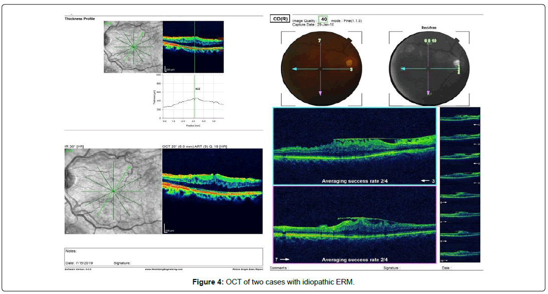
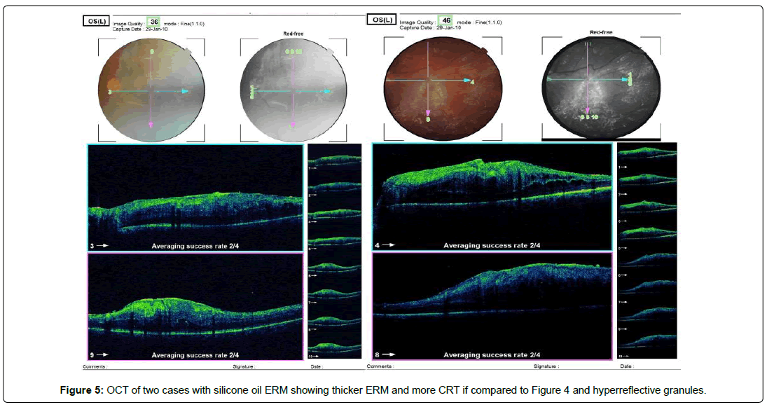
Whereas, OCT images in SO ERMs revealed thickened ERMs with increased central retinal thickness (CRT) underneath and hyper reflective granules; unlike idiopathic epiretinal membranes (ERMs), which is thinner with less increase in CRT (Figures 4 and 5).
Discussion
Since the last half of the twentieth century, silicone oil had become popular in the treatment of complicated retinal detachment which increased the rate of successful detachment repair. however, silicone oil tamponade has short-term and long-term complications like increased intraocular pressure, cataract, emulsification, proliferative vitreoretinopathy and ERM [5-8].
The current study detected the presence of bi-layered membrane in silicone oil ERM, inner spongy layer towards the vitreous side with granulomatous reaction with foreign body giant cells mainly surrounding silicone oil like granules and an outer membrane toward the retinal side lined by extensive hyperplastic glial cells with underlying extracellular matrix, therefore, preoperative OCT is mandatory to detect the presence of SO ERM before silicone oil removal.
The pathological examination was done during surgery and each layer was peeled separately in order to distinguish between the two layers, in case that the two layers were intermingled with each other in the specimen. H& E staining were able to differentiate between both layers. Whereas, idiopathic ERM showed only one layer formed of membrane lined by glial cells with underlying fibrous tissue.
Previous studies as Errara et al. [9] detected minute hyperreflective areas located intraretinally, subretinally and beneath the ERMs by SD-OCT images of the eyes with SO tamponade. Also, intra retinal SO vacuoles were detected in patients who underwent PPV and ILM peeling with silicone oil tamponade in macular hole surgery by Chung and Spaide [10].
As regard to idiopathic ERM, Smiddy et al. [13] reported the presence of varying proportions of four cell types, retinal pigment epithelium (RPE), fibrous astrocytes, fibrocytes and myofibroblasts. Collagen was the main constituent of the extracellular matrix in idiopathic ERMs [14] which was mostly produced from the RPE, fibroblasts and myofibroblasts. Also, Michels [15] detected that the pre retinal membranes following PPV for RRD were different from idiopathic ERMs which contain about 10% vessels, but nearly all idiopathic ERMs were avascular.
Other studies also detected the presence of ERMs in silicone oil filled eyes as the studies done by Duraani et al. and Junior et al. [11,12] and suggested that this is correlated to the duration of silicone oil inside the eye However, Wickham et al. [16 ] did not find any association between the duration of tamponade and the intensity of inflammation or the persistence of inflammation after silicone oil removal and suggested that the use of silicone oil itself is more important than the duration of intraocular tamponade.
As regard to immunostaining in our study, CD68 immunostaining showed that the macrophages that surrounded the emulsified SO in the SO ERMs indicates phagocytosis of the silicone oil. This is coincident with Heidenkummer et al. [17] and Wickham et al [16] who reported the presence of macrophages in SO ERMs. The same findings were reported by Heidenkummer and Kampik [18,19] in their immunohistochemical study of ERM in PVR cases, which reported the predominance of macrophages in the membranes extracted from the eyes with intraocular silicone oil tamponade.
Also in the SO ERMs study group the mean interval between the last vitreous surgery and SO removal was 9 months. The spongy layer in SO ERM was formed mainly due to the presence of long standing emulsified SO, therefore the emulsified SO is the rational of retinal inflammation and edema which mean that SO ERMs should be removed before the development of the bi-layered membrane. This explains why the thickness of the ERM in silicone filled eyes group in our study was much higher than that of idiopathic ERMs group, this is quite similar to other recent study that correlated the clinical and pathological features of ERM in eyes filled with silicone oil [18].
Surgical removal of the SO ERMs was difficult due to the fragility of the spongy layer and the underlying retina that results from inflammation in contrast to the retinal side which was firm. Diamond dusted scrubber (DDS) was used to help in lifting the spongy layer.
Conclusion
SO ERMs are double layered structures. The long standing intraocular silicone oil results in formation of spongy layer that induces retinal inflammation and makes its surgical removal difficult. Preoperative OCT is essential to detect such membranes, care should be taken to remove both layers and avoid injury of the retina as the retina is fragile and edematous.
Conflict of Interest
Authors certify that they have no conflict of interest with any organization and no financial interest in the subject matter or materials discussed in this manuscript.
Ethical Considerations
Procedures performed involving human participants were according to the ethical committee regulations of Tanta University, which is in consistence with the 1964 Helsinki declaration and its ethical standards. Informed consents were obtained from all the participants of the study.
References
- Jackson TL, Donachie PH, Williamson TH, Sparrow JM, Johnston RL, et al. (2015) Report 4, Epiretinal Membrane. The royal college of ophthalmologists' national ophthalmology database study of vitreoretinal surgery. Retina 35: 1615-1621.
- Mitchell P, SmithW, Chey T, Wang J, Chang A (1997) Prevalence and associations of epiretinal membranes: the Blue Mountains Eye Study, Australia. Ophthalmology 104: 1033-1040.
- Klein R, Klein BE, Wang Q, Moss SE (1994) The epidemiology of epiretinal membranes. Trans Am Ophthalmol Soc 92: 403–430.
- Cibis P A, Becker B, Okun E, Cannan S (1962) The use of liquid silicone in retinal detachment surgery. Arch Ophthalmol 68: 590-599.
- Watzke RC (1997) Silicone retinopiesis for retinal detachment: a long-term clinical evalution. Arch Ophthalmol 77: 185-196.
- Foulks GN, Hatchell DL, Proia AD, Klintworth GK (1991) Histopathology of silicone oil keratopathy in humans. Cornea 10: 129-137.
- Abrams GW, Azen SP, Mc Cuen BW, Flynn HW, Lai MY, et al. (1997) Vitrectomy with silicone oil or long-acting gas in eyes with severe proliferative vitreoretinopathy: results of additional and long-term follow-up: silicone study report 11. Arch Ophthalmol 115: 335-344.
- Honavar SG, Goyal M, Majji AB, Sen PK, Naduvilath T, et al. (1999) Glaucoma after pars plana vitrectomy and silicone oil injection for complicated retinal detachments. Ophthalmology 106: 169-177.
- Errera MH, Liyanage SE, Elgohary M, Day AC, Wickham L, et al. (2013) Using spectral-domain optical coherence tomography imaging to identify the presence of retinal silicone oil emulsification after silicone oil tamponade. Retina 33:1567-1573.
- Chung J, Spaide R (2003) Intraretinal silicone oil vacuoles after macular hole surgery with internal limiting membrane peeling. Am J Ophthalmol 136: 766-767.
- Durrani AK, Rahimy E, Hsu J (2017) Outer Retinal Changes on Spectral-Domain Optical Coherence Tomography Pre-and Post-Silicone Oil Removal. Ophthalmic Surg Lasers Imaging Retina 48: 978-82.
- Júnior M, de Oliveira O, Takahashi WY, Nakashima Y, Takahashi BS, et al. (2007) Optical coherence tomography macular study on eyes filled with silicone oil. Arq Bras Oftalmol 70: 281-285.
- Smiddy WE, Maguire AM, Green WR, Michels RG, de la Cruz Z, et al. (1989) Idiopathic epiretinal membranes: ultrastructural characteristics and clinicopathologic correlation. Ophthalmology 96: 811-821.
- Okada M, Ogino N, Matsumura M, Honda Y, Nagai Y (1995) Histological and immunohistochemical study of idiopathic epiretinal membrane. Ophthalmic Research 27: 118-128.
- Michels RG (1982) A clinical and histopathologic study of epiretinal membranes affecting the macula and removed by vitreous surgery. Trans Am Ophthalmol Soc 80: 580-656
- Wickham LJ, Asaria RH, Alexander R, Luthert P, Charteris DG (2007) Immunopathology of intraocular silicone oil: retina and epiretinal membranes. Br J Ophthalmol 91: 258-262.
- Heidenkummer HP, Messmer EM, Kampik A (1996) Recurrent vitreoretinal membranes in intravitreal silicone oil tamponade: Morphologic and immunohistochemical studies. Ophthalmologe 93: 121-125.
- TanakaY, Toyoda F, Shimmura-Tomita M, Kinoshita N, Takano H, et al. (2018) Clinicopathological features of epiretinal membranes in eyes filled with silicone oil. Clin Ophthalmol 12: 1949-1957.
- Heidenkummer HP, Kampik A (1991) Comparative immunohistochemical studies of epiretinal membranes in proliferative vitreoretinal diseases. Fortschr Ophthalmol 88:219-224.
 Spanish
  Chinese
  Russian
  German
  French
  Japanese
  Portuguese
  Hindi
Hindi 


