Spanish
  Chinese
  Russian
  German
  French
  Japanese
  Portuguese
  Hindi
Hindi Research Article, J Vet Sci Med Diagn Vol: 14 Issue: 1
Prevalence and Financial Significance of Bovine Hydatidosis in Bishofitu Elfora Export Abattoir, Oromia, Central Ethiopia
Yakob Bekele Deda1*, Mesay Tsegaye Demissie2 and Tinsae Endale Mengistu1
1Department of Agricultural Science, Arba-Minch University, Arba Minch, Ethiopia
2Department of Agricultural Science, Borena University, Yabello, Ethiopia
*Corresponding Author: Yakob Bekele Deda
Department of Agricultural Science, Arba-Minch University, Arba Minch, Ethiopia
E-mail: yakob4bekele@gmial.com
Received date: 15 November, 2024, Manuscript No. JVSMD-24-152627; Editor assigned date: 18 November, 2024, PreQC No. JVSMD-24-152627 (PQ); Reviewed date: 21 November, 2024, QC No. JVSMD-24-152627; Revised date: 18 March, 2025, Manuscript No. JVSMD-24-152627 (R); Published date: 25 March, 2025, DOI: 10.4172/2325-9550.1000122
Citation:Deda YB, Demissie MT, Mengistu TE (2025) Prevalence and Financial Significance of Bovine Hydatidosis in Bishofitu Elfora Export Abattoir, Oromia, Central Ethiopia. J Vet Sci Med Diagn 14:1.
Abstract
A cross-sectional study of bovine hydatidosis was carried out on local zebu cattle slaughtered at Bishoftu Elfora Export Abattoir from November 2023 to April 2023 to estimate prevalence, to characterize the cysts and to estimate financial loss caused by the disease. Both ante mortem and post mortem inspections procedures were carried out to obtain data about risk factors and to identify positive animals. Out of 400 systematical randomly selected cattle, 103 (25.75%) of them had hydatid cysts in one or more of their organs. Place of origin and age of animals has shown statistically significant (p<0.05) association within study population. Whereas, body condition scores of the cattle were not statistical significant (p>0.05). Among the positive animals, 43 (41.75%) of them harbored only in their lungs, 12 (11.65%) of them only in their livers and the rest 48 (46.6%) harbored within multiple organs. From 539 cysts obtained, 254 (47.12%), 239 (44.34%) and 46 (8.53) cysts were small sized, medium sized and large sized respectively. A total of 111 cysts were selected for cyst characterizations and result showed that 28 (25.23%) were calcified, 42 (37.84%) were sterile and 41 (36.94%) were fertile. Higher fertility rate was observed in cysts of lungs (37%) whereas, cysts from liver had higher calcification rate (55%). From 41 fertile cysts, 24 (58.54%) cysts were viable and the remaining 17 (41.46%) cysts were non-viable. The estimated financial loss as a result of bovine hydatidosis was 1,651,696.55 ETB. The result showed that the disease is highly prevalent and caused high financial loss in the abattoir. Hence it should be controlled and prevented as to decrease public health hazard, to improve livestock productivity and to reduce financial loss attributed by the disease.
Keywords: Cattle; Fertile; Financial loss; Hydatidosis; Viable
Abbreviations
ETB: Ethiopian Birr; NMSA: National Metrology Service Agency; CSA: Central Statistical Agency;ILCA: International Livestock Research Center for Africa; Kg: Kilogram; Km: Kilometer; Mm: Millimeter; Cm: Centimeter
Introduction
Africa had the highest livestock population in the world. Ethiopia is one of the East African countries with high density of livestock population which had 59.5 million cattle, 30.7 million sheep, 30.2 million goats, 11.01 million equids, 1.21 million camels and 56.53 million poultry population. Cattle were mainly used for meat and milk productions in addition to drought force. Number of cattle slaughtered in the country during 2016/17 period was estimated to be 499,841. However, the slaughter yield was minimal mainly attributed to traditional management practices and disease conditions. Hydatidosis is among the major parasitic diseases contributing to low productivity of meat market due to carcass and organ condemnation [1].
Echinococcosis is caused by adult or larval (metacestode) stages of cestodes belonging to the genus Echinococcus and the family Taenidae. It is recognized as one of the world’s major zoonosis affecting both humans and domestic animals. Different species were identified as Echinococcus granulosus, Echinococcus multilocularis, Echinococcus vogeli and Echinococcus oligarthrus which causes cystic echinococcosis, alveolar echinococcosis, polycystic echinococcosis and polycystic echinococcosis diseases in humans respectively. Echinococcus granulosis is the primary causative agent of hydatidosis in cattle and humans and echnococcosis in dogs. The lifecycle of the worm involves two mammalian hosts: Definitive host and intermediate host. Dogs and wild canids are definitive hosts. Ruminants (cattle, sheep and goat), humans and pigs serve as intermediate hosts [2].
The adult worm resides in the small intestine of the final host and eggs were released through feaces of infected canids. The egg is then ingested by intermediate hosts. Then hatches in the small intestine of the intermediate hosts and penetrates the intestinal wall and moves through the circulatory system into different organs, in particular the liver and lungs. The final hosts get infected by ingesting organs which contain hydatid cysts.
Hydatidosis causes considerable economic losses and public health problems worldwide. The primary source of hydatidosis in humans is by accidental ingestion of oncospheres from the coats of dogs or from vegetables and other foodstuffs contaminated by dog feces. This multihost parasite is prevalent all over the world and annually the economic loss in livestock due to this parasite is significant. In Africa, hydatid disease is reported more common in cattle those are communally owned or are raised of free range and which associated more intimately with domestic dogs. Hydatidosis infection constitutes a significant financial constraint derived from human health costs and livestock production losses. On the other hand, hydatidosis in domestic ruminants inflicts enormous economic loss via decreasing livestock production and condemnation of red and green offals in slaughterhouses [3].
In Ethiopia, hydatidosis has been known and documented as early as 1970’s. Hydatidosis is the major cause of organ condemnation in most Ethiopian abattoirs and slaughterhouses causing huge economic losses and highly prevalent. Some traditional activities could be commonly described as factors substantiating the spread and high prevalence of the diseases in the country. These include the widespread backyard animal slaughtering practice, the corresponding absence of rigorous meat inspection procedures and long-standing habit of feeding dogs with condemned offal. Hence knowledge on the prevalence of hydatidosis and financial loss in zebu cattle would have significant importance in justifying the need of an effective control scheme.
Therefore, the objectives of this study were:
• To estimate the prevalence of bovine hydatidosis and its financial loss in Elfora export abattoir, Bishoftu, Ethiopia.
• To characterize the hydatid cysts and identify some of the risk factors for the disease
Materials and Methods
Study area
The study was conducted at ELFORA export abattoir from November, 2023 to April, 2023. The abattoir is a privately-owned and it engages in exporting mutton, lamb, chevon and edible organs like liver and kidney of small ruminants to Middle East countries. It also supplies bovine meat to the domestic markets. This abattoir is found in Bishoftu town, Oromia regional states. The town is located at 90°N and 400°E with an altitude of 1880 meters above sea level in the central highlands of Ethiopia at 47 km South East of Addis Ababa. It has annual rain fall of 866 mm of which 84% falls during the long rainy season that extends from June to September; and the remaining during the short rainy season that extends from March to May. The mean annual minimum and maximum temperature are 14°C and 26°C, respectively and the mean relative humidity is 63.8% [4].
Study population
Animals which were presented to ELFORA export abattoir came from different regions of Ethiopia such as Wollo, Gondar, Gojjam, Wellega and Ambo. Local Zebu cattle brought to the abattoir for slaughter were considered as a study population for the active abattoir survey. The animals included in the study consist of cattle of different age, body condition and origin. The slaughter cattle were all males.
Study design
A cross sectional study was conducted from November, 2017 to April, 2018 to estimate the prevalence of bovine hydatidosis and its financial loss in cattle slaughtered at ELFORA export abattoir. Both ante mortem and postmortem inspection procedures were carried out during the study period. Animals were approached by systematic random sampling i.e., taking second coming animal slaughtered every other day [5].
Sample size determination
The sample size was determined according to Thrusfield 95% confidence interval and 5% absolute precision. The study was done previously and found a prevalence of 37.4%. Thus, using expected prevalence (37.4%), 360 samples were required (see formula below). However, a total of 400 animals were examined for this study by adding approximately 10% of the required sample to increase the precision as well as to increase precaution not to loss data while processing.
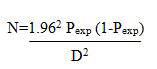
Where N=Required sample size
Pexp=Expected prevalence and
D=Desired absolute precision (0.05)
Study method
Abattoir survey: Ante-mortem and post-mortem examination were conducted by visiting the abattoir three days a week (every other day). During ante-mortem examinations, animals were selected systematically and given identification number by color marker; age, body condition and origin of the animals were recorded on record format/checklist. Age estimation was based on teeth eruption method described by Singh. Animals were grouped into three groups as young (<5 years), adult (5-10 years) and old (>10 years). Study animals were categorized into poor (1-3), medium (4-6) and good (7-9) body condition scores as recommended by Nicholson and Butterworth. For the post-mortem examination the liver, lung, kidney and other visceral organs were inspected for the presence of hydatid cysts by applying meat inspection procedure. During the first stage, visualization and palpation of organs (liver, lung) were carried out. After that, further incisions were made in each doubtful case for the study of the cysts. Cyst distribution and burdens in the affected organ/s were also recorded.
Cyst size, fertility and viability: Cysts obtained were measured for the size determination. Accordingly, cysts were classified as small (>4 cm), medium (4-8 cm) and large (>8 cm) according to Schantz. Hydatid cysts were collected randomly and transported to Addis Ababa University College of Veterinary Medicine and Agriculture, parasitology laboratory for fertility and viability tests. The pressure of the cyst fluid was reduced by using a sterile hypodermic needle. Then cyst was incised with a sterile scalpel blade and the content was poured into a glass Petri dish and examined. The presence of protoscolices either attached to the germinal layer in the form of brood capsule or its presence in the cyst fluid was considered as indicative of fertility [6].
Fertile cysts were further subjected to viability test. A drop of fluid from cyst containing the protoscolices were placed on the microscope glass slide and covered with coverslip and observed for amoeboid like peristaltic movements with 40× objective. For clear vision, a drop of 0.1% aqueous eosin solution was added to equal volume of protoscolices in hydatid fluid on microscope slide with the principle that viable protoscolices should completely or partially exclude the dye while the dead ones take it up. Sterile hydatid cysts are characterized by their smooth inner lining, usually with a slight turbidity of the contained fluid and typical calcified cyst that produced a gritty sound feeling upon incision [7].
Monetary loss analysis: Direct and indirect losses were considered based on estimation of the annual financial losses due to hydatidosis.
Direct loss was calculated on the bases of condemned organs, whereas indirect losses were estimated on the basis of live weight loss due to hydatidosis. Accordingly, the economic values of the loss from organ condemnations were evaluated by considering the following parameters. These include: Market price of each organs labelled by ELFORA export abattoir (lung, liver, spleen, kidney and heart) during study period, and the average annual slaughter rate of cattle at ELFORA export abattoir was 4745 cattle which was estimated from the retrospective data of the last two years and the loss from organs condemned was calculated by using the formula described by Ogunirale as follows:
LOC=(NAS × Ph × Plu × Cplu)+(NAS × Ph × Phr × Cphr)+(NAS × Ph × Pli × Cpli)+(NAS × Ph × Ps × Cps)+(NAS × Ph × Pk × Cpk)
Where: LOC=Loss due to organ condemnation
NAS=Mean number of cattle slaughtered annually
Ph=Prevalence of hydatidosis
Plu=Percent involvement of lung
Cplu=Current price of lung
Phr=Percent involvement of heart
Cphr=Current price of heart
Pli=Percent involvement of liver
Cpli=Current price of liver
Ps=Percent involvement of spleen
Cps=Current price of spleen
Pk=Percent involvement of kidney
Cpk=Current price of Kidney
Likewise, the following parameters were considered to estimate the economic loss encountered from carcass weight losses: Information on the mean price of 1 kg beef at Bishoftu town during the study period which was 210 ETB. The average annual slaughter rate of cattle at ELFORA export abattoir estimated from retrospective data of the last two years. The average carcass weight (dressing percentage) of Ethiopian zebu cattle was 126 kg taken from estimation by ILCA and the average carcass weight loss of 5% due to Hydatidosis. Thus, the economic loss due to denied carcass weight gain was determined as described by Ogunirade using the following formula:
LCWL=NAS × Ph × CPB × 5% × 126 kg
Where: LCWL=Loss from carcass weight loss
NAS=Average number of cattle slaughtered annually
Ph=Prevalence of bovine hyadatidosis
CPB=Current mean price of 1 kg of beef
Total financial loss was calculated by adding direct and indirect financial losses.
TFL=LOC+LCWL
Data analysis
The data obtained were coded and entered into Microsoft excel 2016 and subjected to descriptive statistics and Chi-square in order to assess the magnitude of the difference of comparable variables using SPSS version 25.0. Statistically significant association between variables were considered to exist if the p-value is less than 0.05 [8].
Results
Prevalence
Among the 400 apparently healthy cattle examined during the study period, 103 (25.75%) were infested with hydatid cysts within single or multiple organs involvement. Among the many factors considered, the prevalence was found to be statistically significantly (p<0.05) associated with the age groups and origin of animals. The prevalence among body conditions was found to be statistically insignificant (p>0.05) (Table 1).
| Risk factors | Examined | Positive | Prevalence (%) | Chi-square | P-value |
| Origin | |||||
| Gonder | 102 | 37 | 36.27 | 12.78 | 0.012 |
| Wollo | 109 | 32 | 29.36 | ||
| Ambo | 29 | 6 | 20.69 | ||
| Wellega | 37 | 7 | 18.92 | ||
| Gojjam | 123 | 21 | 17.07 | ||
| Total | 400 | 103 | 25.75 | ||
| Age | |||||
| Young | 42 | 5 | 11.9 | 7.66 | 0.022 |
| Adult | 233 | 57 | 24.46 | ||
| Old | 125 | 41 | 32.8 | ||
| Total | 400 | 103 | 25.75 | ||
| Body condition | |||||
| Poor | 106 | 29 | 27.36 | 0.67 | 0.72 |
| Medium | 251 | 65 | 25.9 | ||
| Good | 43 | 9 | 20.93 | ||
| Total | 400 | 103 | 25.75 | ||
Table 1: Prevalence of hydatidosis in cattle from different origins.
Cyst distributions and characterizations
Overall distribution of cysts in different organs of cattle slaughtered at Bishoftu ELFORA export abattoir was described (Table 2). Of the total 103 cattle positive, 43 (41.75%) had cysts only in lungs followed by 12 (11.65%) in liver, whereas the rest 48 (46.6%) infections involved multiple organs [9].
| Organs | Number of infected | Relative frequency (%) |
| Lung only | 43 | 41.75 |
| Liver only | 12 | 11.65 |
| Lung and liver | 40 | 38.83 |
| Lung and heart | 2 | 1.94 |
| Lung, liver and heart | 3 | 2.91 |
| Lung, liver and kidney | 2 | 1.94 |
| Lung, liver and spleen | 1 | 0.97 |
| Total | 103 | 100 |
Table 2: Cyst distribution in different organs.
From a total of 539 cysts obtained 275 (51.02%), 244 (45.27%), 11 (2.04%), (1.11%) and 3 (0.55%) cysts were located in lungs, liver, heart, kidneys and spleen respectively (Table 3). Cyst size determination revealed that 254 (47.12%), 239 (44.34%) and 46 (8.53%) cysts were small sized, medium sized and large sized respectively.
| Organ | Small cyst | Medium cyst | Large cyst | Cyst burden |
| Lung | 56 (20.36%) | 179 (65.09%) | 40 (14.55%) | 275 (51.02%) |
| Liver | 180 (73.77%) | 58 (23.77%) | 6 (2.46%) | 244 (45.27%) |
| Heart | 9 (81.82%) | 2 (18.18%) | 0 (0.0%) | 11 (2.04%) |
| Kidney | 6 (100%) | 0 (%) | 0 (%) | 6 (1.11%) |
| Spleen | 3 (100%) | 0 (%) | 0 (%) | 3 (0.55%) |
| Total | 254 (47.12%) | 239 (44.34%) | 46 (8.53%) | 539 |
Table 3: Cyst size and cyst burden per organ.
A total of 63 cysts from lung, 45 cysts from liver, 2 cysts from heart and 1 cyst from kidney origin were taken and subjected to cyst fertility test (Table 4). Out of 111 cysts tested for fertility, observation indicated that 37 (58.73%) cysts of lung and 4 (8.89%) cysts of liver origins were fertile. The rest were either sterile or calcified.
| Organ | Calcified (%) | Sterile (%) | Fertile (%) | Total |
| Lung | 3 (4.76) | 23 (36.51) | 37 (58.73) | 63 |
| Liver | 25 (55.56) | 16 (35.56) | 4 (8.89) | 45 |
| Heart | 0 (0.0) | 2 (100) | 0 (0.0) | 2 |
| Kidney | 0 (0.0) | 1 (100) | 0 (0.0) | 1 |
| Total | 28 (25.23) | 42 (37.84) | 41 (36.94) | 111 |
Table 4: Cyst fertility.
A total of 41 fertile cysts originating from lung and liver were tested for viability. The examination indicated that 23 (62.16 %) cysts from lung and 1 (25%) cyst from liver origin were viable and the remaining was non-viable cysts (Table 5).
| Organ | Viable (%) | Non-viable (%) | Total |
| Lung | 23 (62.16%) | 14 (37.84%) | 37 |
| Liver | 1 (25%) | 3 (75%) | 4 |
| Total | 24 (58.54%) | 17 (41.46%) | 41 |
Table 5: Cyst viability.
Financial loss estimation
Organs harboring the hydatid cysts were totally condemned in order to stop the life cycle of the parasite and for aesthetic values. Accordingly, 91 lungs, 58 livers, 5 hearts, 2 kidneys and 1 spleen were totally condemned as shown in Table 6.
Direct financial loss=(4745 × 25.75% × 88.35% × 5)+(4745 × 25.75% × 56.31 × 40)+(4745 × 25.75% × 4.85% × 30)+(4745 × 25.75% × 1.94% × 20)+(4745 × 25.75% × 0.97% × 3)=35,205.54 Birr
Indirect financial loss=4745 × 25.75% × 126 kg × 5% × 210=1,616,491.01 Birr
Total annual financial loss=35,205.54 birr+1616491.01 birr= 1,651,696.55 Birr per annum.
| No. | Organ | Condemned | Percent involvement | Price per unit in Birr |
| 1 | Lung | 91 | 88.35 | 5 |
| 2 | Liver | 58 | 56.31 | 40 |
| 3 | Heart | 5 | 4.85 | 30 |
| 4 | Kidney | 2 | 1.94 | 20 |
| 5 | Spleen | 1 | 0.97 | 3 |
| Total | 157 |
Table 6: Organ condemned and price of organs.
Discussion
In the present study the prevalence of Bovine hydatidosis in Bishoftu ELFORA export abattoir was found to be 25.75% which is comparable with reported prevalence of 24.5% in Sebeta, 24.1% in Debra Tabor, 20.5% in Arbaminch, 25.7% in South Omo and 22.1% in Tigray. It was less than the previous findings of 50.78% in Dodola, 37.4% in Bishoftu and 54% in Adama.
On other hand this finding is higher than the findings by Haftu and Kebede which is 11.88% in Bako, 15.20% in Birre-Sheleko and Dangila by Kebede et al., and 17.4% in Kombolcha. The variation in prevalence of hydatidosis could be attributed to the differences in seasonal variation, geographical locations and strain differences. Besides these, factors like difference in culture, social activity and attitude to dog in different regions might have contributed to this variation.
The variation (P-value<0.05) in the prevalence of hydatidosis in relation to origin of animals could be influenced by factors such as dog population and livestock-dog contact, environmental and climatic conditions, livestock husbandry practices and cultural and religious attitudes towards dogs in places of origin of the study animals. The finding was supported by the findings of Mekuria and Hirpassa, Debas and Ibrahim and Nasr and Pal.
In this study, a significant variation was observed in the rates of infections between age groups where adult and old animals were highly infected. This is in agreement with the findings of Zewdu et al., and Belina et al. This could be mainly due to the fact that aged animals have longer exposure time to eggs of E. granulosus in addition to weaker immunity to combat against the infection.
The differences in the occurrence of the hydatid cysts in different body condition was found statistically insignificant. However, infection tend to be higher in poor (27.36%) followed by medium (25.90%) and good (20.93%) body conditions. The finding was in agreement with Gebreyonnes and Regasssa et al. The reason for the high occurrence of disease in poor body condition could be moderate to severe infection with the parasite may cause retarded growth and performance, reduced quality of meat and milk as well as reduced live weight.
Lungs and liver were found to be predominantly infected by hydatid cysts than other organs such as spleen, heart and kidneys in this study. The result was in line with previous reports by Asfaw and Afera, Tolossa et al. and Al-Shaibani et al. This is explained by the fact that lungs and livers possess the first great capillary sites encountered by the migrating echinococcus oncosphere which adopt the portal vein route and primarily negotiate hepatic and pulmonary filtering system sequentially before any other peripheral organ is involved. In addition, the lungs were mostly infected than any other organ and this might be due to the fact that ruminants are slaughtered at older age. During this period, the liver capillaries are dilated and most cysts directly pass to the lungs.
Lung had higher number of medium and large sized cysts than other organs. Additionally, higher rate of fertile cysts were detected among cysts of lungs which have subjected to fertility tests. This could be attributed to relative soft consistency of lungs and fertility of the cysts which allows easier development of the cyst. On contrast liver had higher number of small sized and calcified cysts. This might be associated with the limited growth of the cyst pressure due to the immune response of the host since the organ bears relatively higher number of reticuloendothelial cells and abundant connective tissue which encapsulates the cyst within a fibrous wall up to 13 mm thick.
In this study annual financial loss due to organ condemnations and carcass weight decrement was estimated and found to be 1,651,696.55 ETB. The estimation was comparable with the findings of 1,848,849.765 ETB at Kombolcha ELFORA industrial abattoir and 1,160,932.4 ETB at Bishoftu ELFORA export abattoir. The slight difference observed attributed to differences in prevalence of the disease and the average price of organs and 1 kg of beef in the study period and area.
Conclusion
The study showed that the disease is prevalent in animals slaughtered at Bishoftu ELFORA export abattoir. The financial loss because of organ condemnations and carcass weight reductions is high in the abattoir. Age and geographical locations plays a role in the prevalence of the disease. Large and medium size cysts were commonly found in the lung than other organs and higher number of fertile cysts also found in the lungs. Liver was found to harbor, unlike lung, calcified and small cysts. Based on the above conclusions the following recommendations were forwarded:
• Infected organs should be burned or buried both in modern abattoir and backyard slaughterhouses to break the life cycle of the disease.
• Preventing the access of dogs especially stray dogs to slaughter houses is crucial to reduce the prevalence of the diseases.
• The public should be given awareness about the disease through media, educational systems and campaigns.
• Higher financial loss should need due attention and should be minimized by preventing and controlling the disease.
Acknowledgment
First and foremost, I honor and respect the GOD for his inspiration and keeping me health during my stay at Haramaya University. My special and sincere gratitude goes to my advisor, Dr Biressaw Serda for his encouragement, support and continual guidance and understandings throughout my work. I also acknowledge my parents and my friends for their encouragement, advice and support. Special thanks go to my father Tsegaye Demissie and my mother Tizita Mohammed for their help, moral support and inspiration.
It is also my pleasure to extend my deep appreciation to Bishoftu ELFORA Export Abattoir and Addis Abebe University College of Veterinary Medicine and Agriculture staffs for their technical and material support.
References
- Adem F, Addis M (2015) Prevalence and monetary loss of hydatidosis in apparently healthy slaughtered cattle at Elfora Export Abattoir, Ethiopia. World Appl Sci J 33: 1784-1792.
- Al-Shaibani I, Saad FA, Al-Mahdi H (2015) Cystic echinococcosis in humans and animals at Dhamar and Taiz governorates, Yemen. Int J Curr Microbiol App Sci 4: 596-609.
- Arene FO (1985) Prevalence of hydatid cysts in domestic livestock in the Niger Delta. Trop Anim Health Prod 17: 3-5.
[Crossref] [Google Scholar] [PubMed]
- Asfaw A, Afera B (2014) Prevalence of hydatid cyst in cattle at municipal abbatoir of Shire. J Vet Sci Technol 5: 186.
- Belina D, Fekadu G, Zegaye E, Belina S (2015) Bovine hydatidosis: Prevalence, public health and its economic significance in and around Harar, Ethiopia. J Veter Med Animal Health 7: 18-26.
- Craig PS, McManus DP, Lightowlers MW, Chabalgoity JA, Garcia HH, et al. (2007) Prevention and control of cystic echinococcosis. Lancet Infect Dis 7: 385-394.
[Crossref] [Google Scholar] [PubMed]
- Cringoli G, Rinaldi L, Musella V, Veneziano V, Maurelli MP, et al. (2007) Geo-referencing livestock farms as tool for studying cystic echinococcosis epidemiology in cattle and water buffaloes from Southern Italy. Geospatial Health 2: 105.
[Crossref] [Google Scholar] [PubMed]
- Debas E, Ibrahim N (2013) Prevalence and economic importance of hydatidosis in cattle slaughtered at North Gonder Elfora abattoir. Eur J Appl Sci 5: 29-35.
- Fuller GK, Fuller DC (1981) Hydatid disease in Ethiopia: Clinical survey with some immunodiagnostic test results. Am J Trop Med Hyg 30: 645-652.
[Crossref] [Google Scholar] [PubMed]