Case Report, J Spine Neurosurg Vol: 9 Issue: 1
Spinal Epidural Hematoma after Lumbar Catheter Insertion in a Patient Who Underwent TEVAR for an Aneurysm of the Descending Aorta
Kostov M1, Lazareska M2, Bushinoska J3, Dodevski A4 and Asani E1*
1University Clinic of Neurosurgery, Clinical Center “Mother Teresa”, Skopje, Republic of North Macedonia
2University Clinic for Radiology, Clinical Center “Mother Teresa”, Skopje, Republic of North Macedonia
3Univeristy Clinic of Anesthesiology, Reanimation and Intensive Care, Clinical Center “Mother Teresa”, Skopje, Republic of North Macedonia
4Institute of Anatomy, Medical Faculty, “University Ss. Cyril and Methodius”, Skopje, Republic of North Macedonia
*Corresponding Author: Asani Elmedina
University Clinic of Neurosurgery
Clinical Center “Mother Teresa”
Skopje, Republic of North Macedonia
Tel: 0038970643604
E-mail: elmedina_asani@hotmail.com
Received: December 31, 2019 Accepted: January 22, 2020 Published: January 29, 2020
Citation: Kostov M, Lazareska M, Bushinoska J, Dodevski A, Asani E (2020) Spinal Epidural Hematoma after Lumbar Catheter Insertion in a Patient Who Underwent TEVAR for an Aneurysm of the Descending Aorta. J Spine Neurosurg 9:1.
Abstract
We present a case of spinal epidural hematoma after Lumbar catheter insertion in a patient who underwent TEVAR for an aneurysm of the descending aorta. Spinal epidural hematoma (SEH) is a relatively rare entity and has been reported to occur in patients who receive anti-coagulant therapy, have bleeding disorders, or after traumatic needle insertion. In our case, the epidural hematoma occurred 4 hours after catheter insertion, in a patient who received anticoagulant and antiplatelet medication.
In these cases, early detection of symptoms and rapid diagnosis are an imperative. Our patient’s diagnosis was done 5 hours after symptom development and it consisted of thoracolumbar MRI that showed a spinal hyper-acute epidural hematoma extending from T9-T10 to L4-L5 and the affected segments of the spinal cord and conus medullar showed edema, most significantly at L1-L2 levels.A neurosurgical consult was done immediately after obtaining the results and within six hours after neurosurgical symptoms occurred, the patient underwent spinal decompression, the hematoma was evacuated and the patient regained motor function in her legs. During her stay at the cardiac ICU, the doctors administrated to our patient antiplatelet medication alongside low-molecular-weight heparin (LMWH)–inj. Clexane 40 I.U., against our recommendations. Due to this, our patient developed re-bleeding and we had to perform a second surgery, to do a more extensive spinal decompression and to evacuate the hematoma. Neurologic rehabilitation after the second surgery was difficult, but with extensive physical therapy, we have managed to achieve some improvement, with motor response 2\5 on the left leg and 3\5 on the right leg.
Although anticoagulants and\or antiplatelet medication are a must in endovascular procedures and are considered safe to use during epidural analgesia, special attention and care to epidural hematoma should be given, especially in cases when an epidural catheter placement is needed.
Keywords: Spinal epidural hematoma; Catheter; Aneurysm
Introduction
Spinal epidural hematoma (SEH) is a relatively rare entity first described by Jackson [1]. Spinal hematoma is the accumulation of blood in the potential space between the dura and bone. It can be a complication of neuroaxial anaesthesia techniques, especially in those patients with a deranged coagulation profile due to systemic diseases (e.g. hepatic diseases, renal failure) or anticoagulant therapy. It is more common in the patients treated with anticoagulants, thrombocytopenia, or in patients with alcoholic liver disease.
There have been many studies that have described the occurrence of spinal epidural hematomas associated with the placement and removal of Lumbar catheters [2–6]. Due to the association between the duration of hematoma-induced spinal cord compression and the degree of neurological symptoms [7], prompt diagnosis and surgical treatment are critically important determinants of neurological recovery. As the progression of severe symptoms generally occurs after an epidural catheter has been placed for more than 6 hours [8].
Here we are presenting a case of spinal epidural hematoma after Lumbar catheter insertion in a patient who underwent TEVAR for an aneurysm of the descending aorta.
Case Report
We present a case of a 66-year-old female with a previous history of hypertension and diabetes mellitus type II, status post bilateral adnexectomy, was admitted at the University Clinic for Cardiac surgery after a previously diagnosed an aneurysm of the descending aorta during a routine cardiac ultrasonography. A cardiac CT scan was performed that showed an aneurysm of the descending aorta (Figure 1).
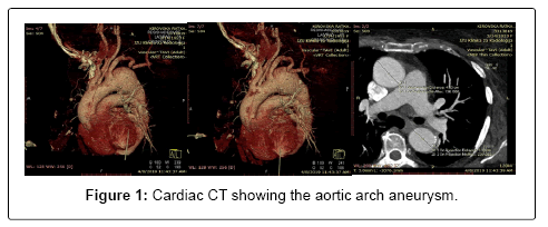
Figure 1: Cardiac CT showing the aortic arch aneurysm.
She was scheduled for an elective procedure-thoracic endovascular aortic repair (TEVAR). Preoperative checkups were performed:
The screening tests of hemostasis were as follows: platelet count 221–10 exp9/L, hematocrit–40%,
Prothrombin time PT)-10.7, Activated partial thromboplastin time (aPTT))-24.7, Thrombin time (TT)-22.7, INR- 0.97
Upon admission at the cardiac surgery wing, she was prepped and underwent surgery to perform a subclavian to carotid transposition (SCT) as an adjunct for TEVAR. The surgical procedure underwent without any complications and she was admitted at the University Clinic of Cardiology for further treatment. Pre intervention bold work-up showed a normal CBC, biochemical profile showed normal liver function tests, urea and creatinine were within normal values, glucose levels were 9.51 mmol/L, cardiac enzymes were within normal ranges and virus markers were negative. Cardiac ultrasonography revealed normal dimensions of the left ventricle with normal global systolic function, moderate hypertrophy of the interventricular septum, slightly enlarged left atrium, right atrium and ventricle within normal dimensions, valvulary apparatus of the heart normal according to age, no pericardial effusion and dilated ascending aorta up to 40 mm. ECG-Gated Cardiac CT was performed that showed a fusiform aneurysm of the descending aorta with maximal diameter less than 6 cm.
After all the necessary pre-interventional analysis the patient underwent TEVAR procedure. Before the procedure an Lumbar drain was placed to drain at 10 mm Hg (15 cm H2O) for 24 hours. The patient during the intervention was in supine position, epidural anesthesia was administered and access was gained through the femoral vein and a Valiant™ thoracic stent graft with the Captivia™ delivery system was inserted and the aneurysm was repaired. During the procedure, an occlusion of the left common carotid artery occurred and a Dynamic 8.0x56 mm stent was placed. The procedure when without any further complications and the patient was transferred at the cardiac ICU for further observation and treatment. After the procedure, the patient received 600 mg of Clopidogrel and 100 mg of Aspirin. Six hours after the intervention, the patient complained about pain in her lower extremities with decreased ability to move. There was blood in her Lumbar drainage catheter. The initial check up from the anesthesiologist revealed a decreased motor function on the lower extremities, and MRI of the lumbar spine was ordered and corticosteroid therapy was initiated and the Lumbar drainage catheter was removed.
Lumbar MRI revealed a spinal hyper-acute epidural hematoma extending from T9-T10 to L4-L5, the affected segments of the spinal cord and conus medullar show edema, most significantly at L1-L2 levels (Figures 2 and 3A,B).
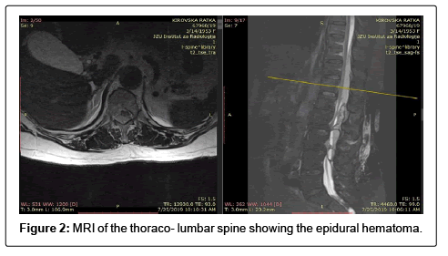
Figure 2: MRI of the thoraco- lumbar spine showing the epidural hematoma.
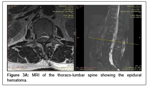
Figure 3A: MRI of the thoraco-lumbar spine showing the epidural hematoma.
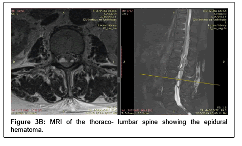
Figure 3B: MRI of the thoraco- lumbar spine showing the epidural hematoma.
After obtaining the MRI results, the cardiologist asked for a neurosurgical consult. Neurological status revealed motor response 1\5 on the left leg indicating plegia of the extremity and 2\5 on the right leg with minimal movements–severe paresis, decreased sensibility to touch, more pronounced on the left leg. An emergent indication for surgery was put forth and the patient was transferred at the PHO University Clinic of Neurosurgery Mother Teresa, Skopje. After pre-op preparation, the patient was brought to the operation room, intubated and positioned in a modified genu-pectoral position. After careful preparation and draping of the surgical field, we performed a midline incision of the skin on the lumbar region of the spine, proceeded with careful dissection of the soft tissues, at L3 level there were tissue defects consistent with multiple puncture in order to place the Lumbar drainage. A double laminectomy L2 and L3 levels was performed, the epidural hematoma was successfully evacuated and local hemostasis was achieved, a drain was placed and the soft tissues were sutured accordingly (Figure 4).
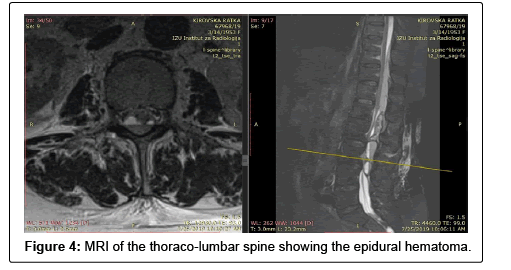
Figure 4: MRI of the thoraco-lumbar spine showing the epidural hematoma.
After the surgery, in cooperation with the cardiologists, the patient was transferred at the cardiac ICU to be monitored and receive further treatment, with recommendation that her anticoagulant therapy to be restarted 24 hours after the surgery and for it to be consisted of low-molecular-weight heparin (LMWH), for the patient to be on an antibiotic therapy at least 24 hour and administration of corticosteroid therapy. Neurological status after surgery improved significantly with motor response 3\5 on the left leg and 4\5 on the right leg (Figure 5).
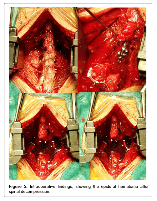
Figure 5: Intraoperative findings, showing the epidural hematoma after spinal decompression.
24 hours after the surgery, at the cardiac ICU, the patient received antiplatelet medication such as Clopidogrel 75 mg and Aspirin 100 mg alongside of low-molecular-weight heparin (LMWH)–inj. Clexane 40 I.U. 48 hour after surgery, the patients neurological symptoms worsened rapidly, she developed complete paraplegia on her lower extremities and complete loss of sensitivity.
We did an MRI of the thoraco-lumbar spine that revealed rebleeding from T9-T10 to L4-L5; the hematoma was with its larger diameter at the site of the laminectomy involving the surrounding soft tissues. The hematoma had significant compressive effect on the spinal cord and the conus medullaris (Figure 6).
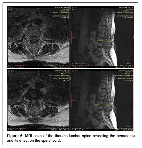
Figure 6: MRI scan of the thoraco-lumbar spine revealing the hematoma and its effect on the spinal cord.
Immediately after obtaining the MRI results, the patient was transferred at the ICU of the Neurosurgery Clinic and underwent second surgery to evacuate the hematoma. After pre-op preparation, the patient was brought to the operation room, intubated and positioned in a modified genu-pectoral position. After careful preparation and draping of the surgical field, the previous midline incision of the skin was continued above up to T9 level of the spine, meticulous dissection of the soft tissues was performed, and we proceeded with T11 laminectomy and partial L4 laminectomy, we evacuated the hematoma and achieved local hemostasis, we inserted two drains in the epidural space.
After the surgery, the patient was admitted at the neurosurgical ICU, received corticosteroid and symptomatic therapy and was under continuous cardiac monitoring. During her stay at the ICU her condition remained stable, she received Low-molecular-weight heparin (LMWH)–inj. Clexane 60 I.U. twice a day, recommended by a transfusiologist, the drains were removed on the second postoperative day and her incision site was healing well, with no signs of infection or dehiscence. She remained at the ICU for three days and then was transferred at the stationary ward of our hospital.
During her hospitalization at our stationary ward, she underwent physical therapy, received the proper therapy and daily wound dressings. In order to gradually restart her antiplatelet medication, we did the aspirin and clopidogrel resistance tests as well as and platelet function testing, with results within the normal ranges. One week after the surgery we reduced her low-molecular-weight heparin (LMWH)–inj. Clexane 60 I.U. to once a day for three days and after that we started administering Clopidogrel 75 mg once a day.
Two weeks after the second surgery we did a control MRI scan of the thoraco-lumbar spine that shoved complete evacuation of the hematoma, with hypersignal at the level of conus medullaris and lower thoracic parts of the spinal cord consistent with myelopathy changes or edema (Figure 7).
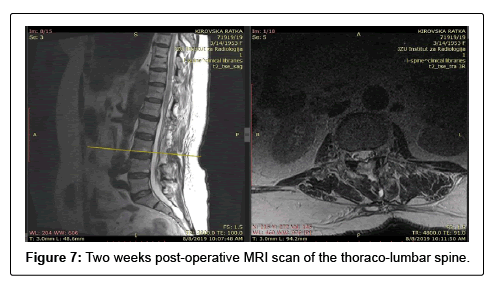
Figure 7: Two weeks post-operative MRI scan of the thoraco-lumbar spine.
The patient was discharged from our hospital three weeks after the surgery in an excellent general condition and slightly improved motor function on the right leg with remaining paralysis of the left leg and minor regain in sensibility in both legs, the surgical incision was healing properly, with no signs of infection. The patient started physical therapy a week after discharge from our hospital and just three months of extensive physical therapy, within the permitted limits for her cardiac conditions, she managed to stand with the help of orthosis.
Regular checkups at the outpatient clinic show improvements in her neurological status (Figure 8).
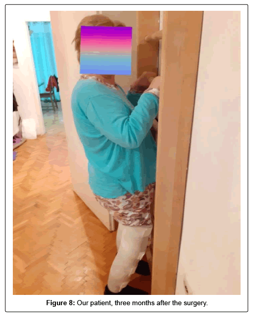
Figure 8: Our patient, three months after the surgery.
Discussion
Our literature research shows that catheter-associated spinal epidural hematomas can occur during catheter placement or removal with reported incidence from 2 to 20 cases per 100,000 Lumbar procedures [9-11]. The hemorrhage into the spinal canal frequently occurs in the epidural space due to epidural venous plexus rupture (Baston venous plexus), however arterial hemorrhage may also occur [12,13]. Spinal hematoma can be seen quickly with arterial bleeding and can lead to neural trauma and ischemia. However, when etiology is the needle or the Lumbar catheter, such as is in our case, the spinal hematoma may become symptomatic after a few hours or days, suggesting that the bleeding is not arterial. Cases of epidural hematomas have been reported to occur in patients who receive anticoagulant therapy, have bleeding disorders, or after traumatic needle insertion. In our case, the epidural hematoma occurred 4 hours after catheter insertion, in a patient who received anticoagulant and antiplatelet medication. In these cases, early detection of symptoms and rapid diagnosis are an imperative. Our patient’s diagnosis was done 5 hours after symptom development and it consisted of thoracolumbar MRI that showed a spinal hyper-acute epidural hematoma extending from T9-T10 to L4-L5, the affected segments of the spinal cord and conus medullar show edema, most significantly at L1-L2 levels. A neurosurgical consult was done immediately after obtaining the results and within six hours after neurosurgical symptoms occurred, the patient underwent spinal decompression, the hematoma was evacuated and the patient regained motor function in her legs with motor response 3\5 on the left leg and 4\5 on the right leg. During her stay at the cardiac ICU, the doctors administrated to our patient antiplatelet medication, alongside of Low-molecular-weight heparin (LMWH)–inj. Clexane 40 I.U., against our recommendations. Due to this, our patient developed re-bleeding and we had to perform a second surgery, to do a more extensive spinal decompression to evacuate the hematoma. Neurologic rehabilitation after the second surgery was difficult, but with extensive physical therapy, we have managed to achieve some improvement, with motor response 2\5 on the left leg and 3\5 on the right leg [14-16].
This case serves to emphasize the fact that both careful monitoring and effective multicenter treatment strategy is an imperative for adequate treatment for these patients. The use of anticoagulant and antiplatelet medication therapy after endovascular procedures and epidural catheter placement should be done carefully, and the preprocedural and post-procedural management should be meticulous.
Conclusion
Although anticoagulants and\or antiplatelet medication are a must in endovascular procedures and are considered safe to use special attention and care to epidural hematoma should be given, especially in cases when an Lumbar catheter placement is needed. Any signs or symptoms suggesting spinal cord compression, requires immediate actions to decompress the spinal canal and this diagnosis of Lumbar or spinal hematoma should be kept in mind in the differential diagnosis while using epidural catheters and anticoagulation. To note that dural puncture, direct trauma to the spinal cord, transient neuropathy and infection are also common complications among others of epidural catheters but are not the subject of this work.
References
- Jackson R (1869) Case of spinal apoplexy. Lancet 2: 5-6.
- Kaneda T, Suzuki T, Takeda J (2006) A Case of Acute Spinal Epidural Hematoma after Abdominal Aortic Aneurysm Operation. J Exp Clin Med 31: 45-48.
- Mahapatra S, Chandrasekhara NS, Upadhyay SP (2016) Spinal epidural haematoma following removal of epidural catheter after an elective intra-abdominal surgery. Indian J Anaesth 60: 355–357.
- Gulur P, Tsui B, Pathak R, Koury KM, Lee H (2015) Retrospective analysis of the incidence of epidural haematoma in patients with epidural catheters and abnormal coagulation parameters. Br J Anaesth 114: 808–811.
- Bateman BT, Mhyre JM, Ehrenfeld J, Kheterpal S, Abbey KR, et al. (2013)The risk and outcomes of epidural hematomas after perioperative and obstetric epidural catheterization: a report from the Multicenter Perioperative Outcomes Group Research Consortium. Anesth Analg 116: 1380–1385.
- Rosero EB, Joshi GP (2016) Nationwide incidence of serious complications of epidural analgesia in the United States. Acta Anaesthesiol Scand 60: 810–820.
- Wulf H (1996) Epidural anaesthesia and spinal haematoma. Can J Anaesth 43: 1260–1271.
- Kreppel D, Antoniadis G, Seeling W (2003) Spinal hematoma: a literature survey with meta-analysis of 613 patients. Neurosurg Rev 26: 1–49.
- Rosero EB, Joshi GP (2016) Nationwide incidence of serious complications of epidural analgesia in the United States. Nationwide incidence of serious complications of epidural analgesia in the United States. Acta Anaesthesiol Scand 60: 810–820.
- Li SL, Wang DX, Ma D (2010) Epidural hematoma after neuraxial blockade: a retrospective report from China. Anesth Analg 111: 1322–1324.
- Moen V, Dahlgren N, Irestedt L (2004) Severe neurological complications after central neuraxial blockades in Sweden 1990-1999. Anesthesiology 101: 950–959.
- Vandermeulen EP, Van Aken H, Vermylen J (1994) Anticoagulants and spinal–epidural anesthesia. Anesth Analg 79: 1165–1177.
- Moen V, Dahlgren N, Irestedt L (2004) Severe neurological complications after central neuraxial blockades in Sweden 1990–1999. Anesthesiology 101: 950–959.
- Moussallem C, Helou A, El-Yahchouchi C, Bou Ghosn R (2006) Epidural Catheters, Anticoagulation and the risk of Spinal Hematoma: A Review of Literature. The Internet Journal of Orthopedic Surgery 6: 1-4.
- Goswami D, Das J, Deuri A, Deka AK (2011) Epidural haematoma: Rare complication after spinal while intending epidural anaesthesia with long-term follow-up after conservative treatment. Indian J Anaesth 55: 71-73.
- Domenicucci M, Mancarella C, Santoro G, Dugoni DE, Ramieri A (2017) Spinal epidural hematomas: personal experience and literature review of more than 1000 cases. J Neurosurg Spine 27: 198–208.
 Spanish
  Chinese
  Russian
  German
  French
  Japanese
  Portuguese
  Hindi
Hindi 
