Case Report, J Vet Sci Med Diagn Vol: 12 Issue: 3
Surgical Management of Umbilical Hernia with Hernioplasty Using Monofilament Polypropylene Mesh in Buffalo Calf-A Case Report
Ankush Kumar1*, Bijender Singh1, Neeraj Arora1 and Satbir Sharma2
1Department of Veterinary Surgery and Radiology, Lala Lajpat Rai University of Veterinary and Animal Sciences, Hisar, India
2Department of Veterinary Clinical Complex, Lala Lajpat Rai University of Veterinary and Animal Sciences, Hisar, India
*Corresponding Author: Ankush Kumar
Department of Veterinary Surgery and Radiology, Lala Lajpat Rai University of Veterinary and Animal Sciences, Hisar, India
Tel: 8728862444
E-mail: dr.ankushgill@gmail.com
Received date: 11 August, 2022, Manuscript No. JVSMD-22-71696; Editor assigned date: 15 August, 2022, PreQC No. JVSMD-22-71696 (PQ); Reviewed date: 29 August, 2022, QC No. JVSMD-22-71696; Revised date: 06 January, 2023, Manuscript No. JVSMD-22-71696 (R); Published date: 16 January, 2023, DOI: 10.4172/2324-9005.1000036
Citation: Kumar A, Singh B, Arora N, Sharma S (2023) Surgical Management of Umbilical Hernia with Hernioplasty Using Monofilament Polypropylene Mesh in Buffalo Calf-A Case Report. J Vet Sci Med Diagn 12:1.
Abstract
A six month old male buffalo calf was presented for primary complaint of swelling in the ventro abdominal region since birth. On the basis of history and clinical examination, the case was tentatively diagnosed with umbilical hernia with hernial ring of size 12 cm with reducible hernial content. Under sedation and local infiltration of anesthesia the hernia was repaired by performing hernioplasty using monofilament polypropylene mesh. Severely adhered hernial sac with the hernial ring was resected before the implantation of the mesh. Post-operative treatment includes administration of antibiotic, analgesic and anti-inflammatory medications with regular antiseptic dressing of the wound. The animal had shown uneventful recovery with complete healing of the skin wound without any post-operative complication.
Keywords: Buffalo, Calf, Hernioplasty, Mesh, Polypropylene, Umbilical hernia
Introduction
When the contents of body cavity are protruded through a weak spot of body wall which may be from a normal anatomical or accidental opening, which does not completely fulfill its physiological function, characterized as hernia [1]. Hernias can be either congenital or acquired. In new born, umbilicus consists of remnants of umbilical vessels that transport blood between fetus and its mother and urachus (tube like structure which connects fetal urinary bladder to the placental sac) [2]. Atrophy of this structure occurs immediately after birth and remnants of urachus are remained in the abdomen. Doijode defined omphalocoele (umbilical hernia) as the displacement of complete organ or part of organ through a defect in the abdominal wall at the region of umbilicus with intact skin. Failure of closure of the umbilical ring appropriately at birth leads to development of umbilical hernia. This can be congenital or acquired at birth. Many factors predisposes for acquired umbilical hernias, like excess force applied during traction of fetus during dystocia, breaking the cord too close to the body wall, improper manual cutting of cord rather than to break it on its own at birth [3-7]. Some systemic conditions like diarrhea, umbilical abscess and constipation causes weakening of abdominal muscles due to increase in the abdominal pressure resulting in hernia. During the first 2 months of life, umbilical infections are associated with risk of umbilical hernia in calves. The herniated content may include omentum, intestine, abomasum or a combination of these, depending on the size of hernia ring [8].
Case Presentation
Surgical correction of umbilical hernia can be done mainly through two techniques, namely herniorrhaphy and hernioplasty. Although open herniorrhaphy is the most common method of veterinary treatment, recurrence is common after herniorrhaphy and seen throughout the year [9]. The present paper reports a case of umbilical hernia in calf which was successfully managed with hernioplasty using monofilament polypropylene mesh.
Case history and clinical observations
A Murrah buffalo male calf with age of six months was presented to the Veterinary Clinical Complex of LUVAS, Hisar, India, with a history of swelling in the ventro abdominal region since birth which had increased in size with successive increase in age of the animal. The animal had shown normal appetite, urination and defecation but occasionally used to kick the abdomen [10]. On palpation, there was a hernial ring of size 12 cm with reducible hernial content in the umbilical region. The physiological parameters were found within the normal clinical range. On the basis of history and clinical examination, the case was tentatively diagnosed with umbilical hernia. Since the size of the hernial ring was greater than 10 cm so it was decided to correct the umbilical hernia with open surgical method using monofilament polypropylene mesh?
Treatment
The animal was kept on pre-operative off feed and off water for 24 hours and 12 hours respectively. The animal was sedated using xyalzine hydrochloride at 0.01 mg/ Kg body weight diluted in 1 ml of distilled water intravenously and atropine sulphate was injected at 0.04 mg/kg body weight intramuscularly to control salivation followed by restraining in dorsal recumbency. The animal was administered adequate fluid therapy to correct any dehydration and acid base imbalances. The surgical site was shaved, scrubbed with Povidone Iodine 10% solution and draped followed by desensitization of the site by infiltration of 2% lignocaine hydrochloride solution [11]. An elliptical incision was given through the skin followed by dissection of fibrous adhesions and removal of devitalized tissue to expose the hernial ring. After pushing the hernia content inside the abdominal cavity, the herniated sac was incised carefully to avoid any damage to the viscera (Figures 1 and 2). Due to the presence of severe adhesions of the hernial sac with the hernial ring the sac was resected out (Figure 3). Before implantation of the polypropylene mesh, the fibrosed edges of the hernia ring was refreshed to reduce chances of suture dehiscence between the implant and the ring and also to promote the faster healing. A sterilized monofilament polypropylene mesh of size 15×15 cm was used for the hernia repair. Four simple interrupted sutures were placed at uniform distance to fix the mesh with the hernial ring muscular layer and the peritoneum along the hernial ring circumference at its position followed by application of Ford interlocking suture pattern in between those four interrupted sutures to cover the whole circumference of the ring (Figure 4). Polyglactin 910 (Vicryl®) # 2 was used for suturing of the polypropylene mesh with the hernial ring. After mobilization of the subcutaneous tissue, simple continuous suture pattern was used for apposition of the subcutaneous tissue over the mesh (Figure 5). The skin was closed by applying horizontal mattress suture with silk # 2 (Figure 6).
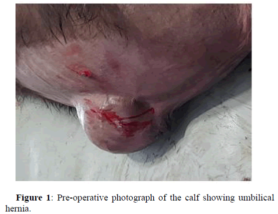
Figure 1: Pre-operative photograph of the calf showing umbilical hernia.
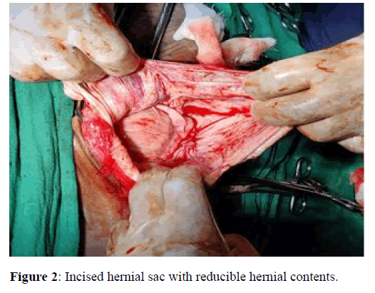
Figure 2: Incised hernial sac with reducible hernial contents.
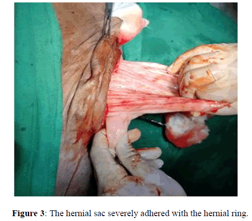
Figure 3: The hernial sac severely adhered with the hernial ring.
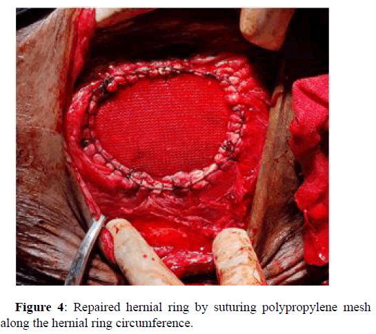
Figure 4: Repaired hernial ring by suturing polypropylene mesh along the hernial ring circumference.
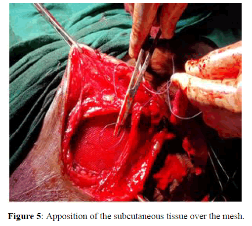
Figure 5: Apposition of the subcutaneous tissue over the mesh.
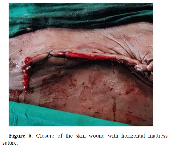
Figure 6: Closure of the skin wound with horizontal mattress suture.
Post-operative treatment included administration of cefotaxime sodium 1 gm and meloxicam at 0.4 mg/kg body weight intramuscularly for 5 days along with antiseptic dressing of the skin wound with povidone iodine 2% solution daily for 15 days. The owner was advised to reduce roughage feeding for first 10 days and then gradually shift the animal to normal diet.
Discussion
Umbilical hernias are the most common congenital defects found in calves and have been postulated to be related to heritable factors or in association with umbilical inflammation. Hernial contents seldom get strangulated with symptoms of pain and intestinal obstruction. There are more chances of development of adhesions between the abdominal structures and the reverted peritoneum in longstanding hernias. Kassem reported that there was drastic reduction in the recurrence rates for all hernias treated with tension free technique as compared to tissue repairs and made it possible to reconstruct large ventral defects that were previously irreparable. Monofilament mesh has lower risk of infection as compare to multifilament mesh because bacteria can get trapped in the smaller pores of multifilament mesh, making it hard for immune cells to get to those microbes.
Conclusion
In this case report, the umbilical hernia with hernial ring of 12 cm in diameter and severe adhesions of the hernial sac with the hernial ring in a six months old calf was successfully managed with hernioplasty using monofilament polypropylene mesh along with the administration of antibiotic, analgesic and anti-inflammatory medications with regular antiseptic dressing of the skin wound. The animal had shown uneventful recovery with complete healing of the skin wound without any post-operative complication. The skin sutures were removed on 14th post-operative day.
References
- Brown CN, Finch JG (2010) Which mesh for hernia repair? Ann R Coll Surg Engl 92:272–278.
[Crossref] [Google Scholar] [PubMed]
- Doijode V (2019) Umbilical hernia in ruminant calves: A review. Pharma Innovation J 8:164-167.
- Jaman MM, Mishra P, Rahman M, Alam MM (2018) Clinical and laboratory investigation on the recurrence of the umbilical hernia after herniorrhaphy in bovine calves. J Bangladesh Agric Univ 16:464-470.
- Kassem MM, El-Kammar MH, Korittum AS, Abdel-Wahed AA (2014) Using of polypropylene mesh for hernioplasty in calves. Alex J Vet Sci 40:112-117.
- Prasad CK, Maruthi ST, Sagar RS, Belakeri P, Mahantesh MT (2017) Surgical management of intestinal evisceration in neonatal calves. Intas Polivet 18:346-347.
- Rings DM. (1995) Umbilical hernias, umbilical abscesses and urachal fistulas. Surgical considerations. Vet Clin North Am Food Anim Pract 11:137-148.
[Crossref] [Google Scholar] [PubMed]
- Ron M, Tager‐Cohen I, Feldmesser E, Ezra E, Kalay D, et al. (2004) Bovine umbilical hernia maps to the centromeric end of Bos taurus autosome. Anim Genet 35:431-437.
[Google Scholar] [PubMed]
- Steenholdt, C. and Hernandez, J (2004) Risk factors for umbilical hernia in Holstein heifers during the first two months after birth. J Am Vet Med Assoc 224:1487-1490.
[Crossref] [Google Scholar] [PubMed]
- Sutradhar BC, Hossain MF, Das BC, Kim G, Hossain MA (2009) Comparison between open and closed methods of herniorrhaphy in calves affected with umbilical hernia. J Vet Sc 10:343-347.
[Crossref] [Google Scholar] [PubMed]
- Trent AM. (1987) Surgical management of umbilical masses in calves. Bovine Practitioner 22:170-173.
- Virtala AM, Mechor GD, Grohn YT, Erb HN (1996) The effect of calfhood diseases on growth of female dairy calves during the first 3 months of life in New York State. J Dairy Sci 79:1040-1049.
[Crossref] [Google Scholar] [PubMed]
 Spanish
  Chinese
  Russian
  German
  French
  Japanese
  Portuguese
  Hindi
Hindi 
