Spanish
  Chinese
  Russian
  German
  French
  Japanese
  Portuguese
  Hindi
Hindi Review Article, J Pulm Med Vol: 2 Issue: 1
The New Generation of the ExVivo Lung Perfusion Systems
Mohamed SA Mohamed*
Deutz Kalker Street 118, 50679 Cologne, Germany
*Corresponding Author : Mohamed SA Mohamed
Deutz Kalker Street 118, 50679 Cologne, Germany
Tel: +4915214736574
E-mail: mohammed. shehatta1@gmail.com
Received: January 25, 2016 Accepted: February 24, 2017 Published: March 03, 2017
Citation: Mohamed MSA (2017) The New Generation of the Ex-Vivo Lung Perfusion Systems. J Pulm Med 1:2.
Abstract
Background: The ex vivo lung perfusion (EVLP) technique is a method of preserving the lung graft between death of the donor and transplantation. Although many centers are applying the technique in their lung transplant programs and reporting good and promising results, there are some problems that impede the perfectionism of the technique.
Objective: In this paper, the author sheds the light on some of those problems and provides some innovative solutions for them. Methods: A new system for EVLP that allows a complete antegrade perfusion of the pulmonary and bronchial arteries, and provides the potential for longer ex vivo graft preservation may potentiate the benefits of the technique.
Hypothesis: The time passes slower within a stronger gravitational field. Accordingly, preserving the graft within an increased gravity might allow for longer preservation periods. Fortunately, other advantages of hypergravity have been reported, which might favor the attenuation of inflammation and the improvement of graft regeneration.
Conclusion: Although being in need of the experimental realization, the reported data on the cellular levels show the potential of the new system to provide better and longer ex vivo graft preservation.
Keywords: Ex-vivo lung perfusion; Lung transplant; Dual-EVLP; Hypergravity in organ preservation
Introduction
At the moment lung transplant is the sole curative therapeutic intervention for end-stage pulmonary diseases, such as chronic obstructive pulmonary disease, idiopathic pulmonary fibrosis, pulmonary hypertension, cystic fibrosis, sarcoidosis, bronchiectasis and lung cancer [1]. Unfortunately, saving the lives through this intervention is limited by the availability of the required grafts. In addition, lung transplant has the lowest long term post-transplant graft surviaval rate among the other solid organs [2,3]. Moreover, the most of the available grafts are declined because of unsuitability, either in general or for the particular patient [4].
The ex vivo lung perfusion (EVLP) technique was developed trying to solve the problems of the general insuitability, where the graft could be reconditioned, monitored and rendered acceptable [5,6]. The principle of the ex vivo perfusion technique relies on the use of a hyperoncotic perfusate, which washes out the inflammatory cells in the vasculature, blood clots, and waste products and, in turn, potentially reduces the ischemia-reperfusion injury (IRI). In addition, EVLP allows the resolution of the pulmonary edema, monitoring the airway and pulmonary vascular pressures and the oxygenation capacity [5,6].
Although many centers are applying the technique in their lung transplant programs and reporting good and promising results, there are some problems that impede the perfectionism of the technique. In this paper, the author sheds the light on some of those problems and provides some innovative solutions for them.
Perfusion through the bronchial arteries (dual-EVLP)
The value of the reconstruction of the bronchial arteries during lung transplantation, as well as its surgical applicability, was previously documented, where it was found to protect the pulmonary endothelium and type II pneumocytes in the early phase after transplant [7]. The bronchial blood flow was also found to be important for the vitality of the airways, the fluid balance of pulmonary tissue, and the metabolic functions of the lungs [8]. Accordingly, the inclusion of the bronchial arteries in the ex vivo lung graft perfusion is expected to provide significant functional improvement. However, all the in-use EVLP systems and protocols concentrate on the perfusion through the pulmonary circulation, which is the main lung vasculature, neglecting the bronchial vasculature that receives 3-5% of the cardiac output, under physiological conditions. This might create the problem that, part of the graft tissue can manifest the hazards of the ischemic-reperfusion injury, increasing the chance for post-transplant graft failure and rejection. To solve this problem, the dual- EVLP system was suggested.
Thus, a dual- EVLP system has been reported that was designed for the ex vivo perfusion of rat heart-lung blocks [9], where the original pulmonary circuit, used for the perfusion through the pulmonary artery, is used, in addition to a collateral circuit, which is used for the perfusion through the bronchial arteries. However, the collateral circuit is subjected to another pump to maintain a target flow of 3- 5% of cardiac output, in comparison to the 40% - 100% target flow in the pulmonary circuit. This system was experimentally studied, where the results of the study confirmed the expected significant attenuation of the cytokine production and the positive impact on the grafts [9].
However, a major risk in that system lies in the application of the retrograde flow through the aorta, which might have some hazardous effects on the vascular endothelium. The retrograde perfusion of the aorta to fill the bronchial arteries may not mimic the physiological perfusion filling, considering the change in the distribution of the maximal and minimal points of shearing forces, to which the endothelial cells of the retrieved vessels will be exposed during perfusion (Figure 1). Such alterations of shearing stress are thought to result in differential genetic expression and impaired endothelial functions in humans [10].

Figure 1: Diagrammatic representative model for the change of the shearing stress on the wall of bronchial arteries by the change of the direction of the aortic filling. In case of anti-grade flow (green arrow) point A and point B are subjected to different levels of shearing forces, which is the physiology. However, in the case of retrograde flow (blue arrow), the situation would be reversed, which might be associated with differential genetic expression. Though these differential changes in the shearing forces affect the arterial stem that gives rise to the bronchial artery rather than the bronchial artery itself, the three dimensional nature of the vascular system and flow has the potential to transmit corresponding changes to the walls of the bronchial arteries.
The increased shearing rate was found to increase the expression of endothelial adhesion molecules and the reactive oxygen species-producing enzymes (such as NADPH oxidase), as well as to decrease the expression of endothelial NO synthase [11,12]. In addition, the change of the shearing forces may constitute the initial stimulus for myointimal hyperplasia, which might result in anastomosis stenosis later after transplantation (Figure 1). To solve this problem, the following EVLP system is introduced.
The model
The Shehata - EVLP system is composed of a sterile box to enclose the graft, where the remnant of left atrium excised with the lung graft is to be reconstructed around a special cannula, connected to a pressure detector and adjustor, and connected to the pulmonary circuit. Perfusate flows through the circuit from a reservoir, and be subjected to a centrifugal pump (pulsatile or continuous flow), temperature adjustor, membrane gas exchanger, and leukocytes and cytokine filters (Figure 2).

Figure 2: Diagrammatic representation of Shehata's ex vivo lung perfusion system. The deoxygenated perfusate flows through the pulmonary vasculature to return oxygenated through the reconstructed left atrium. The oxygenated perfusate flows to both the pulmonary and the bronchial circuits, to be deoxygenated again before entering the pulmonary vasculature.
1. Ventilator
2. Box enclosing the lung graft
3. The reconstructed left atrium around a special cannula, supplemented with pressure electrode and connected to the perfusion circuit
4. The perfusion circuit
5. Centrifugal pump (pulsatile or continuous flow)
6. Temperature adjustor
7. Membrane gas exchanger
8. Leukocytes and cytokines filters
9. The pulmonary artery stump connected to the circuit through a special cannula and a pressure electrode to measure and adjust the pulmonary artery pressure
10. The collateral circuit, connected to the stumps of the bronchial arteries, using special cannulas
11. The source of the gravitational field, placed under or around the graft.
The perfusate then flows through the pulmonary artery, which is connected to the circuit using a special cannula. A pressure measuring electrode is to be connected to the lumen of the pulmonary artery, in order to measure and control its pressure. An in-line gas analyzer and saturation probes are to be included, for repetitive assessment of perfusate parameters. In addition, the tracheal stump is to be connected to a ventilator using an appropriate tube (Figure 2). A collateral circuit, subjected to another pump and a similar set of filters, is introduced. This circuit is to be connected directly to the stumps of the bronchial arteries (Figure 3), avoiding any stage of retrograde flow.

Figure 3: Diagrammatic representation of the method of the inclusion of the bronchial arteries into the collateral circuit of Shehata's dual-EVLP system. The bronchial arteries would be secured as proximal as possible during the retrieval of the graft from the donor, and would be connected using special cannulas to the collateral circuit, in a similar way to that of the pulmonary artery stump.
In the contrary to the other dual-EVLP system, where the bronchial arteries are perfused through the retrograde filling of the descending aorta, in Shehata's system, the bronchial arteries are to be secured as proximal as possible during the retrieval of the graft from the donor, and are to be connected using special cannulas to the collateral circuit, in a similar way to that of the pulmonary artery stump (Figure 3). This would not be technically impeded by the anatomical variations in the origins of the bronchial arteries, and would ensure the avoidance of the retrograde flow through the aorta.
The need for longer ex vivo graft preservation
The longest period of successful EVLP was reported by the Toronto team and lasted for 12-18 hours after 18 hours of cold static preservation (total of 30-36 hours). Longer preservation might result either in graft deterioration or the potential of unsuccessful transplant [3]. To significantly extend the preservation period, an additional modification of the system can be applied, which relies on the relative relationship between gravity and time, which was introduced by Albert Einstein, stating that the time passes slower within the increased gravity field [13].
Gravitational time dilation (GTD), which simply refers to the observation that the stronger the gravity, the slower the time passes, is one of the predictions of the general relativity theory. GTD was first tested in Pound–Rebka experiment that measures the redshift of light moving in a gravitational field [13]. Accordingly, the equivalence principle says that time runs slower at lower altitudes in a gravitational field. The GTD equation is expressed as following:
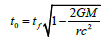
Where:
• t0 is the proper time for a slow-ticking observer within the gravitational field
• tf is the coordinate time for a fast-ticking observer at large distance from the massive object
• G is the gravitational constant
• M is the mass of the object creating the gravitational field
• r is the radial coordinate of the observer
• c is the speed of light, and
• rs = 2GM/C2 is the Schwarzschild radius of M.
The principle of the relationship between gravity and time is also confirmed in the special relativity theory of Einstein [13,14], where the graft preservation within a strong gravitational field might have the potential to slow down the time passing by the graft, allowing for relatively longer time of preservation, which has the potential to increase the number of performed transplants.
Although the effects of hypergravity on the human lung was previously investigated, showing that the effects are mainly related to the perfusion- ventilation matching, where the lung diffusing capacity was found to decrease by 33% when the gravity is 3 times the magnitude of the gravitational force at the surface of the earth (3G), these effects were significantly dependent on the lung position, where the application of 5G has been associated with 46% and 25% decrease in the lung diffusing capacity in the supine and prone positions, respectively. The application of hypergravity (3G) results also in the increase of lung tissue volume by about 38%, which is related to the increased blood sequestration in the dependent parts that are closer to the source of gravity [15].
In the case of EVLP, the application of hypergravity is ex vivo, which enables the control of any perfusion- ventilation mismatching through the controlled ventilation, controlled perfusion pumping, the frequent graft repositioning and or the use of a uniformly applied gravitational field. Accordingly, the principle itself could be safe and applicable.
Effects of hypergravity on lung graft during ex vivo perfusion
The reveal of the precise effects of hypergravity on the graft needs preclincal and clinical studies, however, previously published results can give us a provisional conclusion. Versari et al. [16], Spisni et al. [17], and Sumanasekera et al. [18,19] studied the effects of hypergravity for 24-48 hours on the primary human umbilical vein endothelial cells (HUVEC) and reported the following:
• ↑ migration
• ↑ NO
• Altered distribution of actin fibers
• ↑ cav-1 (tumor supressor gene)
• ↑ distribution of caveolae in the cell interior
• ↑ COX-2, NO, and PGI2 production
• ↓ angiogenesis (through a pathway not involving apoptosis)
• ↓ MAPK phosphorylation
• ↑ Paracellular permeability
• ↓ Transendothelial electrical resistance
• ↓ MAPK activation
• ↓ Extracellular barrier function
Morbidelli et al. [20], and Koyama et al. [21], tested the effects of hypergravity on the bovine aortic endothelial cells and reported the following;
• Modified integrin distribution
• Reorganization of cytoskeletal network
• ↓ genes controlling vasoconstriction and inflammation
• ↓ Proapoptotic signals
• ↑ ATP release
• ↑ actin reorganization via RhoA activation and FAK phosphorylation
• ↑ cell proliferation and migration
Meanwhile, Monici et al. [22], tested the effects of hypergravity on the bovine coronary venular endothelial cells and reported the following;
• ↓ Proapoptotic genes (FADD, Fas, Fas-L)
• ↑ Antiapoptotic gene NF-κB
• Change in cytoskeleton organization
• Alteration of cell energy metabolism
More recently, Grosse et al. [23], and Wehland et al. [24], used the human endothelial cells (line EA.hy926) to test the effects of hypergravity and vibration, and reported no significant changes on gene expression and morphology of the cells.
Although these experiments involved various techniques for the application of hypergravity (various sources, strengths and continuity), the overall results confirm the marked cytoskeletal upregulation and the enhanced cellular migartion and proliferation. In addition to the decreased apoptosis, the increased nitric oxide production, the increased expression of tumor supressor genes, the decreased expression and signaling of the proinflammatory pathways, and the enhanced ATP release. Moreover, the decreased extracellular endothelial barrier functions might help the rapid and effective resolution of the pulmonary edema [25].
Under the influence of the ischemic/anoxic – perfusion/ reoxygenation injury, the exposure to hypergravity (6G) protected the bovine aortic endothelial cells during the first 24 hours, where the exposed cells retained higher viability than the control non-exposed cells. While smooth muscle cells required time to adapt for hypergravity, so that, the hypergravity worsened their anoxia/reoxygenation injury in the acute phase followed by being protective within 48 hours, the rat lung fibroblasts behaved similar to the endothelial cells, where the hypergarvity showed significant protection [26].
Those experiments reffered the protective effects of hypergravity to the increased induction of the heat shock protein family that have been known to protect the cells against wide range of stresses [26]. Accordingly, the application of hypergarvity can be benefitial from many aspects.
Methods for generating artificial gravity
Rotation: A rotating object can produce the feeling of gravity in its inside. The rotation drives any object inside the object toward the hull, thereby giving the appearance of a gravitational pull directed outwards. The artificial gravity by rotation behaves in some ways similarly to the natural gravity, but has the following effects [27]:
• Centrifugal force: unlike the natural gravity, which pulls towards a center, a rotational gravity pushes away from the axis of rotation. Artificial gravity levels vary proportionately with the distance from the center of rotation.
• The Coriolis effect: which gives an apparent force that acts on objects that move relative to a rotating reference frame.
Linear acceleration: Linear acceleration can force objects inside the accelerating object in the opposite direction of the acceleration [27].
Magnetism: A similar effect to gravity can be created through the use of extremely powerful magnets, such as neodymium magnet [27,28].
The selection of the most appropriate method for the generation of the gravitational field and its incorporation in the system will be further studied and performed. However, the idea of introducing a gravitational field in the EVLP - system is firstly introduced and registered in this system (Figure 2).
Description of the Shehata's EVLP system with this modification
In addition to the above described components of the innovative dual - EVLP system, the graft containing box is to be placed on or within a gravity source (e.g. strong magnets) to create a strong uniform gravitational field (for example 5G or more) (Figure 2).
Conflicts of Interest
The intellectual properties and the system included and described in this manuscript belong solely to the author. All rights are registered and protected under the author's name. Reproduction or use of any of the included intellectual properties requires the written permission of the author. No funding was provided for the development of this work. The author welcomes funding cooperation for experimental and clinical studies.
References
- Kotloff RM, Thabut G (2011) Lung transplantation. Am J Respir Crit Care Med 184: 159-171.
- Pomfret EA, Sung RS, Allan J, Kinkhabwala M, Melancon JK, Roberts JP (2008) Solving the organ shortage crisis: the 7th annual American Society of Transplant Surgeons' State-of-the-Art Winter Symposium. Am J Transplant 8: 745-752.
- Cypel M, Keshavjee S (2013) Strategies for safe donor expansion: donor management, donations after cardiac death, ex-vivo lung perfusion. Curr Opin Organ Transplant 18: 513-517.
- Ware LB, Wang Y, Fang X, Warnock M, Sakuma T, et al. (2002) Assessment of lungs rejected for transplantation and implications for donor selection. Lancet 360: 619-620.
- Steen S, Sjoberg T, Pierre L, Liao Q, Eriksson L, et al. (2001) Transplantation of lungs from a non-heart-beating donor. Lancet 357: 825-829.
- Cypel M, Yeung JC, Hirayama S, Rubacha M, Fischer S, et al. (2008) Technique for prolonged normothermic ex vivo lung perfusion. J Heart Lung Transpl 27: 1319-1325.
- Nowak K, Kamler M, Bock M, Motsch J, Hagl S, et al. (2002) Bronchial Artery Revascularization Affects Graft Recovery after Lung Transplantation. Am J Respir Crit Care Med 165: 216-220.
- Kentaro N, Norihisa S, Yugo T, Bhama J, Kobayashi H et al. (2014) Hydrogen preconditioning during ex vivo lung perfusion improves the quality of lung grafts in rats. Transplantation 98: 499-506.
- Tanaka Y, Noda K, Isse K, Tobita K, Maniwa Y, et al. (2015) A Novel Dual Ex Vivo Lung Perfusion Technique Improves Immediate Outcomes in an Experimental Model of Lung Transplantation. Am J Transplant 15: 1219-1230.
- Thijssen DH, Dawson EA, Tinken TM, Cable NT, Green DJ (2009) Retrograde Flow and Shear Rate Acutely Impair Endothelial Function in Humans. Hypertension 53: 986-992.
- Hwang J, Ing MH, Salazar A, Lassègue B, Griendling K, et al. (2003) Pulsatile versus oscillatory shear stress regulates NADPH oxidase subunit expression: implication for native LDL oxidation. Circ Res 93: 1225-1232.
- Piergiorgio Tozzi (2007) Sutureless anastomoses; secrets for success. Steinkopff Verlag, Darmstadt.
- A. Einstein (1908) Über das Relativitätsprinzip und die aus demselben gezogenen Folgerungen. Jahrbuch der Radioaktivität und Elektronik 4: 411-462.
- Pound RV, Rebka GA (1959) Gravitational Red-Shift in Nuclear Resonance. Phys Rev Lett 3: 439-441.
- Malin Rohdin (2004) Effects of gravity and posture on the human lung. Thesis, department of physiology and pharmacology. Karolinska Institute, Stockholm, Sweden.
- Versari S, Villa A, Bradamante S, Maier JAM (2007) Alterations of the actin cytoskeleton and increased nitric oxide synthesis are common features in human primary endothelial cell response to changes in gravity. Biochim Biophys Acta 1773: 1645-1652.
- Spisni E, Blanco MC, Griffoni C, Santi S, Riccio M, et al. (2003) Mechanosensing role of caveolae and caveolar constituents in human endothelial cells. J Cell Physiol 197: 198-204.
- Sumanasekera WK, Zhao L, Ivanova M, Dwight D, Noisin EL et al. (2006) Effect of estradiol and dihydrotestosterone on hypergravity-induced MAPK signaling and occludin expression in human umbilical vein endothelial cells. Cell Tissue Res 324: 243-253.
- Sumanasekera WK, Sumanasekera GU, Mattingly KA, Dougherty SM, Keynton RS, et al. (2007) Estradiol and dihydrotestosterone regulate endothelial cell barrier function after hypergravity-induced alterations in MAPK activity. Am J Phys Cell Phys 293: C566-C573.
- Morbidelli L, Marziliano N, Basile V, Pezzatini S, Romano G, et al. (2009) Effect of hypergravity on endothelial cell function and gene expression. Microgravity Sci Tec 21: 135-140.
- Koyama T, Kimura C, Hayashi M, Watanabe M, Karashima Y, et al. (2009) Hypergravity induces ATP release and actin reorganization via tyrosine phosphorylation and RhoA activation in bovine endothelial cells. Pflugers Arch 457: 711-719.
- Monici M, Marziliano N, Basile V, Romano G, Conti A, et al. (2006) Hypergravity affects morphology and function in microvascular endothelial cells. Microgravity Sci Tec 18: 234-238.
- Grosse J, Wehland M, Pietsch J, Ma X, Ulbrich C, et al. (2012) Short-term weightlessness produced by parabolic flight maneuvers altered gene expression patterns in human endothelial cells. FASEB J 26: 639-655.
- Wehland M, Ma M, Braun M, Hauslage J, Hemmersbach R, et al. (2013) The impact of altered gravity and vibration on endothelial cells during a parabolic flight. Cell Physiol Biochem 31: 432-451.
- Maier JAM, Cialdai F, Monici M, Morbidelli L (2015) The Impact of Microgravity and Hypergravity on Endothelial Cells. BioMed Research International 2015: 434803.
- McCloud H, Pinks Y, Harris Hookefi SA, Melhado CD, Sanfords GL (1997) Hypergravity Alters the Susceptibility of Cells to Anoxia-Reoxygenation Injury. URC97152.
- Clément G, Bukley A (2007) Artificial Gravity. Springer-Verlag, New York, USA.
- Jacob F (2010) Handbook of Modern Sensors: Physics, Designs, and Applications. Springer-Verlag (4th Edtn) New York, USA.