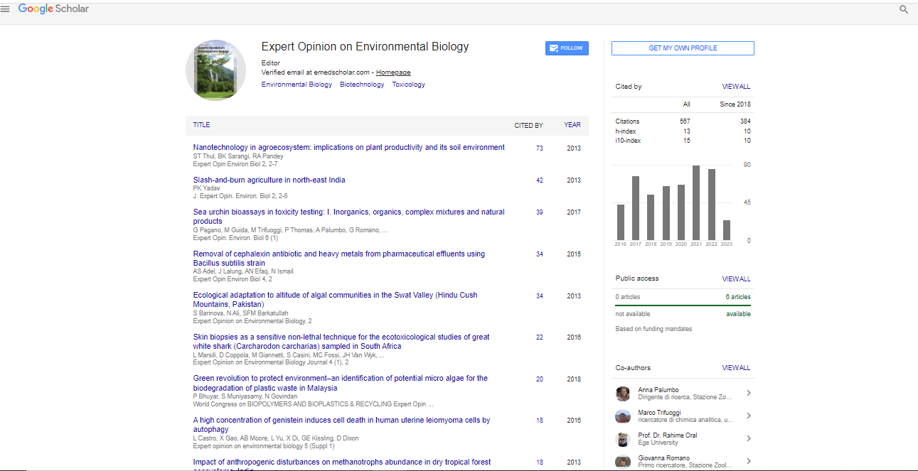
Review Article, Expert Opin Environ Biol Vol: 6 Issue: 1
Keywords |
| Sea urchin; Fertilization; Embryogenesis; Cytogenetic
abnormalities; Redox alterations |
Introduction |
| Sea urchins have proven to be extremely helpful in providing
scientific insight in a number of biological disciplines, including
physiology, embryology, biochemistry and genetics, in studies dating
as far back as the late 19th century to the early 20th century [1-6]. Those
studies provided a body of knowledge that became foundation in
what we know in basic cellular events such as mitosis with the first
reports on chromosomes [2], fertilization and embryogenesis [3-5],
with implications well beyond echinoderm biology and thereafter
translated into general biology and medicine. Relatively few reports
on the exposures of sea urchin embryos to various chemicals were
available, and focused primarily on the influence of some agents in
modulating normal embryogenesis, rather than on investigating any
adverse effects of chemicals, e.g. lithium-induced “animalization”
[6,7]. |
| In the wave of the early concern in radiobiology, drug toxicity,
and environmental pollution, sea urchins were first utilized in
pioneering studies evaluating the effects of sperm irradiation [8],
marine pollution [9], pharmaceuticals and pesticides [10-12], and
the action mechanisms of carcinogens [13,14], in bioassays viewed
as complementary to the classical murine models. Those studies
dating up to 1970’s set the grounds for continuing investigations up
to present. |
| The body of literature of toxicity testing in the sea urchin
model includes reports focused on a number of morphological
and/or molecular endpoints assessing adverse effects. This prevents
attempting extensive comparisons (e.g. scaling EC50’s) in-between
several agents. Though with this limitation, the present review may
both provide an unprecedented archive of sea urchin toxicity testing
reports and a useful contribution in future study design. |
| Inorganic agents |
| Seawater acidification: As shown in Table 1, pH changes were
initially studied in sea urchin embryos and sperm as a subject
of bioassay standardization, or for their possible implications in
freshwater systems, related to the recognized phenomenon termed
acid rain [15]. Early reports found that developing embryos under
slightly decreased pH conditions, in the order of 0.5 pH units,
underwent damage to embryogenesis and mitotic aberrations, with
stronger effects when treatment was performed before hatching
than in later developmental stages (blastula/gastrula) [16,17]. When
sea urchin sperm were exposed to pH changes, fertilization was
inhibited by either pH decrease (<7) or increase (>8.5). Furthermore,
transmissible damage to the offspring of low pH-exposed sperm was
observed, both in terms of developmental defects and of decreased
mitotic activity [18,19]. It should be noted that slightly decreased pH
(such as 7.5) enhanced fertilization success, as confirmed in a more
recent report [20]. A series of studies by Stumpp et al. [21-24] showed
that elevated seawater pCO2, closely resembling the expected values
by the end of this century, altered the expression of 26 representative
genes important for metabolism, calcification and ion regulation,
along with impacting growth and resulting in developmental delay
of echinoid larvae. Adult sea urchins (P. lividus) were tested by Lewis
et al. [25] for their sensitivity to micromolar copper levels in ambient
vs. acidified seawater, resulting in DNA damage and oxidative stress
responses, beyond the mere data of metal speciation. |
 |
Table 1: Reports on inorganics-associated effects on sea urchin early life stages. |
| Altogether, the available literature on the effects of pH decrease in
sea urchin bioassays corroborates the of global concern about ocean
acidification. |
| Other inorganics: A number of inorganics (Cd, As, Cr, Pb, Cl,
Ag, Mn, La, Ce, Gd) were tested for their adverse effects on sea urchin
development, fertilization success, mitotic activity, as well as other effects
as induction of apoptosis, calcification defects, DNA damage and/or
increasing production of reactive oxygen species (ROS) amongst others
[26-59] (Table 1). Groups of inorganics were tested in an extensive body
of studies providing comparative toxicity datasets for various agents
[40,59]. This was the case for early studies [41,42] testing groups of
inorganics as Ag, Cd, Cr, Hg and Pb; other studies reported on the effects
of individual inorganics vs. their mixtures [43,54]. |
| It has been reported that developmental impairment due to
treatment of fertilized eggs with Cd and Mn [29,38] involved the
action of the physiological messenger nitric oxide [47]. Moreover,
maternal exposure to these metal ions impaired reproduction and
progeny fitness [48]. |
| We have reported on the comparative toxicities in P. lividus
embryos and sperm of light rare earth elements (REEs) (Y, La, Ce,
Nd, Sm and Gd) that induced developmental defects and mitotic
abnormalities in exposed embryos, offspring damage following sperm
treatment, and redox anomalies (ROS production, lipid peroxidation,
and nitric oxide release) [58,59]. A current study of heavy REEs (Dy,
Ho, Er, Yb and Lu) suggests more severe toxicity of heavy vs. light
REEs. |
| Nanomaterials: As shown in Table 2, a number of studies have
been focused on the effects of nanomaterials (NM) in sea urchin
early life stages. The growing interest in the multi-fold applications
of NM has led to concerns on their possible health effects in several
biological models, including sea urchins [60-73]. To date, the
reported effects of NM in sea urchin early life stages have included
multiple responses in sea urchin embryos following exposures to
carbon nanoparticles, including developmental defects, abnormal
biomineralization, and altered gene expression [60,61]. Other
adverse effects have been reported in terms of chemosensitization,
increased multidrug resistance, and redox changes following
exposures of sea urchin embryos to Zn and Co oxide nanoparticles
[62-65]. Silica nanoparticles were found to increase developmental
defects while inhibiting fertilization success, and led to the decrease
of cholinesterase activity [67]. TiO2 nanoparticles were found to
enhance phagocyte TLR/p38 MAPK signalling pathway by activating
an internalization mechanism [72]. Altogether, sea urchin bioassays
have been useful in revealing adverse effects after exposure to NM. It
should be recognized, however, that a substantial body of literature,
beyond the scope of this review, points to a number of beneficial effects of NM, especially in medical and agricultural applications. Ad
hoc investigations are warranted to critically re-evaluate beneficial vs.
harmful effects associated with NM exposures. |
|
Table 2: Reports on nanoparticle-associated effects on sea urchin early life stages. |
| Organic agents |
| As shown in Table 3, an extensive body of literature is available
regarding the effects on sea urchin early life stage after exposures
to organic agents. Since early studies [13,14,74] and up to recent
reports [75,76] polycyclic aromatic hydrocarbons (PAHs) and
their hydroxylated derivatives have been investigated for their
bioaccumulation in sea urchin embryos and/or for induction of
developmental defects. Comparisons of the relative toxicities of select
PAHs vs their model mixtures when subjected to fluorescent light
exposure were also investigated in recent literature [76]. Other studies
reported on the relative toxicities of low molecular-weight aromatic
hydrocarbons (benzene and styrene) and their derivatives [77,78]. |
|
Table 3: Reports on the effects of organics, complex mixtures and natural products on sea urchin early life stages. |
| Extensive studies have focused on the effects of pharmaceuticals
on sea urchin embryogenesis and fertilization since early reports
[10,11,79-82] and up to recent studies [83-94]. The available
database comprehends reports on several classes of agents including
antibiotics and antiseptics [79-82], antineoplastic drugs [84-87,89],
teratogens [10,11,86,88,93], anti-inflammatory drugs, hormones or
hormone-like agents [88,92], anesthetics [94-96], cannabinoids [97-99], and antioxidants [86,100]. Altogether, the currently available
literature on the adverse effects of several pharmaceuticals in sea
urchin bioassays contributes to mechanistic information about druginduced
toxicity to early life stages, warranting further investigations
and risk assessment. |
| Pesticides and metal organics: Since a pioneering paper by Bresch
and Arendt [12], several pesticides have been investigated in sea
urchin early life stages. As shown in Table 3, a number of studies have
reported on halogen, nitrogen, and pyrethroid derivative pesticides
for their effects on sea urchin early development, fertilization, and offspring quality [101-120]. Several studies found embryo- and
spermiotoxicity of chlorinated pesticides [101-109], showing
differences in toxicity as a function of number of chlorine atoms per
molecule [102,103]. Organophosphate pesticides were found to affect
sea urchin metamorphosis [110], embryo- and spermiotoxicity, along
with induction of offspring damage following sperm exposure [111]. |
| In some cases pesticide preparations include organic and
inorganic components, as in the case of R6 fungicide, a mixture of an
acetamide pesticide, cymoxanil (CYM) and cupric oxychloride (Cu-
OCl) [120]. The commercial mixture resulted in toxicity that failed to
appear following exposure to the two mixture components alone. A
mixture prepared from analytical grade CYM and Cu-OCl also failed
to induce toxicity, a result that suggested the occurrence of more toxic
components in the formulation of the commercial mixture [120]. Thus, a critical reappraisal may be suggested in studies where toxicity
is commonly evaluated on pure chemicals, while disregarding the
effects induced by the technical-grade counterpart. |
| Either as antifouling agents or as pesticides, a number of
metallorganic compounds have been evaluated for their toxicity
to sea urchin early life stages (Table 3). Most of these reports have
focused on organic tin derivatives used as antifouling agents, while
some studies were aimed at comparing toxicities of individual agents
vs. their mixtures [121-131]. |
| Complex mixtures: An extensive body of literature refers to the
effects of complex mixtures in sea urchin early life stages, since the
1970’s [132,133]. As shown in Table 3, a number of studies have been
published on the effects of complex mixtures in sea urchin bioassays [132-160], including crude oil and oil fractions; spill-treating agents;
refinery wastewater effluent; pulp mill effluent; bauxite manufacturing
by-products; storm water and parking lot runoff; leather tanning
effluents; landfill leachate; pisciculture effluents, and alum-coagulated
municipal wastewater. This body of literature is not confined to studies
of mixtures directly involved in marine pollution, as in the cases for
industrial sludge and effluents [141-146,148-151,153,158], or landfill
leachate [155]. Thus, one may recognize multiple applications of sea
urchin bioassays in toxicity evaluation of complex mixtures both
impacting on marine and on freshwater or terrestrial environments. |
| Another major application of sea urchin bioassays in testing
complex mixture toxicity refers to sediment toxicity evaluations. This
relevant subject is omitted in the present review and will be exposed
in a subsequent paper. |
| Natural products: An established body of literature in sea
urchin bioassays has focused on the effects of a number of natural
products (NP) on sea urchin early life stages. The major goals of this
research line have been devoted to either characterizing some natural
products for their mitotic activity and their potential applications as
pharmaceuticals, or as potential stressors to marine ecosystems. |
| Sea urchin bioassays were successfully used for assay-guided
isolation of natural products from marine diatoms [161]. A body
of literature has been devoted to these molecules, characterized as
polyunsaturated aldehydes (PUAs), showing antimitotic and proapoptotic
activity inducing malformations in developing embryos
[162-168]. Interestingly, the effect was recorded when PUAs were
added before or soon after fertilization, while they were almost
ineffective when added at 40 minutes post fertilization onwards
[167]. Molecular studies of differential gene expression induced by
PUA treatments revealed that sea urchins activated an orchestrated
defense system involving HSP70 as key stress response mediator
[166] and showed that this effect is modulated by the messenger
nitric oxide [165]. Analogous to PUA, diatom-derived oxylipins and
hydroxyacids were reported recently to affect sea urchin development
by antimitotic and pro-apoptotic mechanisms [169,170]. |
| Other studies have reported on mitotoxic or other effects of
natural products on sea urchin early development. Extracts from
several biota, including algae, plants, and sponges were found to
exert mitotic arrest in sea urchin cell division [171-175]. Terpenic
compounds were obtained from sponges and corals providing
evidence for mitotoxicity and embryotoxicity [176,177] and other
effects, such as inhibition of DNA polymerases [178] and nucleic acid
biosynthesis [179]. Two reports focused on the action mechanisms
of β-amyloid in sea urchin early development [180,181]. A subtle
reproductive impairment through nitric oxide-mediated mechanisms
was reported in sea urchins from a site in the Gulf of Naples affected
by (Ostreopsis cf. ovata) [182]. |
| Sea urchin bioassays: Background and perspectives: After
the early laboratory studies of sea urchin bioassays in 1970’s, some
methodology reviews were published in testing developmental
damage, spermiotoxicity and transmissible damage to sperm
offspring, by either treating early life stages or adult sea urchins [183-
185]. |
| A summarized scenario of assay timing and outcomes that are
tested for in embryo exposure is depicted in Figure 1. The commonly
used embryotoxicity protocol starts at zygote stage (10 min postfertilization)
ending with the observation of developmental defects at pluteus larval stage, 48 to 72 hrs after fertilization, according to
species and culture temperature. In order to elucidate the most
sensitive developmental stage to the action of a given xenobiotic,
embryo exposure may be confined to early, pre-hatching stages,
with prevailing mitotic activity, or to post-hatching stages (blastula
and gastrula), leading to larval differentiation. Thus, for example,
allopurinol, some antioxidants (L-methionine and N-acetylcysteine)
and diatom aldehydes [86,100,168] only induced developmental
defects following early (pre-hatching) exposures. By contrast,
cadmium and chlorobenzenes were only effective when administered
to post-hatching embryos [26,77], while lead exerted significantly
stronger effects following post-hatching compared to pre-hatching
exposure [34]. |
|
Figure 1: Schematic view of timing and developmental stages in sea urchin
embryos cultured at 18°C (e.g. P. lividus) showing sensitive stages to mitotoxic
agents vs. embryo-selective agents (i.e. affecting later stages in embryogenesis).
Abbreviations: N: Normally developed plutei; P1: Malformed pluteus; P2:
Developmental arrest at abnormal blastula/gastrula stage. |
| The major embryological endpoints evaluated in larval cultures
are the frequencies of pluteus malformations (P1), pre-larval arrest
(P2), as shown in Figure 1. The most commonly scored endpoint,
as a function of xenobiotic concentration, is the frequency of
developmental defects (%DD)=(P1+P2). Another scored endpoint
consists of the observation of dead plutei and dead pre-larval (or prehatching)
embryos that point to acute effects; however, these effects
are not usually observed in testing sub-acute developmental toxicity,
confined to %DD. |
| In order to perform cytogenetic analysis, evaluating inhibition of
mitotic activity and/or induction of mitotic aberrations in Carnoy’s
fluid, cleaving embryos (approx. 5 hrs post-fertilization) are fixed
and subsequently stained by acetic carmine allowing for observation
of chromosomes, then scoring the frequencies of active mitoses per
embryo, embryos lacking mitotic figures, and the frequencies of
mitotic aberrations. The most commonly observed mitotic aberrations
include anaphase bridges, lagging chromosomes, multipolar spindles,
and scattered chromosomes (Figure 2). |
|
Figure 2: Main cytogenetic endpoints include decreased mitotic activity
(mitoses per embryo) and mitotic aberrations. A. Anaphase bridge; B.Lagging
chromosomes; C. Multipolar spindle; D. Scattered chromosomes. |
| Aside embryological and cytogenetic endpoints, a number
of biochemical or other molecular alterations may be detected in
xenobiotic-exposed sea urchin embryos and larvae. This was the case
for the induction of redox anomalies, with changes in ROS production,
oxidative and nitrosative stress endpoints, and oxidative DNA
damage [39,48,49,59,89,90]. Other reported endpoints, evaluated in
testing developmental toxicity in sea urchin embryogenesis, included
calcification defects [23,37-39], induction of multi-drug resistance [64] and MAPK-mediated signalling pathway [72], and production
of the endogenous antioxidant, ovothiol [186]. |
| Sea urchin sperm are exposed to agents for assigned time
intervals varying upon species, e.g. 60 min for Paracentrotus lividus
or 10 min for Sphaerechinus granularis. The endpoints measured
following sperm exposures to xenobiotics include fertilization
success and offspring damage, expressed as developmental defects
and/or altered redox endpoints (Figure 3). Changes in fertilization
success are mostly evaluated in zygotes by formalin fixation shortly
after fertilization by a number of authors [49,95,96,114,183,184].
We follow - and recommend - a different procedure, by reading
fertilization rate starting from 1 hr post-fertilization on live and
cleaving embryos, which are then allowed to undergo further
development up to pluteus stage. Indeed, reading fertilization rate in
fixed zygotes is confined to the appearance of fertilization membrane,
which may be more or less evident and lead to imprecise evaluation,
or to fixation artifacts. Otherwise, reading fertilization rate in
cleaving embryos is both supported by evidence for fertilization
membrane and by on-going cleavage. Thus, the observation of
any transmissible damage from sperm to offspring is warranted.
The induction of developmental defects in the offspring of sperm
exposed to xenobiotics has been reported in a number of studies
[19,31,33,43,44,54,58,59,89,90,111,143-145,148-151]. The finding of
developmental defects or embryonic larval mortality following sperm
exposures may be attributed to induction of dominant lethals, an
effect referred to since early genetics studies up to date [187,188]. |
|
Figure 3: Sperm pretreatment experiments, timing and observation endpoints:
a) Changes in fertilization success; b) Offspring damage, detected as: b1)
Developmental defects; b2) Altered redox endpoints. |
Conclusions |
| The body of literature on the use of sea urchin in toxicity testing
encompasses an extensive number of xenobiotics and mixtures that are
relevant both in environmental science and in general pharmacology and
toxicology. Well beyond the current database and the on-going research,
the appearance of new pollutants, pharmaceuticals and occupational
agents, along with the need for characterizing widespread complex
mixtures, open the field for further research efforts and future regulation
utilizing sea urchin bioassays in current and forthcoming work. |
| Beyond their half-century historical background, sea urchin bioassays represent a thriving tool in future investigations and health
risk assessment, unconfined to marine environment and in prospect
evaluation of novel xenobiotics. |
References |
- Boveri Th (1907) Cell studies VI: The development of dry sea urchins. A contribution to the theory of fertilization and the theory of the nucleus. Jena Time Naturw 43: 1-292.
- Baltzer F (1909) The chromosomes of Strongylocentrotus lividus and Echinus microtuberculatus. Arch f Zellf 2.
- Lillie FR (1921) The effects of copper salts on the fertilization reaction in Arbacia and a comparison of mercury effects. Biol Bull 41: 125-143.
- Mazia D (1937) The release of calcium in Arbacia eggs on fertilization. J Cell Comp Physiol 10: 291-304.
- Runnstrom J (1950) The problems of fertilization as elucidated by work on sea urchins. Harvey Lect Series 46: 116-152.
- Rulon O (1946) Modifications of sand-dollar development by exposure to lithium chloride and sodium thiocyanate before and after fertilization. Physiol Zool 19: 58-66.
- Lallier R (1955) Animalization of sea urchin eggs by zinc and cadmium salts. Exp Cell Res 8: 230-231.
- Rustad RC (1959) Induction of multipolar spindles by single x-irradiated sperm. Experientia 15: 323-324.
- Kobayashi N (1971) Fertilized sea urchin eggs as indicatory material for marine pollution bioassay, preliminary experiments. Publ Seto Mar Biol Lab 18: 379-406.
- Hagström BE, Lönning S (1973) The sea urchin egg as a testing object in toxicology. Acta Pharmacol Toxicol (Copenh) 1: 3-49.
- Hagström BE, Lönning S (1976) Teratogenic effect of tolbutamide on the development of the sea urchin embryo (Paracentrotus lividus Lamarck). Experientia 32: 744-746.
- Bresch H, Arendt U (1977) Influence of different organochlorine pesticides on the development of the sea urchin embryo. Environ Res 13: 121-128.
- Bresch H, Spielhoff R, Mohr U, Barkemeyer H (1972) Use of the sea urchin egg for quick screen testing of the biological activities of substances. I. Influence of fractions of a tobacco smoke condensate on early development. Proc Soc Exp Biol Med 141: 747-752.
- De Angelis E, Giordano GG (1974) Sea urchin egg development under the action of benzo(a)pyrene and 7,12-dimethylbenz(a)anthracene. Cancer Res 34: 1275-1280.
- Likens GE, Bormann FH (1974) Acid rain: a serious regional environmental problem. Science 184: 1176-1179.
- Kimura H, Shiroya T, Aoyama T, Shima A (1981) Effect of low pH treatment and agents causing mitotic delay on intracellular pH of sea urchin eggs. J Radiat Res 22: 160-164.
- Pagano G, Cipollaro M, Corsale G, Esposito A, Ragucci E, et al. (1985) pH-induced changes in mitotic and developmental patterns in sea urchin embryogenesis. I. Exposure of embryos. Teratog Carcinog Mutagen 5: 101-122.
- Pagano G, Cipollaro M, Corsale G, Esposito A, Ragucci E, et al. (1985) pH-induced changes in mitotic and developmental patterns in sea urchin embryogenesis. II. Exposure of sperm. Teratog Carcinog Mutagen 5: 113-122.
- Cipollaro M, Corsale G, Esposito A, Ragucci E, Staiano N, et al. (1986) Sub-lethal pH decrease may cause genetic damage to eukaryotic cell. A study on sea urchins and Salmonella typhimurium. Teratog Carcinog Mutagen 6: 275-288.
- Moulin L, Catarino AI, Claessens T, Dubois P (2011) Effects of seawater acidification on early development of the intertidal sea urchin Paracentrotus lividus (Lamarck 1816). Mar Pollut Bull 62: 48-54.
- Stumpp M, Wren J, Melzner F, Thorndyke MC, Dupont ST (2011a) CO2 induced seawater acidification impacts sea urchin larval development I: Elevated metabolic rates decrease scope for growth and induce developmental delay. Comp Biochem Physiol A Mol Integr Physiol 160: 331-340.
- Stumpp M, Dupont S, Thorndyke MC, Melzner F (2011b) CO2 induced seawater acidification impacts sea urchin larval development II: gene expression patterns in pluteus larvae. Comp Biochem Physiol A Mol Integr Physiol 160: 320-330.
- Stumpp M1, Hu MY, Melzner F, Gutowska MA, Dorey N, et al. (2012) Acidified seawater impacts sea urchin larvae pH regulatory systems relevant for calcification. Proc Natl Acad Sci USA 109: 18192-18197.
- Stumpp M, Hu MY, Casties I, Saborowski R, Bleich M, et al. (2013) Digestion in sea urchin larvae impaired under ocean acidification. Nat Clim Change 3: 1044-1049.
- Lewis C, Ellis RP, Vernon E, Elliot K, Newbatt S, et al. (2016) Ocean acidification increases copper toxicity differentially in two key marine invertebrates with distinct acid-base responses. Sci Rep 6: 21554.
- Pagano G, Esposito A, Giordano GG (1982) Fertilization and larval development in sea urchins following exposure of gametes and embryos to cadmium. Arch Environ Contam Toxicol 11: 47-55.
- Bowen WJ 3rd, Engel DW (1996) Effects of protracted cadmium exposure on gametes of the purple sea urchin, Arbacia punctulata. Bull Environ Contam Toxicol 56: 493-499.
- Agnello M, Filosto S, Scudiero R, Rinaldi AM, Roccheri MC (2007) Cadmium induces an apoptotic response in sea urchin embryos. Cell Stress Chaperones 12: 44-50.
- Filosto S, Roccheri MC, Bonaventura R, Matranga V (2008) Environmentally relevant cadmium concentrations affect development and induce apoptosis of Paracentrotus lividus larvae cultured in vitro. Cell Biol Toxicol 24: 603-610.
- Chiarelli R, Agnello M, Bosco L, Roccheri MC (2014) Sea urchin embryos exposed to cadmium as an experimental model for studying the relationship between autophagy and apoptosis. Mar Environ Res 93: 47-55.
- Pagano G, Esposito A, Bove P, de Angelis M, Rota A, et al. (1982) Arsenic-induced developmental defects and mitotic abnormalities in sea urchin development. Mutat Res 104: 351-354.
- Gaion A, Scuderi A, Pellegrini D, Sartori D (2013) Arsenic exposure affects embryo development of sea urchin, Paracentrotus lividus (Lamarck). Bull Environ Contam Toxicol 91: 565-570.
- Pagano G, Esposito A, Bove P, De Angelis M, Rota A, et al. (1983) The effects of hexavalent and trivalent chromium on fertilization and development in sea urchins. Environ Res 30: 442-452.
- Warnau M, Pagano G (1994) Developmental toxicity of PbCL2 in the echinoid Paracentrotus lividus (echinodermata). Bull Environ Contam Toxicol 53: 434-441.
- Hose JE, Di Fiore D, Parker HS, Sciarrotta T (1989) Toxicity of chlorine dioxide to early life stages of marine organisms. Bull Environ Contam Toxicol 42: 315-319.
- Ward TJ, Kramer JR, Boeri RL, Gorsuch JW (2006) Chronic toxicity of silver to the sea urchin (Arbacia punctulata). Environ Toxicol Chem 25: 1568-1573.
- Saitoh M, Kuroda R, Muranaka Y, Uto N, Murai J, et al. (2010) Asymmetric inhibition of spicule formation in sea urchin embryos with low concentrations of gadolinium ion. Dev Growth Differ 52: 735-746.
- Pinsino A, Matranga V, Trinchella F, Roccheri MC (2010) Sea urchin embryos as an in vivo model for the assessment of manganese toxicity: developmental and stress response effects. Ecotoxicology 19: 555-562.
- Pinsino A, Roccheri MC, Costa C, Matranga V (2011) Manganese interferes with calcium, perturbs ERK signaling, and produces embryos with no skeleton. Toxicol Sci 123: 217-230.
- Congiu AM, Calendi E, Ugazio G (1984) Effects of metal ions and CCl4 on sea urchin embryo (Paracentrotus lividus). Res Commun Chem Pathol Pharmacol 43: 317-323.
- Dinnel PA, Link JM, Stober QJ, Letourneau MW, Roberts WE (1989) Comparative sensitivity of sea urchin sperm bioassays to metals and pesticides. Arch Environ Contam Toxicol 18: 748-755.
- Ringwood AH (1992) Comparative sensitivity of gametes and early developmental stages of a sea urchin species (Echinometra mathaei) and a bivalve species (Isognomon californicum) during metal exposures. Arch Environ Contam Toxicol 22: 288-295.
- Pagano G, His E, Beiras R, De Biase A, Korkina LG, et al. (1996) Cytogenetic, developmental, and biochemical effects of aluminum, iron, and their mixture in sea urchins and mussels. Arch Environ Contam Toxicol 31: 466-474.
- Warnau M, Iaccarino M, De Biase A, Temara A, Jangoux M, et al. (1997) Spermiotoxicity and embryotoxicity of heavy metals in the echinoid Paracentrotus lividus. Environ Toxicol Chem 15: 1931-1936.
- Fernández N, Beiras R (2001) Combined toxicity of dissolved mercury with copper, lead and cadmium on embryogenesis and early larval growth of the Paracentrotus lividus sea-urchin. Ecotoxicology 10: 263-271.
- Sanchez-Marin P, Santos-Echeandia J, Nieto-Cid M, Alvarez-Salgado XA (2010) Effect of dissolved organic matter (DOM) of contrasting origins on Cu and Pb speciation and toxicity to Paracentrotus lividus larvae. Aquat Toxicol 96: 90-102.
- Migliaccio O, Castellano I, Romano G, Palumbo A (2014) Stress response to cadmium and manganese in Paracentrotus lividus developing embryos is mediated by nitric oxide. Aquat Toxicol 156: 125-134
- Migliaccio O, Castellano I, Cirino P, Romano G, Palumbo A (2015) Maternal exposure to cadmium and manganese impairs reproduction and progeny fitness in the sea urchin Paracentrotus lividus. PLoS One 10: e0131815.
- Novelli AA, Losso C, Ghetti PF, Ghirardini AV (2003) Toxicity of heavy metals using sperm cell and embryo toxicity bioassays with Paracentrotus lividus (Echinodermata: Echinoidea): comparisons with exposure concentrations in the Lagoon of Venice, Italy. Environ Toxicol Chem 22: 1295-1301.
- Kobayashi N, Okamura H (2004) Effects of heavy metals on sea urchin embryo development. 1. Tracing the cause by the effects. Chemosphere 55: 1403-1412.
- Kobayashi N, Okamura H (2005) Effects of heavy metals on sea urchin embryo development. Part 2. Interactive toxic effects of heavy metals in synthetic mine effluents. Chemosphere 61: 1198-1203.
- Bielmyer GK, Brix KV, Capo TR, Grosell M (2005) The effects of metals on embryo-larval and adult life stages of the sea urchin, Diadema antillarum. Aquat Toxicol 74: 254-263.
- Geffard O, Geffard A, Budzinski H, Crouzet C, Menasria R, et al. (2005) Mobility and potential toxicity of sediment-bound metals in a tidal estuary. Environ Toxicol 20: 407-417.
- Caplat C, Oral R, Mahaut ML, Mao A, Barillier D, et al. (2010) Comparative toxicities of aluminum and zinc from sacrificial anodes or from sulfate salt in sea urchin embryos and sperm. Ecotoxicol Environ Saf 73: 1138-1143.
- Nadella SR, Tellis M, Diamond R, Smith S, Bianchini A, et al. (2013) Toxicity of lead and zinc to developing mussel and sea urchin embryos: critical tissue residues and effects of dissolved organic matter and salinity. Comp Biochem Physiol C Toxicol Pharmacol 158: 72-83.
- Tellis MS, Lauer MM, Nadella S, Bianchini A, Wood CM (2014) The effects of copper and nickel on the embryonic life stages of the purple sea urchin (Strongylocentrotus purpuratus). Arch Environ Contam Toxicol 67: 453-464.
- Rosen G, Rivera-Duarte I, Colvin MA, Dolecal RE, Raymundo LJ, et al. (2015) Nickel and Copper Toxicity to Embryos of the Long-Spined Sea Urchin, Diadema savignyi. Bull Environ Contam Toxicol 95: 6-11.
- Oral R, Bustamante P, Warnau M, D'Ambra A, Guida M, et al. (2010) Cytogenetic and developmental toxicity of cerium and lanthanum to sea urchin embryos. Chemosphere 81: 194-198.
- Pagano G, Guida M, Siciliano A, Oral R, Kocbas F, et al. (2016) Comparative toxicities of selected rare earth elements: sea urchin embryogenesis and fertilization damage with redox and cytogenetic effects. Environ Res 147: 453-460.
- Manno D, Carata E, Tenuzzo BA, Panzarini E, Buccolieri A, et al. (2012) High ordered biomineralization induced by carbon nanoparticles in the sea urchin Paracentrotus lividus. Nanotechnology 23: 495104.
- Carata E, Anna Tenuzzo B, Arnò F, Buccolieri A, Serra A, et al. (2012) Stress response induced by carbon nanoparticles in Paracentrotus lividus. Int J Mol Cell Med 1: 30-38.
- Fairbairn EA, Keller AA, Madler L, Zhou D, Pokhrel S, et al. (2011) Metal oxide nanomaterials in seawater: linking physicochemical characteristics with biological response in sea urchin development. J Hazard Mater 192: 1565-1571.
- Manzo S, Miglietta ML, Rametta G, Buono S, Di Francia G (2013) Embryotoxicity and spermiotoxicity of nanosized ZnO for Mediterranean sea urchin Paracentrotus lividus. J Hazard Mater 254-255: 1-9.
- Wu B, Torres-Duarte C, Cole BJ, Cherr GN (2015) Copper oxide and zinc oxide nanomaterials act as inhibitors of multidrug resistance transport in sea urchin embryos: their role as chemosensitizers. Environ Sci Technol 49: 5760-5770.
- Torres-Duarte C, Adeleye AS, Pokhrel S, Mädler L, Keller AA, et al. (2015) Developmental effects of two different copper oxide nanomaterials in sea urchin (Lytechinus pictus) embryos. Nanotoxicology 8:1-9
- Della Torre C, Bergami E, Salvati A, Faleri C, Cirino P, et al. (2014) Accumulation and embryotoxicity of polystyrene nanoparticles at early stage of development of sea urchin embryos Paracentrotus lividus. Environ Sci Technol 48: 12302-12311.
- Gambardella C, Morgana S, Bari GD, Ramoino P, Bramini M, et al. (2015a) Multidisciplinary screening of toxicity induced by silica nanoparticles during sea urchin development. Chemosphere 139: 486-495.
- Gambardella C, Ferrando S, Morgana S, Gallus L, Ramoino P, et al. (2015b) Exposure of Paracentrotus lividus male gametes to engineered nanoparticles affects skeletal bio-mineralization processes and larval plasticity. Aquat Toxicol 158: 181-191.
- Siller L, Lemloh ML, Piticharoenphun S, Mendis BG, Horrocks BR, et al. (2013) Silver nanoparticle toxicity in sea urchin Paracentrotus lividus. Environ Pollut 178: 498-502.
- Burić P, Jakšić Ž, Štajner L, Dutour Sikirić M, Jurašin D, et al. (2015) Effect of silver nanoparticles on Mediterranean sea urchin embryonal development is species specific and depends on moment of first exposure. Mar Environ Res 111: 50-59.
- Magesky A, Pelletier E (2015) Toxicity mechanisms of ionic silver and polymer-coated silver nanoparticles with interactions of functionalized carbon nanotubes on early development stages of sea urchin. Aquat Toxicol 167: 106-123.
- Pinsino A, Russo R, Bonaventura R, Brunelli A, Marcomini A, et al. (2015) Titanium dioxide nanoparticles stimulate sea urchin immune cell phagocytic activity involving TLR/p38 MAPK-mediated signalling pathway. Sci Rep 5: 14492.
- Kanold JM, Wang J, Brummer F, Siller L (2016) Metallic nickel nanoparticles and their effect on the embryonic development of the sea urchin Paracentrotus lividus. Environ Pollut 212: 224-229.
- Hose JE, Puffer HW, Oshida PS, Bay SM (1983) Developmental and cytogenetic abnormalities induced in the purple sea urchin by environmental levels of benzo(a)pyrene. Arch Environ Contam Toxicol 12: 319-325.
- Bellas J, Saco-Alvarez L, Nieto O, Beiras R (2008) Ecotoxicological evaluation of polycyclic aromatic hydrocarbons using marine invertebrate embryo-larval bioassays. Mar Pollut Bull 57: 493-502.
- Suzuki N, Ogiso S, Yachiguchi K, Kawabe K, Makino F, et al. (2015) Monohydroxylated polycyclic aromatic hydrocarbons influence spicule formation in the early development of sea urchins (Hemicentrotus pulcherrimus). Comp Biochem Physiol C Toxicol Pharmacol 171: 55-60.
- Pagano G, Cipollaro M, Corsale G, Esposito A, Giordano GG, et al. (1988) Comparative toxicities of benzene, chlorobenzene and dichlorobenzenes on sea urchin embryos and sperm. Bull Environ Contam Toxicol 40: 481-488.
- Pagano G, Esposito A, Giordano GG, Hagström BE (1978) Embryotoxic and teratogenic effects of styrene derivatives on sea urchin development. Scand J Work Environ Health 4 Suppl 2: 136-141.
- Kimura H (1975) Effects of caffeine on cleavage delay of sea urchin eggs induced by ethidium bromide or puromycin. J Radiat Res 16: 125-131.
- Schneider EG, Nguyen HT, Lennarz WJ (1978) The effect of tunicamycin, an inhibitor of protein glycosylation, on embryonic development in the sea urchin. J Biol Chem 253: 2348-2355.
- Rosenkranz PG, Gordon DR, Speck WT, Rosenkranz HS (1980) Developmental abnormalities in the American sea urchin induced by povidone-iodine, a widely used vaginal antiseptic. Mutat Res 77: 387-390.
- Czinn SJ, Speck WT, Rosenkranz HS (1981) Abnormalities in the development of the American sea urchin induced by nalidixic acid. Mutat Res 91: 119-121.
- Stephens L, Hardin J, Keller R, Wilt F (1986) The effects of aphidicolin on morphogenesis and differentiation in the sea urchin embryo. Dev Biol 118: 64-69.
- Schatten G, Schatten H, Bestor TH, Balczon R (1982) Taxol inhibits the nuclear movements during fertilization and induces asters in unfertilized sea urchin eggs. J Cell Biol 94: 455-465.
- Madari H, Panda D, Wilson L, Jacobs RS (2003) Dicoumarol: a unique microtubule stabilizing natural product that is synergistic with Taxol. Cancer Res 63: 1214-1220.
- Graillet C, Pagano G, Girard JP (1993) Stage-specific effects of teratogens on sea urchin embryogenesis. Teratog Carcinog Mutagen 13: 1-14.
- Sconzo G, Romancino D, Fasulo G, Cascino D, Giudice G (1995) Effect of doxorubicin and phenytoin on sea urchin development. Pharmazie 50: 616-619.
- Sconzo G, Fasulo G, Romancino D, Cascino D, Giudice G (1996) Effect of retinoic acid and valproate on sea urchin development. Pharmazie 51: 175-180.
- Korkina LG, Deeva IB, Iaccarino M, De Biase A, Oral R, et al. (2000) Redox-dependent toxicity of diepoxybutane and mitomycin C in sea urchin embryogenesis. Carcinogenesis 21: 213-220.
- Pagano G, de Biase A, Deeva IB, Degan P, Doronin YK, et al. (2001) The role of oxidative stress in developmental and reproductive toxicity of tamoxifen. Life Sci 68: 1735-1749.
- Roepke TA, Snyder MJ, Cherr GN (2005) Estradiol and endocrine disrupting compounds adversely affect development of sea urchin embryos at environmentally relevant concentrations. Aquat Toxicol 71: 155-173.
- Kiyomoto M, Kikuchi A, Morinaga S, Unuma T, Yokota Y (2008) Exogastrulation and interference with the expression of major yolk protein by estrogens administered to sea urchins. Cell Biol Toxicol 24: 611-620.
- Reichard-Brown JL, Spinner H, McBride K (2009) Sea urchin embryos exposed to thalidomide during early cleavage exhibit abnormal morphogenesis later in development. Birth Defects Res B Dev Reprod Toxicol 86: 496-505.
- Ribeiro S, Torres T, Martins R, Santos MM (2015) Toxicity screening of diclofenac, propranolol, sertraline and simvastatin using Danio rerio and Paracentrotus lividus embryo bioassays. Ecotoxicol Environ Saf 114: 67-74.
- Hinkley RE Jr, Wright BD (1985) Comparative effects of halothane, enflurane, and methoxyflurane on the incidence of abnormal development using sea urchin gametes as an in vitro model system. Anesth Analg 64: 1005-1009.
- Hinkley RE Jr, Wright BD (1986) Effects of the volatile anesthetic halothane on fertilization and early development in the sea urchin Lytechinus variegatus: evidence that abnormal development is due to polyspermy. Teratology 34: 291-301.
- Schuel H, Schuel R, Zimmerman AM, Zimmerman S (1987) Cannabinoids reduce fertility of sea urchin sperm. Biochem Cell Biol 65: 130-136.
- Schuel H, Berkery D, Schuel R, Chang MC, Zimmerman AM, et al. (1991) Reduction of the fertilizing capacity of sea urchin sperm by cannabinoids derived from marihuana. I. Inhibition of the acrosome reaction induced by egg jelly. Mol Reprod Dev 29: 51-59.
- Chang MC, Schuel H (1991) Reduction of the fertilizing capacity of sea urchin sperm by cannabinoids derived from marihuana. II. Ultrastructural changes associated with inhibition of the acrosome reaction. Mol Reprod Dev 29: 60-71.
- Pagano G, Bonassi S, De Biase A, Degan P, Deeva IB, et al. (1997) L-Methionine induces stage-dependent changes of differentiation and oxidative activity in sea urchin embryogenesis. Pharmacol Toxicol 81: 134-143.
- Adams JA (1983) Effect of PCB (aroclor 1254) on early development and mortality in Arbacia eggs. Water Air Soil Poll 20: 1-5.
- Pagano G, Cipollaro M, Corsale G, Esposito A, Ragucci E, et al. (1985) Comparative toxicities of chlorinated biphenyls on sea urchin egg fertilization and embryogenesis. Mar Environ Res 17: 240-244.
- Trieff NM, Cipollaro M, Corsale G, Esposito A, Ragucci E, et al. (1988) Aroclor 1254 toxicity in sea urchin embryos and gametes. Exp Oncol Life Science Adv 7: 57-64.
- Coteur G, Danis B, Fowler SW, Teyssie JL, Dubois P, et al. (2001) Effects of PCBs on reactive oxygen species (ROS) production by the immune cells of Paracentrotus lividus (Echinodermata). Mar Pollut Bull 42: 667-672.
- Ozretic B, Krajnovic-Ozretic M (1985) Morphological and biochemical evidence of the toxic effect of pentachlorophenol on the developing embryos of the sea urchin. Aquat Toxicol 7: 255-263.
- Green JD, Mwatibo JM, Swartz WJ (1997) The effects of methoxychlor on early sea urchin development. Environ Res 72: 56-64.
- Buznikov GA, Nikitina LA, Bezuglov VV, Lauder JM, Padilla S, et al. (2001) An invertebrate model of the developmental neurotoxicity of insecticides: effects of chlorpyrifos and dieldrin in sea urchin embryos and larvae. Environ Health Perspect 109: 651-661.
- Pesando D, Robert S, Huitorel P, Gutknecht E, Pereira L, et al. (2004) Effects of methoxychlor, dieldrin and lindane on sea urchin fertilization and early development. Aquat Toxicol 66: 225-239.
- Bellas J, Beiras R, Marino-Balsa JC, Fernandez N (2005) Toxicity of organic compounds to marine invertebrate embryos and larvae: a comparison between the sea urchin embryogenesis bioassay and alternative test species. Ecotoxicology 14: 337-53.
- Aluigi MG, Falugi C, Mugno MG, Privitera D, Chiantore M (2010) Dose-dependent effects of chlorpyriphos, an organophosphate pesticide, on metamorphosis of the sea urchin, Paracentrotus lividus. Ecotoxicology 19: 520-529.
- Manzo S, Buono S, Cremisini C (2006) Toxic effects of irgarol and diuron on sea urchin Paracentrotus lividus early development, fertilization, and offspring quality. Arch Environ Contam Toxicol 51: 61-68.
- Wang H, Huang HH, Ding J, Wang YH (2010) Embryotoxicity and teratogenicity of pesticide indoxacarb to sea urchin (Strongylocentrotus intermedius). Water Sci Technol 61: 2733-2739.
- Buono S, Manzo S, Maria G, Sansone G (2012) Toxic effects of pentachlorophenol, azinphos-methyl and chlorpyrifos on the development of Paracentrotus lividus embryos. Ecotoxicology 21: 688-697.
- Hwang J, Suh SS, Chang M, Yun Park S, Ryu TK, et al. (2014) Effects of triclosan on reproductive parameters and embryonic development of sea urchin, Strongylocentrotus nudus. Ecotoxicol Environ Saf 100: 148-152.
- Gharred T, Ezzine IK, Naija A, Bouali RR, Jebali J (2015) Assessment of toxic interactions between deltamethrin and copper on the fertility and developmental events in the Mediterranean sea urchin, Paracentrotus lividus. Environ Monit Assess 187:193.
- Erkmen B (2015) Spermiotoxicity and embryotoxicity of permethrin in the sea urchin Paracentrotus lividus. Bull Environ Contam Toxicol 94: 419-424.
- Arslan OC, Parlak H, Oral R, Katalay S (2007) The effects of nonylphenol and octylphenol on embryonic development of sea urchin (Paracentrotus lividus). Arch Environ Contam Toxicol 53: 214-219.
- Cakal Arslan O, Parlak H (2007) Embryotoxic effects of nonylphenol and octylphenol in sea urchin Arbacia lixula. Ecotoxicology 16: 439-444.
- Ozlem CA, Hatice P (2008) Effects of bisphenol A on the embryonic development of sea urchin (Paracentrotus lividus). Environ Toxicol 23: 387-392.
- Pagano G, Iaccarino M, De Biase A, Meric S, Trieff NM (2001) Factors affecting R6 fungicide toxicity on sea urchin fertilization and development: Roles of exposure routes and mixture components. Hum Exp Toxicol 20: 404-411.
- Marin MG, Moschino V, Cima F, Celli C (2000) Embryotoxicity of butyltin compounds to the sea urchin Paracentrotus lividus. Mar Environ Res 50: 231-235.
- Novelli AA, Argese E, Tagliapietra D, Bettiol C, Ghirardini AV (2002) Toxicity of tributyltin and triphenyltin to early life-stages of Paracentrotus lividus (Echinodermata: Echinoidea). Environ Toxicol Chem 21: 859-864.
- Kobayashi N, Okamura H (2002) Effects of new antifouling compounds on the development of sea urchin. Mar Pollut Bull 44: 748-751.
- Bellas J, Granmo K, Beiras R (2005) Embryotoxicity of the antifouling biocide zinc pyrithione to sea urchin (Paracentrotus lividus) and mussel (Mytilus edulis). Mar Pollut Bull 50: 1382-1385.
- Shim WJ, Hong SH, Agafonova IG, Aminin DL (2006) Comparative toxicities of organotin compounds on fertilization and development of sea urchin (Anthocidaris crassispina). Bull Environ Contam Toxicol 77: 755-762.
- Bellas J (2008) Prediction and assessment of mixture toxicity of compounds in antifouling paints using the sea-urchin embryo-larval bioassay. Aquat Toxicol 88: 308-315.
- Perina FC, Abessa DM, Pinho GL, Fillmann G (2011) Comparative toxicity of antifouling compounds on the development of sea urchin. Ecotoxicology 20: 1870-1880.
- Wang H, Li Y, Huang H, Xu X, Wang Y (2011) Toxicity evaluation of single and mixed antifouling biocides using the Strongylocentrotus intermedius sea urchin embryo test. Environ Toxicol Chem 30: 692-703.
- Xu X, Wang X, Li Y, Wang Y, Wang Y (2011) Acute toxicity and synergism of binary mixtures of antifouling biocides with heavy metals to embryos of sea urchin Glyptocidaris crenularis. Hum Exp Toxicol 30: 1009-1021.
- Tsunemasa N, Tsuboi A, Okamura H (2012) Effects of organoboron antifoulants on oyster and sea urchin embryo development. Int J Mol Sci 14: 421-433.
- Oliveira IB, Beiras R, Thomas KV, Suter MJ, Barroso CM (2014) Acute toxicity of tralopyril, capsaicin and triphenylborane pyridine to marine invertebrates. Ecotoxicology 23: 1336-1344.
- Hagstrom BE, Lonning S (1977) The effects of Esso Corexit 9527 on the fertilizing capacity of spermatozoa. Mar Pollut Bull 8: 136-138.
- Falk-Petersen I-B (1979) Toxic effects of aqueous extracts of ekofisk crude oil, crude oil fractions, and commercial oil products on the development of sea urchin eggs. Sarsia 64: 161-169.
- Fernandez N, Cesar A, Salamanca MJ, DelValls TA (2006) Toxicological characterisation of the aqueous soluble phase of the Prestige fuel-oil using the sea-urchin embryo bioassay. Ecotoxicology 15: 593-599.
- Saco-Alvarez L, Bellas J, Nieto O, Bayona JM, Albaiges J, et al. (2008) Toxicity and phototoxicity of water-accommodated fraction obtained from Prestige fuel oil and Marine fuel oil evaluated by marine bioassays. Sci Total Environ 394: 275-282.
- Leite MB, de Araújo MM, Nascimento IA, da Cruz AC, Pereira SA, et al. (2011) Toxicity of water-soluble fractions of biodiesel fuels derived from castor oil, palm oil, and waste cooking oil. Environ Toxicol Chem 30: 893-897.
- Murado MA, Vázquez JA, Rial D, Beiras R (2011) Dose-response modelling with two agents: application to the bioassay of oil and shoreline cleaning agents. J Hazard Mater 185: 807-817.
- Rial D, Radovic JR, Bayona JM, Macrae K, Thomas KV, et al. (2013) Effects of simulated weathering on the toxicity of selected crude oils and their components to sea urchin embryos. J Hazard Mater 260: 67-73.
- Rial D, Vázquez JA, Menduiña A, García AM, González MP, et al. (2013) Toxicity of binary mixtures of oil fractions to sea urchin embryos. J Hazard Mater 263 Pt 2: 431-440.
- Rial D, Vázquez JA, Murado MA (2014) Toxicity of spill-treating agents and oil to sea urchin embryos. Sci Total Environ 472: 302-308.
- Rueda-Marquez JJ, Levchuk I, Salcedo I, Acevedo-Merino A, Manzano MA (2016) Post-treatment of refinery wastewater effluent using a combination of AOPs (H2O2 photolysis and catalytic wet peroxide oxidation) for possible water reuse. Comparison of low and medium pressure lamp performance. Water Res 91: 86-96.
- Kinae N, Hashizume T, Makita T, Tomita I, Kimura I (1981) Kraft pulp mill effluent and sediment can retard development and lyse sea urchin eggs. Bull Environ Contam Toxicol 27: 616-623.
- Trieff NM, Romana LA, Esposito A, Oral R, Quiniou F, et al. (1995) Effluent from bauxite factory induces developmental and reproductive damage in sea urchins. Arch Environ Contam Toxicol 28: 173-177.
- Pagano G, Meriç S, De Biase A, laccarino M, Petruzzelli D, et al. (2002) Toxicity of bauxite manufacturing by-products in sea urchin embryos. Ecotoxicol Environ Saf 51: 28-34.
- Pagano G, De Biase A, Doronin YK, Iaccarino M, Meric S, et al. (2002) Bauxite manufacturing residues from Gardanne (France) and Portovesme (Italy) exert different patterns of pollution and toxicity to sea urchin embryos. Environ Toxicol Chem 21: 1272-1278.
- Brunori C, Cremisini C, Massanisso P, Pinto V, Torricelli L (2005) Reuse of a treated red mud bauxite waste: studies on environmental compatibility. J Hazard Mater 117: 55-63.
- Bay S, Jones BH, Schiff K, Washburn L (2003) Water quality impacts of stormwater discharges to Santa Monica Bay. Mar Environ Res 56: 205-223.
- De Nicola E, Gallo M, Iaccarino M, Meriç S, Oral R, et al. (2004) Hormetic versus toxic effects of vegetable tannin in a multitest study. Arch Environ Contam Toxicol 46: 336-344.
- Meric S, De Nicola E, Iaccarino M, Gallo M, Di Gennaro A, et al. (2005) Toxicity of leather tanning wastewater effluents in sea urchin early development and in marine microalgae. Chemosphere 61: 208-217.
- De Nicola E, Meric S Cheggour M, Della Rocca C, Di Gennaro A, et al. (2007) Wastewater toxicity of tannin- vs. chromium-based leather tanneries in Marrakesh, Morocco. Arch Environ Contam Toxicol 53: 321-328.
- De Nicola E, Meric S, Gallo M, Iaccarino M, Della Rocca C, et al. (2007) Vegetable and synthetic tannins induce hormesis/toxicity effects in sea urchin early development and in algal growth. Environ Poll 146: 46-54.
- Greenstein D, Tiefenthaler L, Bay S (2004) Toxicity of parking lot runoff after application of simulated rainfall. Arch Environ Contam Toxicol 47: 199-206.
- Kobayashi N, Okamura H (2005) Effects of heavy metals on sea urchin embryo development. Part 2. Interactive toxic effects of heavy metals in synthetic mine effluents. Chemosphere 61: 1198-1203.
- Bellas J (2007) Toxicity of the booster biocide Sea-Nine to the early developmental stages of the sea urchin Paracentrotus lividus. Aquat Toxicol 83: 52-61.
- Byrne M, Oakes DJ, Pollak JK, Laginestra E (2008) Toxicity of landfill leachate to sea urchin development with a focus on ammonia. Cell Biol Toxicol 24: 503-512.
- Carballeira C, Ramos-Gomez J, MartÃn-DÃaz L, DelValls TA (2012) Identification of specific malformations of sea urchin larvae for toxicity assessment: application to marine pisciculture effluents. Mar Environ Res 77: 12-22.
- Rial D, Santos-Echeandía J, Álvarez-Salgado XA, Jordi A, Tovar-Sánchez A, et al. (2016) Toxicity of seabird guano to sea urchin embryos and interaction with Cu and Pb. Chemosphere 145: 384-393.
- Guida M, Pagano G, Della Rocca C, Meric S (2014) Toxicity evolution of alum-coagulated municipal wastewater to sea urchin embryogenesis and fertilization. Desalination Water Treatment 52: 3004-3011.
- Nobre CR, Santana MF, Maluf A, Cortez FS, Cesar A, et al. (2015) Assessment of microplastic toxicity to embryonic development of the sea urchin Lytechinus variegatus (Echinodermata: Echinoidea). Mar Pollut Bull 92: 99-104.
- Díaz-Garduño B, Rueda-Marquez JJ, Manzano MA, Garrido-Perez C, Martin-Diaz ML (2016) Are combined AOPs effective for toxicity reduction in receiving marine environment? Suitability of battery of bioassays for wastewater treatment plant (WWTP) effluent as an ecotoxicological assessment. Mar Environ Res 114: 1-11.
- Miralto A, Barone G, Romano G, Poulet SA, Ianora A, et al. (1999) The insidious effect of diatoms on copepod reproduction. Nature 402: 173-176.
- Moubax I, Bontemps-Subielos N, Banaigs B, Combaut G, Huitorel P, et al. (2001) Structure-activity relationship for bromoindole carbaldehydes: effects on the sea urchin embryo cell cycle. Environ Toxicol Chem 20: 589-596.
- Romano G, Russo GL, Buttino I, Ianora A, Miralto A (2003) A marine diatom-derived aldehyde induces apoptosis in copepod and sea urchin embryos. J Exp Biol 206: 3487-3494.
- Romano G, Miralto A, Ianora A (2010) Teratogenic effects of diatom metabolites on sea urchin Paracentrotus lividus embryos. Mar Drugs 8: 950-967.
- Romano G, Costantini M, Buttino I, Ianora A, Palumbo A (2011) Nitric oxide mediates the stress response induced by diatom aldehydes in the sea urchin Paracentrotus lividus. PLoS One 6: e25980.
- Marrone V, Piscopo M, Romano G, Ianora A, Palumbo A, et al. (2012) Defensome against toxic diatom aldehydes in the sea urchin Paracentrotus lividus. PLoS One 7: e31750.
- Varrella S, Romano G, Ianora A, Bentley MG, Ruocco N, et al. (2014) Molecular response to toxic diatom-derived aldehydes in the sea urchin Paracentrotus lividus. Mar Drugs 12: 2089-2113.
- Sartori D, Gaion A (2016) Toxicity of polyunsaturated aldehydes of diatoms to Indo-Pacific bioindicator organism Echinometra mathaei. Drug Chem Toxicol 39: 124-128.
- Ruocco N, Varrella S, Romano G, Ianora A, Bentley MG, et al. (2016). Diatom-derived oxylipins induce cell death in sea urchin embryos activating caspase-8 and caspase 3/7. Aquat Toxicol 176: 128-140.
- Varrella S, Romano G, Ruocco N, Ianora A, Bentley MG, et al. (2016) First morphological and molecular evidence of the negative impact of diatom-derived hydroxyacids on the sea urchin Paracentrotus lividus. Toxicol Sci 151: 419-433.
- Beleneva IA, Shamshurina EV, Eliseikina MG (2015) Assessment of the toxic effect exerted by fluorescent pseudomonads on embryos and larvae of the sea urchin Strongylocentrotus nudus. Ecotoxicol Environ Saf 115: 263-271.
- Biyiti L, Pesando D, Puiseux-Dao S, Girard JP, Payan P (1990) Effect of antibacterial plant flavanones on the intracellular calcium compartment involved in the first cleavage of sea urchin eggs. Toxicon 28: 275-283.
- Hansen E, Eilertsen HC, Ernstsen A, Genevre AM (2003) Anti-mitotic activity towards sea urchin embryos in extracts from the marine haptophycean Phaeocystis pouchetii (Hariot) Lagerheim collected along the coast of northern Norway. Toxicon 41: 803-812.
- Militão GC, Jimenez PC, Wilke DV, Pessoa C, Falcão MJ, et al. (2005) Antimitotic properties of pterocarpans isolated from Platymiscium floribundum on sea urchin eggs. Planta Med 71: 683-685.
- Pagliara P, Caroppo C (2011) Cytotoxic and antimitotic activities in aqueous extracts of eight cyanobacterial strains isolated from the marine sponge Petrosia ficiformis. Toxicon 57: 889-896.
- Grace KJ, Medina M, Jacobs RS, Wilson L (1992) Selective inhibition of cytokinesis in sea urchin embryos by the marine natural product pseudopterolide. Mol Pharmacol 41: 631-638.
- Costa-Lotufo LV, Cunha GM, Farias PA, Viana GS, Cunha KM, et al. (2002) The cytotoxic and embryotoxic effects of kaurenoic acid, a diterpene isolated from Copaifera langsdorffii oleo-resin. Toxicon 40: 1231-1234.
- Mizushina Y, Murakami C, Yogi K, Ueda K, Ishidoh T, et al. (2003) Kohamaic acid A, a novel sesterterpenic acid, inhibits activities of DNA polymerases from deuterostomes. Biochim Biophys Acta 1648:55-61.
- Ponomarenko LP, Terent'eva NA, Krasokhin VB, Kalinovsky AI, Rasskazov VA (2011) Terpenoid metabolites from Spongia spp. and their effects on nucleic acid biosynthesis in sea urchin eggs. Nat Prod Commun 6: 773-776.
- Carrotta R, Di Carlo M, Manno M, Montana G, Picone P, et al. (2006) Toxicity of recombinant β-amyloid prefibrillar oligomers on the morphogenesis of the sea urchin Paracentrotus lividus. FASEB J 20: 1916-1927.
- Pellicanò M, Picone P, Cavalieri V, Carrotta R, Spinelli G, et al. (2009) The sea urchin embryo: a model to study Alzheimer's beta amyloid induced toxicity. Arch Biochem Biophys 483: 120-126.
- Migliaccio O, Castellano I, Di Cioccio D, Tedeschi G, Negri A, et al. (2016) Subtle reproductive impairment through nitric oxide-mediated mechanisms in sea urchins from an area affected by harmful algal blooms. Sci Rep 6: 26086.
- Dinnel PA, Link JM, Stober QJ (1987) Improved methodology for a sea urchin sperm cell bioassay for marine waters. Arch Environ Contam Toxicol 16: 23-32.
- Chapman GA (1995) Sea urchin sperm cell test. In: Fundamentals of Aquatic Toxicology: Effects, Environmental Fate, and Risk Assessment (2nd edn). CRC Press, Taylor & Francis, Washington, DC, USA.
- Pagano G, Cipollaro M, Corsale G, Esposito A, Ragucci E, et al. (1986) The sea urchin: Bioassay for the assessment of damage from environmental contaminants. Association for Standard Testing and Materials, Philadelphia, Penn, USA.
- Castellano I, Migliaccio O, D'Aniello S, Merlino A, Napolitano A, et al. (2016) Shedding light on ovothiol biosynthesis in marine metazoans. Sci Rep 6: 21506.
- Bishop DW (1937) Induction of Dominant Lethal Effects by X-Radiation in Habrobracon. Genetics 22: 452-456.
- Lee WR (2008) Dominant lethal mutations in the honeybee: a perspective 50 years later. Genetics 179: 1-2.
|
 Spanish
  Chinese
  Russian
  German
  French
  Japanese
  Portuguese
  Hindi
Hindi