Case Report, J Surg Clin Pract Vol: 2 Issue: 1
Aneurysmal Bone Cyst of the Upper Cervical Spine in a Child: A Case Report
Kamoun KH1, Jlalia Z1*, Sellami T1, Bouaziz M2, Farah Klibi F3, Jenzri M1 and Daghfous S1
1Departement of pediatric orthopedics Kassab institute, Kasar said, Tunisia
2Departement of Medical radiology Kassab institute, Kasar said, Tunisia
3Departement of Anatomopathology, Kassab institute, Kasar said, Tunisia
*Corresponding Author : Dr Zied Jlalia
Pediatric orthopedic department, Kassab institute for orthopedic surgery, Ksar said 2010 Tunis, Tunisia
Tel : +00 216 21069395
E-mail: zied_j@ yahoo.fr / Jlalia.zied@gmail.com;
Received: February 04, 2018 Accepted: February 22, 2018 Published: March 01, 2018
Citation: Kamoun KH, Jlalia Z, Sellami T, Bouaziz M, Farah Klibi F, et al. (2018) Aneurysmal Bone Cyst of the Upper Cervical Spine in a Child: A Case Report. J Surg Clin Pract 2:1.
Abstract
Aneurysmal bone cysts (ABCs) are benign osteolytic lesion representing 15% of all primary spine tumors. We report a case of voluminous C2 ABCs in 13 years old girl. Clinical symptoms involved cervical pain followed by insidious swelling and stiffness in the back of the neck without any neurological symptoms. Radiology investigation and MRI showed posterolateral expanding trabeculated osteolytic lesion of C2 spinous process with no soft tissue or body involvement. Fluid levels were identified with no spinal cord development. Surgical biopsy confirmed the diagnosis of ABCs. A primary external stabilization by halo cast preceded en bloc surgical resection using a posterior approach. Follow up showed a progressive ossification of posterior elements without recurrence or instability.
Keywords: Cyst; Spine ; Cervical; Bone; Tumor
Introduction
Aneurysmal bone cysts(ABCs) are benign locally aggressive osteolytic lesion accounting for about 9.1% of all bone tumors and only 15% of primary spine tumors [1,2]. This lesions occurs either in thoraco-lombar or in cervical spine [2-5]. Treatment can be challenging specially in the upper cervical spine with instability and neurologic risks. We report a management strategy of voluminous C2 ABCs with primary external stabilization and en bloc resection.
Case Report
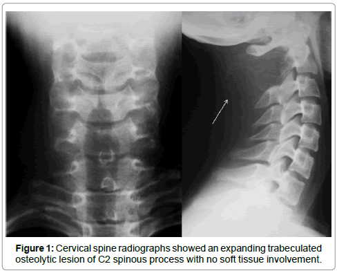
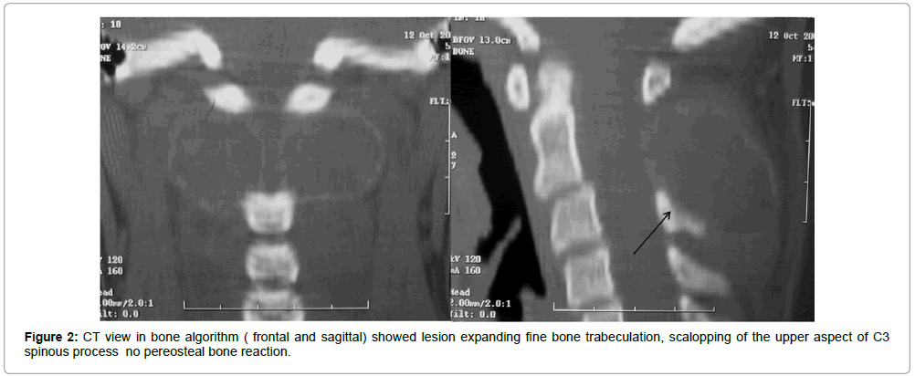
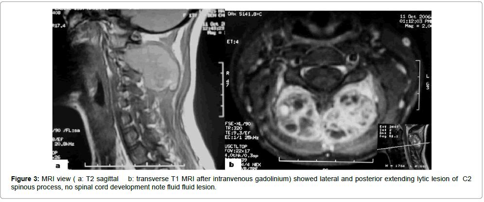
A 13 years old girl with no significant past medical history presented with neck pain , with out any traumatic history . Clinical exam found an adherent posterior cervical swelling with moderate neck limitation motion without torticolis. Neurological examination was normal and no inflammatory profile was found. An X-ray of cervical spine was done , it showed an expanding trabeculated osteolytic lesion of C2 spinous process (Figure 1). CT view showed postero-lateral expanding lesion, scalopping of the upper aspect of C3 spinous process with no pereosteal bone reaction (Figure 2). To better see soft tissue involvement and the marrow, a Magnetic resonance imaging (MRI) was done. The MRI showed C2 spinous process osteolytic lesion, fluid-fluid levels with no spinal cord development (Figure 3). The destruction of the bone and the invasion of the soft parts of the neck necessitated a biopsy
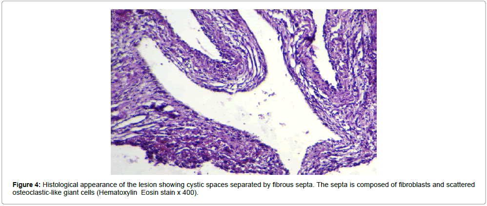
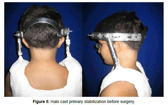
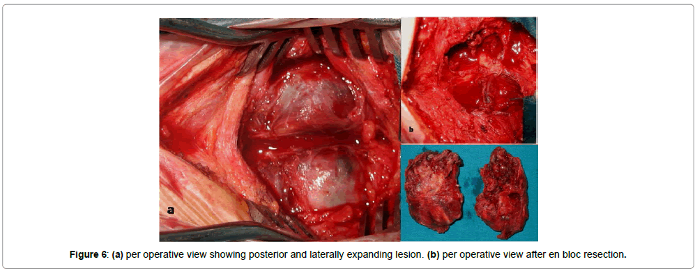
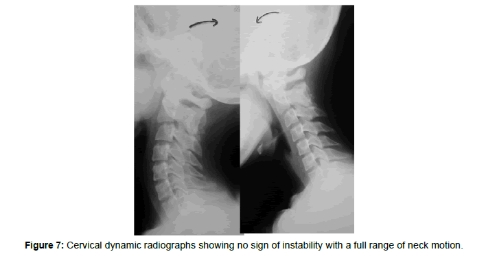
A surgical biopsy was performed by a posterior approach, histological analysis showed cystic spaces separated by fibrous septa. The septa is composed of fibroblasts and scattered osteoclasticlike giant cells confirm ABCs diagnosis (Figure 4). After halo cast immobilization this girl had en bloc cyst resection (Figures 5 and 6). Halo cast was removed 3 months following surgery. Controlled radiographs showed progressive ossification of posterior elements and no instability on dynamic radiographs . No recurrence with 2 years follows up with no longer neck pain (Figure 7).
Discussion
ABCs are actually defined as ‘‘an expanding lesion with blood filled cavities separated by septa of trabecular bone or fibrous tissue containing osteoclast giant cells [1]. This lesion is usually found in long bone [3] and represents only 1.4 % of primary vertebral column tumors [5]; the cervical spine is involved in 30-41 % [6]. This begnin lesion was classified as latent (type 1), active (type 2) or aggressive (type 3) [1], and also as primary or secondary existent lesion such as giant cell tumor, hemangiomas, osteoblastomas, chondroblastomas, and telangiectatic osteosarcomas [1]. ABCs pathogenesis remains unclear. Several hypotheses ranging from a vascular degenerative process to a traumatic reparative process, a genetic translocation was also reported [1]. Spine lesions usually involve vertebral body and posterior elements described as a ‘blown out’ or ‘ballooned’ ‘soap bubble’ to underline the erosion of the cortex of the bone and elevation of the periosteum [7]. The lesion has a trabeculated appearance within flocculent densities surrounded by a ‘shell’ of delicate cortical bone [1]. CT imaging can be helpful in identifying the margins and fluid level of the cyst with high sensitivity and specificity [9]. MRI allows identification of the thin septa dividing the cyst. Biopsy of cervical lesion is indicated in a typical ABCs imaging or if secondary form was suspected [10,11] or if, as in our case, direct lesion accessibility. Biopsy must be carefully planned to be consistent with oncologic principles and not compromise definitive treatment [1]. In long bone, curettage or en bloc excision and grafting of aneurysmal cysts is the mainstay of treatment related to easily accessibility without sequelae [8]. En bloc removal is considered as the gold standard procedure in ABCs treatment which is associated with the lowest rate of local recurrence [8]. This procedure can be very challenging in the cervical spine due to the infiltration of the vertebral artery and the nerve roots [12-14] in addition of intraoperative bleeding and instability. Furthermore, children have the highest incidence of post laminectomy kyphosis because of the wedging change in the cartilaginous portion of the vertebral body and to the viscoelasticity of ligaments in children [15]. In this case anterior fusion might become necessary .
When en bloc excision is technically difficult, we may consider other treatment options as complete curettage, adjuvant selective arterial embolization, radiotherapy, intra-lesional injection of sclerosing agent, cryotherapy or embolization [12,13]. Curettage is generally performed in conjunction with an open biopsy [2]. It can be sufficiently curative in some cases [16], but with highest recurrence rates [4,6] especially in pediatric population . An innovative biopsy technique recently introduced by Reddy KI [16,17], consist in a percutaneous limited curettage procedure targeting various quadrants of the cyst might lead to an ad integrum healing. Selective arterial embolization is often recommended prior to the surgery to reduces peroperative bleeding, but it was also described as a successful sole treatment modality [18] . In this case, this technique may offer a non destabilizing, curative option [19], but on the other hand , ischemic complications are frequent with cervical and thoracic localization above T6 [19] . Despite its efficiency in reducing recurrence rate, Radiotherapy is contraindicated within pediatric population due to its high risk complications such as myelopathy, radiation-induced spinal deformity [20] and secondary sarcomas. Therefore radiation therapy should be reserved for patients with inoperable lesions or a recurrence. Although en bloc removal of the tumor has the lowest rate of local recurrence, it can be technically challenging or impossible particularly in the cervical spine due to the infiltration of the vertebral artery and the nerve roots [10]. Surgery has many risks such as paralysis, intraoperative bleeding, neurological complications, and scarring complications. In addition to the operative risks, children have the highest incidence of post laminectomy kyphosis [8]. In upper cervical spine ,external primary stabilization is an optional procedure before surgical resection.
Conclusion
ABCs remains rare in upper cervical spine in children. There are no specific clinical signs suggestive of the diagnosis of aneurysmal cyst of the cervical spine. Multiple optional treatment are currently available depends on localization accessibility [2]. When en bloc resection is possible, as in our case, halo cast can preceed surgery providing primary stabilization until recovery .This method of temporary immobilization is less combursom than osteosynthesis which leads in most cases to arthrodesis compromizing cervical motion.
References
- Burch S, Hu S, Berven S (2008) Aneurysmal bone cysts of the spine. Neurosurg Clin N Am 19: 41-47.
- Boriani S, De Iure F, Campanacci L, et al. (2001) Aneurysmal bone cyst of the mobile spine: report on 41 cases. Spine 26: 27-35.
- Cottalorda J, Kohler R, Sales de Gauzy J, et al. (2004) Epidemiology of aneurysmal bone cyst in children: a multicenter study and literature review. J Pediatr Orthop B 13: 389-394.
- deKleuver M, van der Heul RO, Veraart BE (1998) Aneurysmal bone cyst of the spine: 31 cases and the importance of the surgical approach. J Pediatr Orthop B 7: 286-292.
- Papagelopoulos PJ, Currier BL, Shaughnessy WJ, Sim FH, Ebsersold MJ (1998) Aneurysmal bone cyst of the spine.management and outcome. Spine 23: 621-628.
- Mankin HJ, Hornicek FJ, Ortiz-Cruz E, et al. (2005) Aneurysmal bone cyst: a review of 150 patients. J Clin Oncol 23: 6756-6762.
- Mahnken AH, Nolte-Ernsting CC, Wildberger JE, et al. (2003) Aneurysmal bone cyst: value of MR imaging and conventional radiography. Eur Radio l13: 1118-1124.
- Casabianca L, Journé A, Mirouse G, Zerah M, Moulies D, et al. (2015) Solid aneurysmal bone cyst on the cervical spine of a young child. Eur Spine J 24: 1330-1336.
- Keenan S, Bui-Mansfield LT (2006) Musculoskeletal lesions with fluid–fluid level: a pictorial essay. J Comput Assist Tomogr 30: 517-524.
- Kitamura T, Ikuta K, Senba H, Komiya N, Shidahara S (2013) Long-term follow-up of a case of aneurysmal bone cyst in the lumbar spine. Spine J 13: 55-58.
- Bret P, Confavreux C, ThouardH, et al. (1982) Aneurysmal bone cyst of the cervical spine: report of a case investigated by computed tomographic scanning and treated by a two-stage surgical procedure. Neurosurgery 10: 111-115.
- Rai AT, Collins JJ (2005) Percutaneous treatment of pediatricaneurysmal bone cyst at C1: a minimally invasive alternative: a case report. Am J Neuro radiol 26: 30-33.
- Zileli M, Isik HS, Ogut FE, Is M, Cagli S, et al. (2013) Aneurysmal bone cysts of the spine. Eur Spine J 22: 593-601.
- Gladden ML Jr, Gillingham BL, Hennrikus W, Vaughan LM (2000) Aneurysmal bone cyst of the first cervical vertebrae in achild treated with percutaneous intralesional injection of calcitoninand methylprednisolone A case report. Spine 25: 527-530.
- Yasuoka S, Peterson HA, Laws ER Jr, MacCarty CS (1981) Pathogenesis and prophylaxis of postlaminectomy deformity of the spine after multiple level laminectomy: difference betweenchildren and adults. Neurosurgery 9: 145-152.
- Garg S, Mehta S, Dormans JP (2005) Modern surgical treatment of primary aneurysmalbone cyst of the spine in children and adolescents. J Pediatr Orthop 25: 387-392.
- Reddy KI, Sinnaeve F, Gaston CL, Grimer RJ, Carter SR (2014) Aneurysmal bone cysts: do simple treatments work? Clin Orthop Relat Res 472: 1901–1910.
- Konya A, Szendroi M (1992) Aneurysmal bone cysts treated bysuperselective embolization. Skeletal Radiol 21: 167-172.
- Amendola L, Simonetti L, Simoes CE, Bandiera S, De IureF,Boriani S (2013) Aneurysmal bone cyst of the mobile spine: thetherapeutic role of embolization. Eur Spine J 22: 533–541.
- Mayfield JK, Riseborough EJ, Jaffe N, Nehme ME (1981) Spinaldeformity in children treated for neuroblastoma. J Bone Joint Surg Am 63: 183-193.
 Spanish
  Chinese
  Russian
  German
  French
  Japanese
  Portuguese
  Hindi
Hindi 