Research Article, J Genes Proteins Vol: 1 Issue: 1
Antibacterial Mechanism of Bacteriolyses of Bacterial Cell Walls by Zinc(â…¡) Ion Induced Activations of PGN Autolysins, and DNA damages
Ishida T*
Life and Environment Science Research Division, Japan
*Corresponding Author : Ishida Tsuneo
2-3-6, Saido, Midoriku, Saitama city, Saitama prefecture 336-0907, Japan
Tel: +048-881-3970
E-mail: ts-ishida@ac.auone-net.jp
Received: December 13, 2017 Accepted: December 19, 2017 Published: December 26, 2017
Citation: Ishida T.(2017) Antibacterial Mechanism of Bacteriolyses of Bacterial Cella Wlls by Zinc(Ⅱ) Ion Induced Activations of PGN Autolysins, and DNA damages. J Genes Proteins 1:1.
Abstract
Bacteriolyses and destructions of bacterial cell walls by zinc (�?�) ion solutions were investigated against Staphylococcus aureus and Escherichia coli. Bacteriolysis of S. aureus peptidoglycan (PGN) cell wall by zinc ion solution is due to the inhibition of PGN elongation by the activations of PGN autolysins of amidases. The other, bacteriolysis and destruction of E. coli cell wall are caused by the destruction of outer membrane structure due to degradative enzymes of lipoproteins at N- and C-terminals, and also inhibition of PGN elongation is dependent on the activities of PGN hydrolases and autolysins of amidase and carboxypeptidase-transpeptidase. Zinc ions induced ROS such as O2-, H2O2, ・OH, OH-producing in bacterial cell wall occur oxidative stress. DNA damages may be due to Zn ion complex formation by Zn2+ substitution into hydrogen bonds in DNA base pairs.
Keywords: Zinc ions; PGN cell wall; Outer membrane lipoproteins; Bacteriolysis; Degradation; Biosynthesis and autolysin; Reactive oxygen species (ROS); DNA base-pairs
Introduction
Silver, copper, and zinc of transition metals have highly antibacterial activities and are utilized as chemotherapy agents. Recently, antibacterial activities of zinc and its complexes call attention to potential treatments such as prevention of serious diseases [1], exploitation during bacterial pathogenesis [2,3], virus counter- measure [4], anti-cancer and anti-tumor cells [5]. Halo inhibitory tests against Staphylococcus epidermidis were carried out various sulfate solutions, in which the result was also obtained that the antibacterial effect of zinc ions was the highest in the sulfate system with halo inhibitory large zone for zinc ion solutions [6]. In this study, considering such as the highest antibacterial activity for Zn2+ ions obtained from halo inhibitory tests of metallic sulfate solutions, the processes of bacteriolyses and destructions by antibacterial activities of Zn2+ ions are analyzed and considered against thick peptidoglycan(PGN) layer cell wall, and outer membrane-connecting thin PGN layer cell wall. Further, the bacteriolytic mechanisms by zinc (Ⅱ) ions solutions have been revealed against Gram-positive S. aureus and Gram-negative E. coli cell walls, with additional DNA damage by Zn2+-DNA base pairs interactions. These Zn2+-proteins and Zn2+- DNA interactions are considered to enhance the persistence in several forms of Zn-ligand bonds (O, S, NH3, H2O, OH, SCH3, and H ligands) with Zn coordination chemistry and molecular orbital theory.
Analyses of Bacterial Cell Walls, PGN Biosyntheses and Autolysins, and the action sites of PGN syntheses and autolysins
The surface envelop cell structures of S. aureus as representative of Gram-positive bacterium and E. coli as representative of Gramnegative bacterium, molecular structures of these cell walls, molecular structure of PGN, and PGN biosyntheses and autolysins were searched in detail. Further, the reaction and the behavior of metallic ions and bacteria cell, molecular bonding manner, and zinc ion characteristics also were searched.
Molecular structure of S. aureus and E. coli cell walls, and action sites of PGN biosyntheses of both transglycosylase TG and transpeptidase TP; TG/TP and PGN autolysins
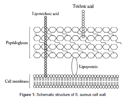
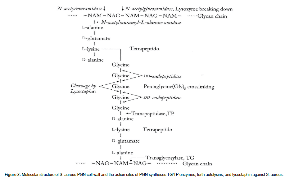
S. aureus surface layer consists of teichoic acids, lipoteichoic acids, and thick PGN cell wall [6]. The schematic structure of S. aureus cell wall is shown in Figure 1, in which the molecular structure of S. aureus PGN cell wall and the action sites of TG/TP synthesis enzymes, PGN forth autolysins, and lysostaphin are represented in Figure 2.
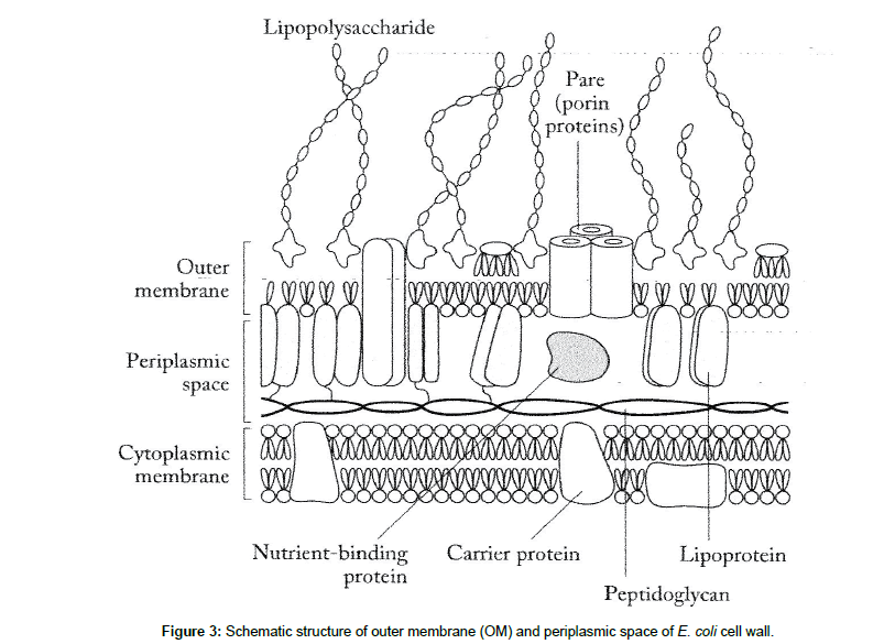
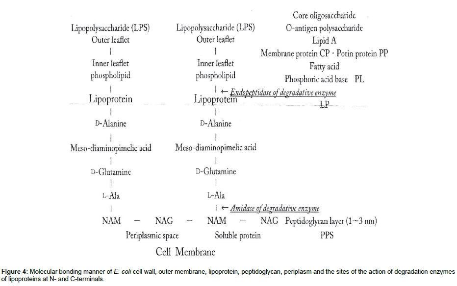
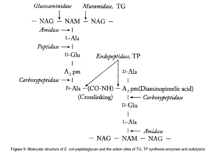
The other, E. coli cell wall consists of lipid A, lipopolysaccharide, porin proteins, outer membrane of lipoprotein, and thinner 2-7nm PGN layer in 30-70nm periplasmic space [7]. Figure 3 shows the schematic structure of E. coli, in which the molecular bonding manner of E. coli cell wall and periplasmic PGN, and the action sites of the hydrolases and degradative enzymes of lipoproteins are represented in Figure 4. Furthermore, Figure 5 shows E. coli PGN synthetic enzymes TG/TP and the action sites of the autolysins such as amidase, peptidase, carboxy- peptidase, etc. Interactions of PGN molecular structure, synthesis, autolysin molecular structure, synthesis, and autolysin influence in any event the bacteriolysis of bacterial cell walls (Figures 1-3).
Discussions on bacteriolyses by zinc ions
Bacteriolysis and destruction of S. aureus PGN cell wall by zinc ions: For the sake of growth of S. aureus PGN cell wall, there is necessarily required for the adequate balance between PGN biosynthesis and PGN autolysin. When the balance was broken to be imbalanced, bacteriolysis and destruction of the cell wall should become to occur.
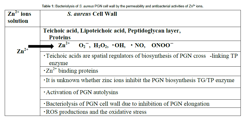
PGN biosynthesis enzymes of transglycosy-lase TG and transpeptidase TP: Wall teichoic acids are spatial regulators of PGN cross-linking biosynthesis TP [8]; however, it is not explicit whether zinc ions could inhibit both TG and TP enzymes of the PGN.
Inhibition of PGN elongation due to the activations of autolysins: Zn2+ binding Rv3717 showed no activity on polymerized PGN and but, it is induced to a potential role of N-Acetylmuramyl-Lalanine Amidase [9], PGN murein hydrolase activity and generalized autolysis; Amidase MurA [10] (Figure 4).
Lytic Amidase LytA [11], enzymatically active domain of autolysin LytM [12], Zinc-dependent metalloenzyme AmiE [13] as prevention of the pathogen growth, and Lysostaphin-like PGN hydrolase and glycylglycine endopeptidase LytM [14]; The activations of these PGN autolysins could be enhanced the inhibitions of PGN elongation simultaneously, with bacteriolysis and destruction of S. aureus PGN cell wall. O2ï¼Âand H2O2 permeates into membrane and cytoplasm, and then, DNA molecular is damaged by oxidative stress [15] (Figure 5).
Production of reactive oxygen species (ROS) against S. aureus: For the penetration of zinc ions to PGN cell wall, the ROS production such as superoxide anion radical O2ï¼Â, hydroxyl radicalï½¥OH, hydrogen peroxide H2O2 occurred from superoxide radical O2ï¼ molecular [16].
O2 + e- + H+ →ï½¥HO2
ï½¥HO2 → H+ + O2
ï½¥O2ï¼Â+ 2H+ + e- → H2O2
H2O2 + e- → HOï¼Â+ï½¥OH
ï½¥OH + e- + H+ → H2O
2H+ + 2ï½¥O2ï¼Â→ H2O2 + O2
H2O →ï½¥OH + ï½¥H + e- → H2O2
From above-mentioned results, the bacteriolytic process of S. aureus PGN cell wall is shown in Table 1.
Bacteriolysis and destruction of E. coli cell wall by zinc ions
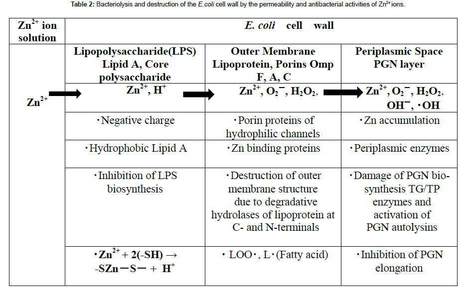
Permeability of zinc ions into E. coli cell wall: E. coli cell wall is comprised of lipopolysaccharide (LPS), lipoproteins (LP), and peptidoglycan (PGN) thinner layer within periplasmic space. When permeability of zinc ions in the E. coli cell wall, highly anionic LPS with hydrophobic lipid A, core polysaccharide, O-polysaccharide, inhibition of LPS biosynthesis may be possibility to occur [17].
The OmpA, OmpC, OmpF porins of lipoproteins have metallic cation selective and hydrophilic membrane crossing pore, to be effective for zinc transfer [18]. Zinc (Ⅱ) ions reactive with -SH base, and then generate H2. Zinc bivalent is unchangeable as 4-coodinated -SZnï¼ÂSï¼Âbond.
Zn2++2 (-SH) → -SZn(Ⅱ)ï¼ÂS +2H+
Destruction of E .coli cell wall outer membrane structure by hydrolases of lipoproteins at C- and N-terminals: ZnPT (zinc pyrithione) and Tol (Tol proteins)-Pal (Protein-associated lipoprotein) complex are antimicrobial agents widely used, however, it has recently been demonstrated to be essential for bacterial survival and pathogenesis that outer membrane structure may be destroyed [19,20].
Inhibition of PGN elongation due to the damage of PGN synthesis enzyme of zinc-protein amidase in periplasmic space, and the activities of PGN autolysins: The zinc-induced decrease of protein biosynthesis led to a partial disappearance of connexin-43 of protein synthesis in neurons [21], but it is unknown whether PGN biosynthesis is inhibited. Further, it is also unclear whether the both TG/TP should be inhibited by the zinc ions [22-24].
The other, zinc ions were accumulated in E. coli periplasmic space, in which the zinc ions are spent to the activation of bacteriolysis of the cell wall. Zinc depending PGN autolysin, amidase PGRPs [25], zinc metalloenzymes AmiD [26], amidase zinc-containing amidase; AmpD [27], zinc-present PGLYRPs [28] serve to be effective for the PGN autolysins. It is particularly worth noting that enhancement of the activities of autolysins is characterized on PGN carboxypeptidasetranspeptidase IIW [29] requiring divalent cations. Accordingly, the inhibition of PGN elongation had occurred by zinc ion induced activities of PGN hydrolases and autolysins.
ROS production and oxidative stress against E. coli: Zinc ions reacted with -SH, and H+ generates. In E. coli, free radicals O2ï¼Â, OHï¼Â, ï½¥OH and H2O2 are formed as follows [30]:
O2 + e →O2ï¼Â
2O2ï¼Â+ 2H+ → H2O2 + O2
O2ï¼Â+ H2O2 → OHï¼Â+ ï½¥OH + O2ï¼Â
In cell wall, reacting with polyunsaturated fatty acids:
LH + OHï½¥ → Lï½¥ + HOH
Lï½¥ + O2 → LOOï½¥
LH + LOOï½¥ → Lï½¥ + LOOH
Zinc-containing Peptidoglycan Recognition Proteins (PGRPs) induce ROS production of H2O2, O2ï¼Â, HOï½¥, and then the ROS occur the oxidative stress, and killing by stress damage [31].
As above-mentioned, the bacteriolysis and the destruction of E. coli cell wall in process of the permeability and antibacterial activities of zinc ions are summarized in Table 2.
Damage of DNA base-pairs
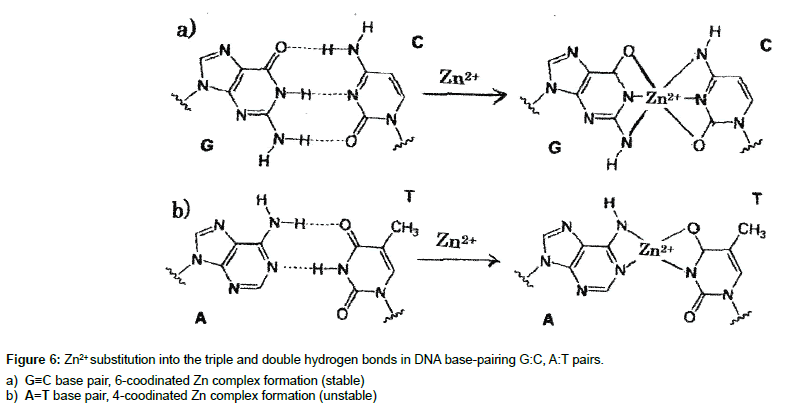
Zn2+ ion induced occurrence of generations of ROS and hydrogen peroxide H2O2 in bacterial cells damages DNA, in which formation of DNA damage resulting from a release of catalytic binding of zinc ion to DNA with generation of ï½¥OH radicals, and by reaction of H2O2 with the metal produces the strand breaks in DNA as well as DNA base-pairs modifications and deoxyribose fragmentation. Transfer of Zn2+ ions into hydrogen bonds in DNA base-pairing
G (guanine) ≡ C (cytosine) and A (adenine) = T (thymine) pairs occurs by Zn2+ ion substitution shown in Figure 6. Thus, it may be considered that DNA damages due to Zn-complex within DNA basepairs G≡C, A=T is formed in the hydrogen bonds. A=T base pairs are less stable than G≡C base pairs in Zn-DNA [32]. These considerations in the Zn-DNA base-pairs interactions must account for Zn2+-ligands (6-coordination and 4-coodination complex formations) in DNA base pairs of G≡C and A=T, according to coordinated chemistry and molecular orbital theory [33,34].
Conclusion
(1) Bacteriolysis and destruction of S. aureus PGN cell wall, in which wall teichoic acids control PGN synthesis cross-linking TP, are due to the inhibition of PGN elongation by the activities of PGN autolysins; amidase AmiA and AmiE, PGN hydrolase Lysostaphinlike endopeptidase (Glycine cutting).
(2) Bacteriolysis and destruction of E. coli cell wall are due to the damage of LPS biosynthesis, destructing of outer membrane structure by degrading of lipoprotein at C-, N-terminals, owing to inhibitions of PGN formations by activities of PGN autolysins of amidase and carboxypeptidase.
(3) By the penetration of zinc ion into S. aureus cell wall, production of O2, H+, H2O2, ONOOï¼Âoccurs against S. aureus. The other, in E. coli cell wall, the productions of O2ï¼Â, H+ in outer membrane, and H2O2, OHï¼Â, ï½¥OH in periplasmic space occur. These ROS and H2O2 give the damages cell membrane proteins and DNA molecular in cytoplasm.
(4) DNA damages due to zinc ion complex formation within DNA base-pairs G≡C, A=T may be occurred in cytoplasm of bacterial cells.
References
- AM Grabrucker, M Rowan, C Garner (2011) Brain-Delivery of Zinc-Ions as Potential Treatment for Neurological Diseases: Mini Review. Drug Deliv Lett 1: 13-23
- Ma L, Terwilliger A, Maresso AW (2015) Iron and Zinc Exploitation during Bacterial Pathogenesis. Metallomics 7: 1542-1554.
- Faiz U, Butt T, Satti L, Hussain W, Hanif F (2011) Efficacy zinc as an antibacterial agent against enteric bacterial pathogens. J Ayub Med Coll Abbottabad 23: 8-21.
- Suara RO, Crowe JE Jr. (2004) Effect of Zinc Salts on Respiratory Syncytial Virus Replication. Antimicrob Agents Chemother 48: 783-790.
- Gumulec J, Masarik M, Krizkova S, Adam V, Hubalek J, et al. (2011) Insight to physiology and pathology of zinc (II) ions and their actions in breast and prostate carcinoma. Curr Med Chem 18: 5041-5051.
- Tsuneo Ishida (2017) Halo-inhibitory susceptibility tests of the nitrate and sulfate solutions and antibacterial activities of transition metals ions. Japanese Journal of Medical Prevention 11: 93-99.
- Thomas J Silhavy, Daniel Kahne, Suzanne Walker (2014) The Bacterial Cell Envelope, Cold Spring Harb Perspect Biol 2: 1-14.
- Atilano ML, Pereira PM, Yates J, Reed P, Veiga H, et al. (2010) Teichoic acid are temporal and spatial regulators of peptidoglycan cross-linking in S. aureus. PNAS 107: 18991-18996.
- Prigozhin DM, Mavrici D, Huizar JP, Vansell HJ, Alber T (2013) Structural and Biochemical Analyses of Mycobacterium tuberculosis N-Acetylmuramyl-L-alanine Amidase Rv3717 Point to a Role in peptidoglycan Fragment Recycling. J Biological Chemistry 288: 31549-31555.
- Carroll SA, Hain T, Technow U, Darji A, Pashalidis P, et al. (2003) Identification and Characterization of a Peptidoglycan Hydrolase, MurA, ofListeria monocytogenes, a Muramidase Needed for Cell Separation. J Bacteriology 185: 6801-2003.
- Mellroth P, Sandalova T, Kikhney A, Vilaplana F, Hesek D, et al. (2014) Structural and Functional Insights into Peptidoglycan Access for the Lytic Amidase LytA ofStreptococcus pneumonia. Am Society Micro15:1-10.
- Jagielska E, Chojnacka O, Sabała I (2016) LytM Fusion with SH3b-Like Domain Expands Its Activity to Physiological Conditions. Microbial Drug Resistance 23: 461-468.
- Zoll S, Pätzold B, Schlag M, Götz F, Kalbacher H, et al. (2010) Structural Basis of Cell Wall Cleavage by a Staphylococcal Autolysin. PloS Pathogens 6: 1-13.
- Ramadurai L, Lockwood KJ, Nadakavukaren MJ, Jayaswal RK (1999) Characterization of a chromosomally encoded glycyglycine endopeptidase of S. aureus. Microbiology 145: 801-808.
- Morina F, Vidovic M, Kukavica B, Jovanović SV (2015) Induction of peroxidase isoforms in the roots of two Verbascum ThapsusL.populations is involved in adaptive responses to excess Zn2+ and Cu2+. Botanica SERBICA 39: 151-158.
- Gaupp R, Ledala N, Somerville GA (2012) Staphylococal response to oxidative stress. Front Cell Infect Microbiol 2: 1-8.
- Langley S, Beveridge TJ (1999) Effect of O-Side-Chain-LPS Chemistry on Metal Binding. Appl Environ Microbiol 65: 489-498.
- Blindauer CA (2015) Advances in the molecular understanding of biological zinc transport. Chem Commun 51: 4544-4563.
- Dinning AJ, Al-Adham IS, Austin P, Charlton M, Collier PJ (1998) Pyrithione biocide interactions with bacterial phospholipid head groups. J Appl Microbiol 85: 132-140.
- Godlewska R1, Wiśniewska K, Pietras Z, Jagusztyn-Krynicka EK (2009) Peptidoglycan- associated lipoprotein(Pal) of Gram-negative bacteria: function, structure, role in pathogenesis and potential application in immunoprophylaxis. FEMS Microbiol Lett 298: 1-11.
- Alirezaei M, Mordelet E, Rouach N, Nairn AC, Glowinski J (2002) Zinc-induced inhibition of protein synthesis and reduction of connexin-43 expression and intercellular communication in mouse cortical astrocytes. Eur J Neurosci 26: 1037-1044.
- Egan AJ, Biboy J, van't Veer I, Breukink E, Vollmer W (2016) Activities and regulation of peptidoglycan synthases, Philos Trans R Soc Lond B Biol Sci doi: 10.1098/rstb.2015.0031.
- Singh SK, SaiSree L, Amrutha RN, Reddy M (2012) Three redundant murein endo- peptidases catalyse an essential cleavage step in peptidoglycan synthesis of E. coli K12. Mol Microbiol 86: 1036-1051.
- Ramachandran V1, Chandrakala B, Kumar VP, Usha V, Solapure SM (2006) Screen for Inhibitors of the Coupled Transglycosylase-Transpeptidase of Peptidoglycan Biosynthesis in E. coli, Antimicrobial Agents. Antimicrob Agents Chemother 50: 1425-1432.
- Ivanna Rivera, Rafael Molina, Mijoon Lee, Shahriar Mobashery, Juan A Hermoso (2016) Orthologous and Paralogous AmpD Peptidoglycan Amidases from Gram-Negative Bacteria. Microb Drug Resist 22: 470-476.
- Pennartz A, Généreux C, Parquet C, Mengin-Lecreulx D, Joris B (2009) Substrate-Induced Inactivation of the E.coli AmiD N-Acetylmuramoyl-L-Alanine Amidase Highlights a New Strategy To Inhibit This Class of Enzyme. Antimicrob Agents Chemother 53: 2991-2997.
- Carrasco-López C, Rojas-Altuve A, Zhang W, Hesek D, Lee M, et al. (2011) Crystal Structures of Bacterial Peptidoglycan Amidase AmpD and an Unprecedented Activation Mechanism. J Biol Chem 286: 31714-31722.
- Wang M, Liu LH, Wang S, Li X, Lu X, et al. (2007) Human Peptidoglycan Recognition Proteins Require Zinc to Kill Both Gram-Positive and Gram-negative Bacteria and Are Synergistic with Antibacterial Peptides. J Immunol 178: 3116-3125.
- DasGupta H, Fan DP (1979) Purification and Characterization of a Carboxy- peptidase-Transpeptidase of Bacillus megaterium acting on the Tetrapeptide Moiety of the Peptidoglycan. J Biol Chem 254: 5672-5682.
- Kashmiri ZN, Mankar SA (2014) Free radicals and oxidative stress in bacteria. Int J Curr Microbiol App Sci 3: 34-40.
- Kashyap DR, Rompca A, Gaballa A, Helmann JD, Chan J, et al. (2014) Peptidoglycan Recognition Proteins Kill Bacteria by Inducing Oxidative, Thiol and Metal Stress. PLOS Pathogen 7: 1-17.
- Wang AH, Hakoshima T, van der Marel G, van Boom JH, Rich A (1984) AT base pairs are less stable than GC base pairs in Z-DNA. Cell 37: 321-331.
- Amin EA, Truhlar DG (2008) Zn Coordination Chemistry: Development of Benchmark Suites for Geometries, Dipole Moments, and Bond Dissociation Energies and Their Use to Test and Validate Density Functionals and Molecular Orbital Theory. J Chem Theory Comput 4: 75-85.
- Cerasi M, Ammendola S, Battistoni A (2013) Competition for zinc binding in the host-pathogen. Front Cell Infect Microbiol 3: 1-6.
 Spanish
  Chinese
  Russian
  German
  French
  Japanese
  Portuguese
  Hindi
Hindi 