Short Communication, J Womens Health Issues Care Vol: 11 Issue: 9
Can Contrast-Enhanced Spectral Mammography (CESM) Reduce the Number of Unnecessary Breast Biopsies? Analysis of 593 Cases of Patients with Suspected Breast Cancer
Katarzyna Steinhof-Radwanska1*, Anna grazynska1, Anna barczykgutkowska1, Mateusz winder1, Andrzej lorek2 and Agnieszka bobola3
1Department of Radiology and Nuclear Medicine, Medical University of Silesia, Katowice, Poland
2Department of Oncological Surgery, Medical University of Silesia, Katowice, Poland
3Department of Oncology and Radiotherapy, Medical University of Silesia, katowice, Poland
*Corresponding Author: Katarzyna Steinhof-Radwańska, Department of Radiology and Nuclear Medicine, Medical University of Silesia, Medyków 18, 40-514Katowice, Poland, Tel: +48695404695; E-mail: kasia.steinhof@gmail.com
Received date: 24 July, 2022, Manuscript No. JWHIC-22-70150; Editor assigned date: 27 July, 2022, PreQC No. JWHIC-22-70150 (PQ); Reviewed date: 10 August, 2022, QC No. JWHIC-22-70150; Revised date: 04 October, 2022, Manuscript No. JWHIC-22-70150 (R); Published date: 11 October, 2022, DOI:10.4172/2325-9795.1000417
Citation: Radwańska KS, Grażyńska A, Gutkowska AB, Winder M, Lorek A, Bobola A (2022) Can Contrast-Enhanced Spectral Mammography (CESM) Reduce the Number of Unnecessary Breast Biopsies? Analysis of 593 Cases of Patients with Suspected Breast Cancer. J Womens Health Issues Care 11:9.
Abstract
Breast cancer is the most frequently diagnosed malignancy, with a frequency of 22.8% of all new cancer incidence rates in Poland.
Introduction
Breast cancer is the most frequently diagnosed malignancy, with a frequency of 22.8% of all new cancer incidence rates in Poland. Contrast Enhanced Spectral Mammography (CESM) is a relatively new method used in breast cancer diagnosis, which involves the phenomenon of neoangiogenesis of cancerous tumours, allowing contrast enhancement in the areas of vessel proliferation in the background of the surrounding breast tissue. In recent years, the number of mammography centers using CESM on a daily basis has increased. Typically, CESM is used to evaluate patients with suspected focal lesions in whom conventional Mammography (MG) and additional Ultrasound examinations (US) do not allow a definitive diagnosis. CESM is particularly useful in the diagnosis of dense breasts (ACR categories C, D), where cancer detection is difficult due to the lower sensitivity of conventional mammography [1-2].
Our study aimed to assess whether it is possible to reduce the number of unnecessarily performed breast Core Needle Biopsies (CNB) in case of lesions that did not undergo post-contrast enhancement while using Contrast Enhanced Spectral Mammography (CESM). An additional aim of the study was to calculate the potential financial savings from performing a CESM instead of a biopsy.
Description
Patients and methods
547 patients with 593 breast lesions detected in ultrasonography and classic mammography were enrolled in the retrospective study.
All patients before the biopsy underwent CESM examination. The CESM results have been compared with the gold standard in the diagnosis of breast cancer, which is histopathological examination.
Sensitivity, specificity, negative and positive predictive values of CESM in detecting breast cancer was calculated. Changes that were not enhanced after intravenous contrast administration in CESM subtraction images were classified as ones for which CNB could be omitted.
Then the possible financial profit resulting from the withdrawal from CNB and CESM implementation was calculated. For the study, the average cost of one CNB and one CESM was set at 170 euros and 65 euros (conversion from PLN to EUR) [3].
CESM examination
All CESM examinations were carried out with a digital mammography device dedicated to performing dual-energy CESM acquisitions (SenoBright, GE Healthcare, 3000 N. Grandview Blvd., Waukesha, WI, USA).
An intravenous injection of 1.5 mL/kg of body mass of a non-ionic contrast agent was performed. The exposure pair (low and high energy) was performed automatically. Specific image processing of low energy and high energy images was done to obtain subtraction images to highlight contrast enhancement and suppress structured noise due to fibroglandular breast tissue.
Rhodium anode material was used for all acquisitions, with molybdenum and rhodium filters with kVp ranging from 26 to 32 used for low energy acquisitions. The total duration of the examination was usually around 10 min.
The analysis includes 593 breast lesions diagnosed in 547 women. In the studied group cancer was detected in 327 (55.14%) lesions and in 256 (43.17%) cases benign lesions were confirmed by histopathological examination and at least 12 months of observation.
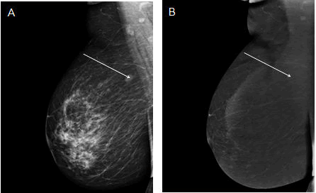
In 428 (72.2%) lesions changes of increased vascularization were detected, while in the remaining 165 (27.8%) lesions the CESM result was negative. Taking the CESM enhanced result as the criterion of malignancy, the method shows differentiation of benign and malignant lesions in the breast: Sensitivity of 97.86%, specificity of 59.4%; PPV 74.76%; NPV 95.76% (Table 1).
| Histopathology result | ||||
|---|---|---|---|---|
| Malignant | Benign | |||
| Contrast | Enhanced | 320 | 108 | PPV: 74.76% |
| Enhancement | Non-enhanced | 7 | 158 | NPV: 95.76% |
| Sensitivity: 97.86% | Specificity: 59.4% | |||
Table 1: Comparison of lesions clinical characteristics and the results of the spectral mammography analysis.
The 165 changes did not present post-contrast enhancement in CESM. 158 (95.76%) of these lesions were verified as benign in the histopathological examination and only 7 as malignant (4.24%). Table 2 presents the individual histopathological diagnoses of changes that were not enhanced with post-contrast enhancement [4-5].
| Lesion | Quantity (% of all non-enhanced lesions) |
|---|---|
| Fibrotic sclerosis | 124/165 (75.15%) |
| Fibroadenoma | 18/165 (10.9%) |
| NST | 2/165 (1.2%) |
| DCIS | 5/165 (3.03%) |
| Papilloma | 1/165 (0.61%) |
| LCIS | 6/165 (3.6%) |
| Other | 9/165 (5.45%) |
| NST-non-specific type cancer | |
Note: DCIS-pre-invasive ductal carcinoma, LCIS-Lobular Carcinoma in situ, Other-focal apocrine metaplasia, atheroma, usual ductal hyperplasia, microglandular hyperplasia, cyst, phyllodes tumour
Table 2: Individual non-enhanced lesions depending on the histopathological results.
The estimated cost of 165 core needle biopsies was 28 050 euros and CESM was only 10 725 euros. By performing only CESM without CNB the potential financial savings would be 17,325 euros (61.76%) (Figure 1).
Conclusion
• The lack of post-contrast enhancement in CESM is a good indicator of benign character.
• CEM is an emerging modality that may provide critical information in a number of clinical scenarios. Today, CEM is most commonly used to evaluate disease extent in patients with contraindications to MRI. It is also increasingly being used in the diagnostic setting for patients recalled from screening. However, as the interest in CEM grows, additional studies are needed to further understand the role of CEM in breast imaging.
• Thus, unnecessary biopsies can be avoided and cost of diagnostics in breast cancer patients reduced.
References
- Strigel RM, Burnside ES, Elezaby M, Fowler AM, Kelcz F, et al. (2017) Utility of BI-RADS assessment category 4 Subdivisions for screening breast MRI. AJR Am J Roentgenol 208:1392–1399.
[Crossref] [Googlescholar] [Indexed]
- Ghosh K, Melton LJ, Suman VJ, Grant CS, Sterioff S, et al. (2005) Breast biopsy utilization: A population-based study. Arch Intern medi 165:1593-1598.
- Schonberg MA, Silliman RA, Ngo LH, Birdwell RL, Fein-Zachary V, et al. (2014) Older women’s experience with a benign breast biopsy-A mixed methods study. J Gen Intern Med 29:1631-1640.
[Crossref] [Googlescholar] [Indexed]
- Brett J, Bankhead C, Henderson B, Watson E, Austoker J (2005) The psychological impact of mammographic screening. A systematic review. Psychooncology 14:917–938.
[Crossref] [Googlescholar][Indexed]
- Perry H, Phillips J, Dialani V, Slanetz PJ, Fein-Zachary VJ, et al. (2019) Contrast-enhanced mammography: A systematic guide to interpretation and reporting. AJR Am J Roentgenol 212:222–231.
[Crossref] [Googlescholar][Indexed]
 Spanish
  Chinese
  Russian
  German
  French
  Japanese
  Portuguese
  Hindi
Hindi 