Case Report, J Otol Rhinol Vol: 12 Issue: 4
Ectopic Tooth in Orbital Floor Causing Diplopia in a Child: An Unusual Presentation
Roshan K Verma* and Jyotika Sharma
Department of Otolaryngology, Government Medical College and Hospital, Chandigarh, India
*Corresponding Author:
Roshan K Verma,
Department of Otolaryngology,
Government Medical College and Hospital,
Chandigarh,
India,
Tel: 9014452996;
E-mail:roshanverma@hotmail.com
Received date: 25 September, 2022, Manuscript No. JOR-23-75536; Editor assigned date: 27 September, 2022, PreQC No. JOR-23-75536 (PQ); Reviewed date: 11 October, 2022, QC No. JOR-23-75536; Revised date: 31 January, 2023, Manuscript No. JOR-23-75536 (R); Published date: 28 February, 2023, DOI: 10.4172/2324-8785.100041
Citation: Verma RK, Sharma J (2023) Ectopic Tooth in Orbital Floor Causing Diplopia in a Child: An Unusual Presentation. J Otol Rhinol 12:1.
Abstract
Ectopic teeth can occur due to a developmental anomaly, iatrogenic cause or a pathological process. Dentigerous cysts surrounding impacted teeth often displace these teeth into ectopic positions. We report a case of ectopic tooth lying in the floor of orbit causing diplopia in the upward gaze. Removal of ectopic tooth resulted in resolution of symptoms. This case is reported to make clinicians aware that diplopia with missing dentition in child should arouse suspicion of ectopic tooth as cause of symptoms.
Keywords: Child, Ectopic tooth, Floor of orbit, Diplopia
Introduction
The development of tooth starts at 6 weeks of intrauterine life. It is a complex process which results from interactions between the ectoderm of oral cavity and the ectomesenchyme. An abnormal interaction during development of tooth can lead ectopic eruption of tooth [1]. It can occur due to abnormal interaction during development of tooth which may be a developmental anomaly, iatrogenic, trauma or due to pathological process.
Ectopic eruption of tooth usually occurs in the oral region and is rarely seen in regions other than the oral cavity. Few reports have reported ectopic teeth in sites such as the mandibular condyle, nasal septum, chin, palate and maxillary sinus [2]. We report a case of 10 year old child who presented to us with symptoms of maxillary sinusitis and diplopia on upward gaze. CT scan showed presence of ectopic tooth in the floor of orbit along with maxillary sinusitis [3]. Removal of ectopic tooth resulted in resolution of symptoms. This case is reported to make clinicians aware that diplopia with missing dentition in child should arouse suspicion of ectopic tooth as cause of symptoms [4].
Case Presentation
A 10 years old male presented with a history of swelling on left side of face below eye for 1 month which was sudden in onset, painless, diffuse, and non-progressive. The child also complained of persistent purulent rhinorrhea of left nasal cavity and double vision on upward gaze.
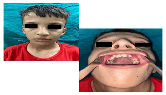
On examination anterior rhinos copy showed purulent discharge in the left side of nasal cavity. Nasal endoscopy showed purulent discharge in the middle meatal area and floor of left nasal cavity. Eye examination had visual acuity of 6/6 on both sides but complained of double vision on upward gaze with restriction of upward movement of left eye. Dental examination showed missing left first premolar tooth (Figure 1).
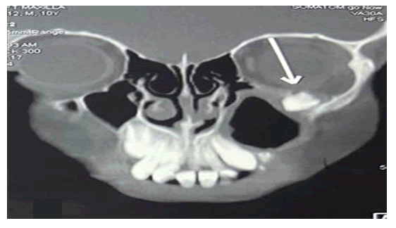
A contrast enhanced Computed Tomography (CT) of nose and paranasal sinuses was done which showed presence of ectopic tooth at the level of floor of left orbit with mucosal thickening along walls of left maxillary sinus (Figure 2).
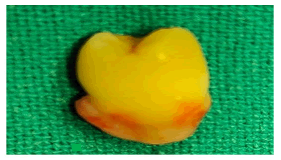
Patient was planned for FESS under general anesthesia. Wide antrostomy was performed and view of the entire maxillary sinus obtained using 0, 30°, 70° endoscopes. Intra operatively, tooth along with the cyst wall around the tooth was removed from the lateral most part of the roof of left maxillary sinus with erosion of floor of orbit. The tooth was noted to be first premolar tooth (Figure 3). Child is still on follow up and his diplopia on upward gaze has improved.
Results and Discussion
The etiology of ectopic tooth is not clear, can be due to iatrogenic tooth displacement, pathologies (cysts or tumors) or developmental anomalies. Un-erupted ectopic teeth usually occur in relation to oral cavity they rarely involve non oral sites though few reported sites include nasal septum, mandibular condyle, coronoid process and the palate, maxilla [5]. Ectopic tooth in orbital floor is even rare and is often with associated pathology. Very few cases of impacted teeth with a dentigerous cyst along the floor of the orbit have been reported in the literature. We report a case of un-erupted ectopic premolar tooth along the floor of orbit along with dentigerous cyst of maxilla in 10 year old child.
Dentigerous cysts are most common developmental cysts arising from un-erupted or impacted teeth. Around 70% of dentigerous cysts are seen in the mandible and 30% arise in the maxilla. Dentigerous cysts can lead to displacement of the impacted teeth into ectopic positions. In maxilla, teeth may be displaced into the maxillary sinus. Most often involved teeth are the mandibular third molar and maxillary canines, followed by mandibular premolars and maxillary third molar [6]. In our case, the displaced tooth was found to be second premolar, which was present at floor of the orbit. Usually, ectopic impacted tooth in association with a dentigerous cyst in maxillary sinus would give rise to ocular problems, notably diplopia and rhinorrhea, facial pain and swelling or delayed tooth eruption [7]. Due to their slow growing nature, they may be diagnosed late or incidentally when radiography is done. Extension into the orbital floor can lead to diplopia or even blindness.
The diagnosis of ectopic tooth is made by radiological investigations such as panoramic radiographs, Cone Beam Computerized Tomography (CBCT) and CT scans. CBCT is useful as it delineates the three dimensional morphology of ectopic tooth, its exact location and proximity to the sinus which helps in adequate surgical planning [8]. In our case, CT scan showed presence of tooth along the orbital floor along with mucosal thickening in the walls of maxillary sinus giving impression of a cyst wall. The treatment of ectopic maxillary tooth is surgical removal along with nucleation of cyst via a Caldwell-Luc procedure or endoscopic approach based on the location [9,10]. Like Di Pasquale P11 and Shermetaro C12 we have also used nasal endoscopes to create a middle meatal antrostomy and deliver the ectopic tooth and its cystic contents. In case of very large dentigerous cysts initial marsupialization followed by enucleation and tooth extraction has been also been advocated. Endoscopic approach has a lesser intraoperative and postoperative morbidity. Postoperative follow up with radiographic examination at regular intervals is mandatory to rule out recurrence.
Conclusion
We report this case because the occurrence of ectopic tooth in maxillary sinus is a rare phenomenon. Moreover, most commonly seen ectopic tooth is a mandibular molar, but in our case, it was a maxillary premolar which was seen at the level of floor of orbit which was causing diplopia in the child. Regular postoperative follow up is important in such cases to rule out any recurrences.
References
- Buyukkurt MC, Tozoglu S, Aras MH, Yolcu U (2005) Ectopic eruption of a maxillary third molar tooth in the maxillary sinus: A case report. J Contemp Dent Pract 6: 104-110.
[Google Scholar] [PubMed]
- Kaya O, Bocutoglu O (1994) A misdiagnosed giant dentigerous cyst involving the maxillary antrum and affecting the orbit: A case report. Aust Dent J 39: 165-167.
[Crossref] [Google Scholar] [PubMed]
- Shetty N, Majid I, Patel RU, Shammam M (2012) An unusual case of tooth in the floor of the orbit: The libyan experience. Case Rep Dent 12: 954.
[Crossref] [Google Scholar] [PubMed]
- Rai A, Rai NJ, Rai MA, Jain G (2013) Transoral removal of ectopic maxillary third molar situated superiorly to maxillary antrum and postero-inferiorly to the floor of orbit. Indian J Dent Res 24: 756-758.
[Crossref] [Google Scholar] [PubMed]
- Garde JB, Kulkarni AU, Dadhe DP (2012) Ectopic tooth in the orbital floor: An unusual case of dentigerous cyst. BMJ Case Rep 20: 266.
[Crossref] [Google Scholar] [PubMed]
- Barbieri D, Cappare P, Gastaldi G, Trimarchi M (2017) Ectopic tooth involving the orbital floor and infraorbital nerve. J Osseointegr 9: 323-325.
- Buyukkurt MC, Omezli MM, Miloglu O (2010) Dentigerous cyst associated with an ectopic tooth in the maxillary sinus: A report of 3 cases and review of the literature. Oral Surg Oral Med Oral Pathol Oral Radiol Endod 109: 67-71.
[Crossref] [Google Scholar] [PubMed]
- Golden AL, Foote J, Lally E, Beideman R, Tatoian J (1981) Dentigerous cyst of maxillary sinus causing elevation of the orbital floor: Report of a case. Oral Surg Oral Med Oral Pathol 52: 133-136.
[Crossref] [Google Scholar] [PubMed]
- Di Pasquale P, Shermetaro C (2006) Endoscopic removal of a dentigerous cyst producing unilateral maxillary sinus opacification on computed tomography. Ear Nose Throat J 85: 747-748.
[Google Scholar] [PubMed]
- Kim DH, Kim JM, Chae SW, Hwang SJ, Lee SH, et al. (2003) Endoscopic removal of an intranasal ectopic tooth. Int J Pediatr Otorhinolaryngol 67: 79-81.
[Crossref] [Google Scholar] [PubMed]
 Spanish
  Chinese
  Russian
  German
  French
  Japanese
  Portuguese
  Hindi
Hindi 


