Spanish
  Chinese
  Russian
  German
  French
  Japanese
  Portuguese
  Hindi
Hindi Research Article, J Appl Bioinforma Comput Biol Vol: 14 Issue: 1
In silico Humanization of Human Tumor Necrosis Factor-A-Specific Immunoglobulin-Domain Antibody (I-DAb): A Case Study
Sampada Kulkarni, Suraj H Shinde, Sandeep and Abhay H Pande*
Department of Biotechnology, National Institute of Pharmaceutical Education and Research (NIPER), S.A.S. Nagar, Punjab, India
*Corresponding Author: Abhay H Pande
Department of Biotechnology, National Institute of Pharmaceutical Education and Research (NIPER), S.A.S. Nagar, Punjab, India
E-mail: apande@niper.ac.in
Received date: 17 November, 2024, Manuscript No. JABCB-24-152747; Editor assigned date: 21 November, 2024, PreQC No. JABCB-24-152747 (PQ); Reviewed date: 05 December, 2024, QC No. JABCB-24-152747; Revised date: 09 April, 2025, Manuscript No. JABCB-24-152747 (R); Published date: 16 April, 2025, DOI: 10.4172/2329-9533.1000298.
Citation:Kulkarni S, Shinde SH, Sandeep, Pande AH (2025) In silico Humanization of Human Tumor Necrosis Factor-A-Specific Immunoglobulin-Domain Antibody (I-DAb): A Case Study. J Appl Bioinforma Comput Biol 14:1.
Abstract
The evolving landscape of antibody therapeutics has enabled the discovery and development of next-gen antibodies i.e., camelid nanobodies or heavy chain-only Immunoglobulin Domain Antibodies (I-DAbs), which have demonstrated high therapeutic potential. To mitigate the risk of immunogenicity in humans, it is frequently necessary to humanize therapeutic antibodies obtained from animal sources. Generally, humanization results in the loss of binding affinities that are restored by introducing back-mutations within the humanized sequence. Here, an exemplified process of humanization and optimization of back-mutations is reported for an anti-human Tumor Necrosis Factor-α (hTNF-α) I-DAb via computational methods. This case study describes the computational humanization of a camelid anti-hTNF-α I-DAb, utilizing an enhanced Back-Mutation (BM) protocol recently published by Sulea 2022. To ease this process, molecular docking using HADDOCK v2.4 and a humanness evaluation tool, T20 analyzer, were additionally incorporated.
Keywords: I-Dab; Nanobody; VHH; Single domain antibody; Antibody humanization; CDR grafting; AlfaFold2; Molecular docking; Humanness score; Back mutation; Back-mutation scoring; Anti-hTNF-α; TNF
Keywords
I-Dab; Nanobody; VHH; Single domain antibody; Antibody humanization; CDR grafting; AlfaFold2; Molecular docking; Humanness score; Back mutation; Back-mutation scoring; Anti-hTNF-α; TNF
Abbreviations
CDR: Complementarity Determining Region; mAbs: Monoclonal Antibodies; I-DAb: Immunoglobulin-Domain Antibody; BM: Back-Mutation; FR: Framework; HACA: Human Anti-Chimeric Antibodies; AbRSA: Antibody Resource for Sequence Analysis PDB: Protein Data Bank; hTNF-α: human Tumor Necrosis Factor-α
Introduction
Owing to the developments in antibody engineering, the size of antigen-binding agents has reduced significantly in recent years. Functional antibodies occurring in the serum of animals belonging to the family Camiledae (llamas, alpacas, etc.) and sharks are completely devoid of light chains. The antigen-binding domains of heavy chainonly antibodies are referred to as heavy chain-only immunoglobulin domain antibodies (I-DAb). I-DAb, also referred to as VHH or single domain antibody or a NANOBODY® is a fully functional unit that can bind to antigens. Figure 1 shows a diagrammatic representation of an I-DAb and summarizes the key characteristics and desirable drug-like properties of I-DAbs. Utilizing these unique attributes, new I-DAb therapies that are superior to conventional antibody therapeutics are now being produced [1].
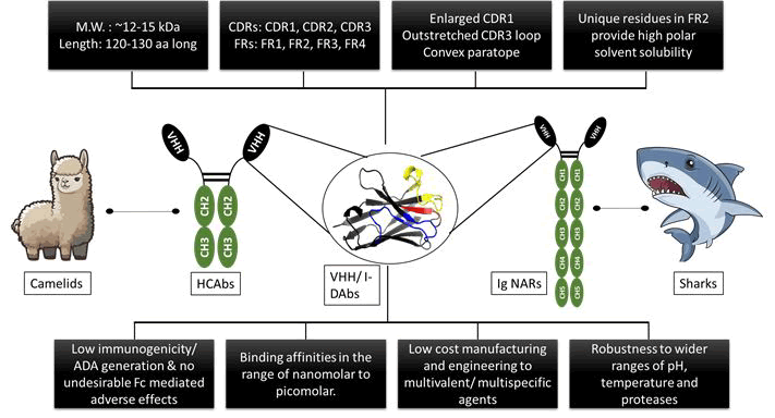
Figure 1: An overview of I-DAb’s distinct qualities that set it apart from other mAb formats, as well as an outline of its advantages.
Despite possessing all the optimal drug-like characteristics, the non-human origin of single domain antibodies creates a bottleneck in its development as a safe therapeutic for human administration. Given that mouse antibody fragments must undergo humanization before being approved for use as human treatments, it stands to reason that camelid-derived nanobodies should likewise successfully complete the humanization process. Humanization is a two-step process; the primary one is reducing immunogenicity and the secondary one is restoring the lost affinity due to CDR grafting. Loss of affinity in humanized variants is restored by back-mutating the original amino acid residues in the framework region. Selection of these critical Back Mutations (BMs) is based on empirical knowledge as well as trial and error as every antibody structure and sequence is unique in its own aspect, resulting in multiple back-mutated variants [2].
Humanization has contributed significantly to the success of mAbs as therapeutics by greatly reducing their immunogenicity. Chimerization and CDR grafting are the foundation of conventional techniques used to lower the likelihood of severe immunogenic reactions to non-human therapeutic antibodies. Resurfacing, shuffling of the framework, computational human content optimization, super humanization, human antibody library screening and phage display and transgenic mouse immunization are examples of more advanced techniques for humanizing an antibody. CDR grafting is a popular technique in which the CDRs of an antibody of non-human origin are transferred onto a human antibody framework. Despite being so widely accepted, the major drawback with the CDR grafting strategy of humanizing antibodies is that the humanized antibodies tend to lose their affinity for their intended targets. This is the result of the extensive sequence modifications within the Framework (FR) during the humanization process. Critical positions within the parent antibody Framework Regions (FRs) govern certain attributes that are believed to impact the antibody affinity. When these are substituted, the binding affinity is negatively affected. But when mutated back, the binding affinity is restored to some extent. There is no straightforward BM protocol for addressing this prominent issue. Residues influencing the binding affinity cannot be back mutated completely, as it will just increase the non-human parent antibody residues in the humanized antibody which will again increase the immunogenicity [3].
The greatest added value of computational techniques in this step comes from offering quick and affordable means of directing experimental procedures. The use of structural modeling can shed light on exposed residues that can be employed in mutagenesis to improve binding, lessen immunogenicity or identify undesirable mutations. Anticipating and mapping the interactions between epitope and paratope using these tools will not only guide the humanization process but also it will be time and cost-effective.
In this case study, an in silico humanization was performed on a camelid anti-hTNF-α I-DAb, by using an enhanced BM protocol. This approach utilized an empirical BM scoring method combined with an improved strategy for BM insertion, aiming to strike a balance in between the lowest possible BMs and the immunogenic potential of the humanized I-DAb.
Materials and Methods
All the tools and servers described and used in this work are open source and free to use.
Generation of anti-hTNF-α I-DAbs
The sequences for llama-derived anti-hTNF-α I-DAbs were procured from CRO service provided by Diaclone SAS, Besancon Cedex, France. The llamas were first immunized with hTNF-α, followed by the isolation of mRNA from them. This mRNA was then subjected to further quantification, cloning, sequencing, insertion in a plasmid and then cultured. With the aid of biopanning, anti-hTNF-α expressing phages were chosen and then amplified. Two rounds of biopanning were done utilizing recombinant hTNF-α protein by ELISA. After the biopanning, the amplified library was screened by using periplasmic extract with the help of ELISA. After the screening, positive clones were selected and further subjected to sequencing. The final seven (S1-S7) candidates were identified as potential binders to the hTNF-α. Among these sequences, ‘S1 I-DAb’ was utilized in the following work of humanization [4].
Prediction of 3D-structure of antigen and antibody using AlphaFold2
The 3D/crystallographic structures of these novel anti-hTNF-α IDabs are unknown. As homology modelling could not be performed in our case, computational protein structure prediction methods such as AlphaFold2 were used. The FASTA sequence of S1 I-DAb was used for the construction of a homology model. To generate a predicted three-dimensional protein structure, AlphaFold2, was employed a deep learning model developed by Google DeepMind, which is based on neural networks. Because it takes into account different structural arrangements during training, AlphaFold2 has an advantage in predicting secondary structures of nanobodies and performs best in predicting the CDR structures of I-DAbs. For building the 3D structure, the amino acid sequences of S1 I-DAb and its humanized variants were submitted one at a time. From the predicted structures for each I-DAb sequence, the topmost scoring relaxed model for individual query sequences was downloaded in .pdb format.
The quality assessment of the best generated models for backbone geometry (torsional angles) and side chain parameters (non-bonded interactions) was done using Ramachandran plot and the ERRAT plot, respectively. The PROCHECK (Ramachandran plot) and ERRAT (ERRAT plot) tools available at the SAVESv6.0 server were used for analyzing the protein structure.
The FASTA sequence of hTNF-α was obtained from Protein Data Bank (PDB) (PDB code: 1TNF) and used for further tasks. Similarly, the comparative model for hTNF-α with higher resolution was built using AlphaFold2 and assessed using PROCHECK and ERRAT tools available at SAVESv6.0 server [5].
All the models were visualized using PyMol v2.5.5.
Antibody sequence annotation
The delineation of the antibody sequence into CDRs and FRs was carried out using Antibody Resource for Sequence Analysis (AbRSA) server. Of all the available antibody numbering schemes (Kabat, Chothia, IMGT), the Kabat numbering system, which is widely accepted due to its accurate annotations of CDRs and FRs, was selected for annotation of S1 I-DAb and used for further computational analysis.
Humanization of antibody using CDR-grafting
To humanize S1 I-DAb by CDR-grafting, the homologous human heavy chain variable region framework was obtained using IMGT Domain Gap Align tool. Homologous human VH framework germlines were obtained against the llama derived S1 I-DAb. Using Clustal omega, the amino acid sequences of the most homologous germlines were aligned with S1 I-DAb. Except for the CDR sequences, all the FRs of S1 I-DAb were replaced with the homologous human VH frameworks of equal length from the alignment. This variant of CDR-grafted humanized I-DAb was referred to as HM00.
Designing of back-mutant humanized I-DAb variants
In this article, the BM protocol particularly designed for camelid nanobodies by Sulea was followed. In the original article by Sulea 2022, residues affecting the binding affinity of a nanobody were identified and ranked according to their potential to influence the same. These affecting residues were grouped according to the different properties (attributes) contributed by them individually to the antibody affinity. A total 8 types of attributes were selected, where each attribute is controlled by a bunch of residues. These attributes were then ranked on the basis of their potential to impact the binding affinity. More is the impact governed by the attribute on the binding affinity, higher is the score allotted to it. Each attribute consists of certain number of residues and every residue has the same score. A single residue may govern more than one attribute. For example, let us take into consideration residue number 98 (Kabat no. 94). This residue contributes to indirectly affecting CDR 3 conformation, Vernier Zone, residue with the propensity of antigen contact, and FR residue adjacent to CDR but not part of Vernier Zone, thus governing three attributes [6].
As these residues could not be added abruptly, the previous ranking and scores would be utilized for the same. These attributes are mapped onto the sequence. The scoring for each residue is calculated based on the total number of attributes governed by it. For example, residue no. 98 (Kabat no. 94) contributes to four different attributes, so the total score for the same is 8. Similarly, this was calculated for other residues and then these residues were ranked on the basis of this score. According to this score each ranked mutation was then sequentially inserted in an incremental order one at a time in the humanized nanobody, thus resulting in back-mutated variants. To exemplify, HM01 contains K98A mutation and its successor HM02 contains K98A as well as W47F BMs.
This same protocol was repeated for S1 I-Dab. These residues governing critical attributes were then mapped on the llama derived S1 I-DAb sequence. The non-conserved residues with respect to the FR were considered and selected and assigned scores to them based on their impact and involvement in affecting S1 I-DAb overall binding affinity. Total BM score for each residue was calculated and then used to rank these mutations accordingly. Then starting from the highest ranked mutation, started back-mutating the residues in incremental order as follows: HM01 contains a single mutation and HM07 consists of 8 mutations altogether. Each mutation was added in sequential order of their ranking.
The residues contributing to soluble expression in of humanized variants were reported for E. coli expression system. These were not taken into consideration as the expression system may be changed as required and adding these residues may increase the immunogenicity. The overall goal is to design humanized back-mutants with minimum no. of required BMs. Excluding the step of considering the residues contributing to the expression, the next BM was added to the HM04 mutant. This resulted in two more variants HM04-27 and HM04-28 [7].
Evaluation of immunogenicity of antibodies
To evaluate the immunogenic potential of I-DAbs, humanness scores were calculated. The term "humanness" related to antibodies refers to the sequence's resemblance to antibodies found in the human repertoire, thus more is the resemblance more is the score and lesser is the immunogenicity. Applying this criterion, the humanness was assessed using the T20 score analyzer from Gao et al., which has been demonstrated to be consistent with the immunogenicity of a related set of clinical antibodies. A score above 85 indicates that the antibody is more human-like, thereby suggesting that it carries a lower immunogenicity risk in humans. The T20 score for S1 I-DAb (wild type, llama derived), HM00 (CDR-grafted S1 I-DAb), HM01 to 07 as well as HM04-27 and HM04-28 (humanized, back-mutated I-DAbs) were calculated using their FASTA sequences.
Molecular docking
To study the impact of the CDR-grafting and the BMs on the binding affinity, molecular docking was performed. To predict the binding affinity between the epitope and the paratope of the binding IDAb structures, HADDOCK v2.4 an online server that generates docking predictions for given structures utilized. HADDOCK v2.4 accepts two or more .pdb files as input and several predicted protein complexes in .pdb format, as well as docking metrics, are returned. In the initial docking of wild type, the active residues of the I-DAb were CDRs according to Kabat definition, while the passive residues were automatically defined. No residues were defined for the antigen, i.e., hTNF-α. The docking pose with the lowest docking score and the lowest Root Mean-Square Deviation (RMSD) value was selected and further used for defining the active residues for the antigen.
CDR residues obtained using the previously defined Kabat numbering scheme were used as active residues to dock the CDRgrafted and back-mutated humanized I-DAb variants. The interacting residues obtained in the previous docking were used for defining active sites for the antigen and the passive residues were automatically defined. The intermolecular interactions were noted down using PDBsum [8].
Results
3D structure prediction of antigen and antibody
For each predicted structure of the I-DAb, the Ramachandran plot and ERRAT plot assessment did not result in any deviation. Hence, there was no need for further refinement of these structures (Supplementary Figure S1 and Table S1). Models for S1 I-DAb and hTNF-α are shown in Figure 2. The CDRs are observed as loops and FRs are observed as β sheets that are anti-parallel with respect to each other. hTNF-α is seen as a timer. Similarly, the homology model of hTNF-α passed all the tests for model quality assessment (Supplementary Figure S2). plDDT scores for all the models have been represented in Supplementary Figure S3.
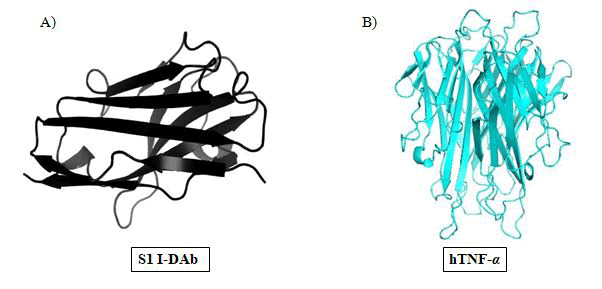
Figure 2: (A) Alphafold 2.0 constructed 3-D model of S1 I-DAb. (B) Homotrimer of hTNF-α built using Alphafold 2.0. Both the models were visualized using PyMol v2.5.5.
Antibody sequence annotation
The Kabat style numbering of the I-DAb sequences using AbRSA resulted in three CDRs and four FRs for each sequence. Figure 3 represents the antibody annotation and CDR identification for S1 IDAb. The CDRs in Kabat numbering were delimited for S1 I-DAb as CDR 1: 31-35, CDR 2: 50-65, and CDR 3: 95-102. Residues 1-30, 36-49, 66-94 and 103-113 were delineated as FR1, FR2, FR3, and FR4 respectively. When this exercise was repeated for the humanized variant, the CDRs and FRs so obtained matched perfectly with the wild type I- DAb (Supplementary Figure S4) [9].
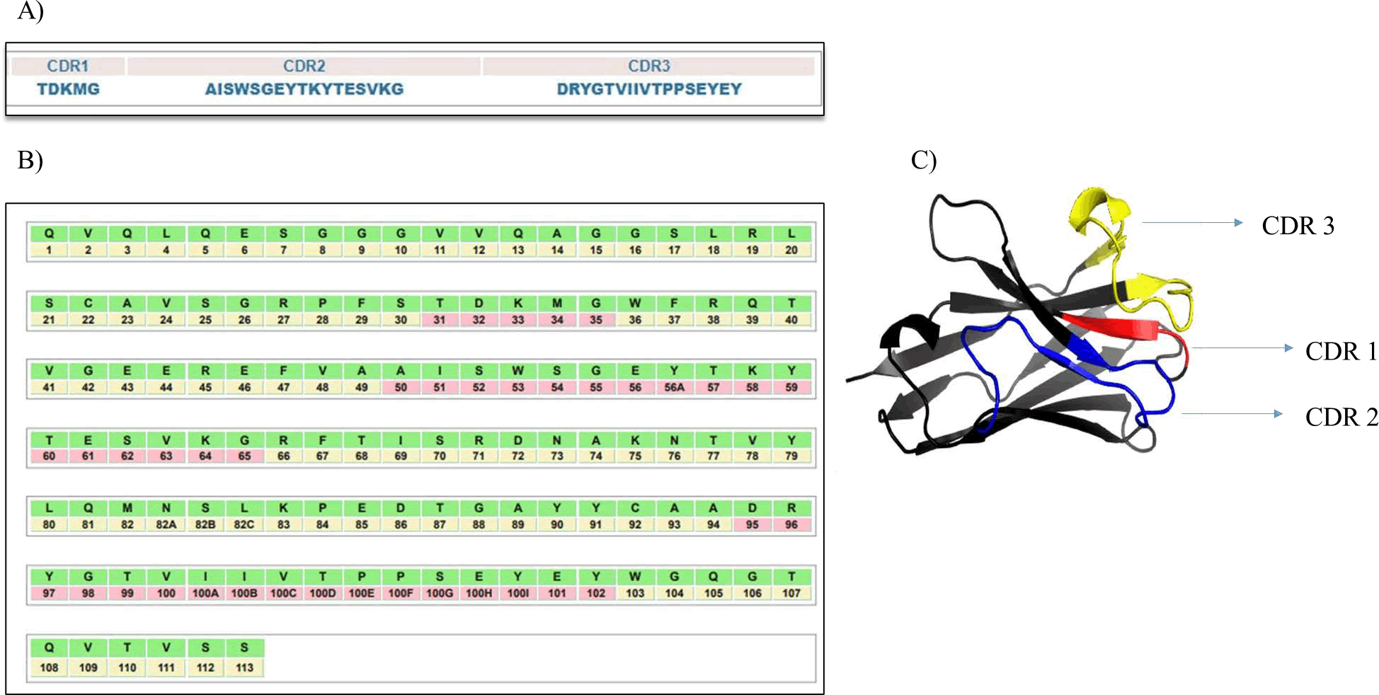
Figure 3: S1 I-DAb sequence annotation as per Kabat definition. (A) CDR1, 2 and 3 demarcated for s1 I-DAb (B) Delineation of CDRs and FRs of S1 I-DAb according to the Kabat convention. CDRs are highlighted in pink color and FRs are highlighted in light yellow color.(C) CDR 1, 2 and 3 residues are highlighted in the model in red color, blue color and yellow color respectively. The model was visualized using PyMol v2.5.5.
Humanization of antibody using CDR-grafting
VH23-03* and J4-01* are the human immunoglobulin germlines that showed the highest sequence similarity with S1 I-DAb (Supplementary Figure S5). After aligning sequences of these germlines with S1 I-DAb, the CDRs were carefully placed onto the germline framework scaffold. The resultant sequence was a successfully CDR-grafted humanized variant of S1 I-DAb. The CDRs and FRs were correctly annotated for the humanized variant. This variant was named as HM00 (Figure 4).
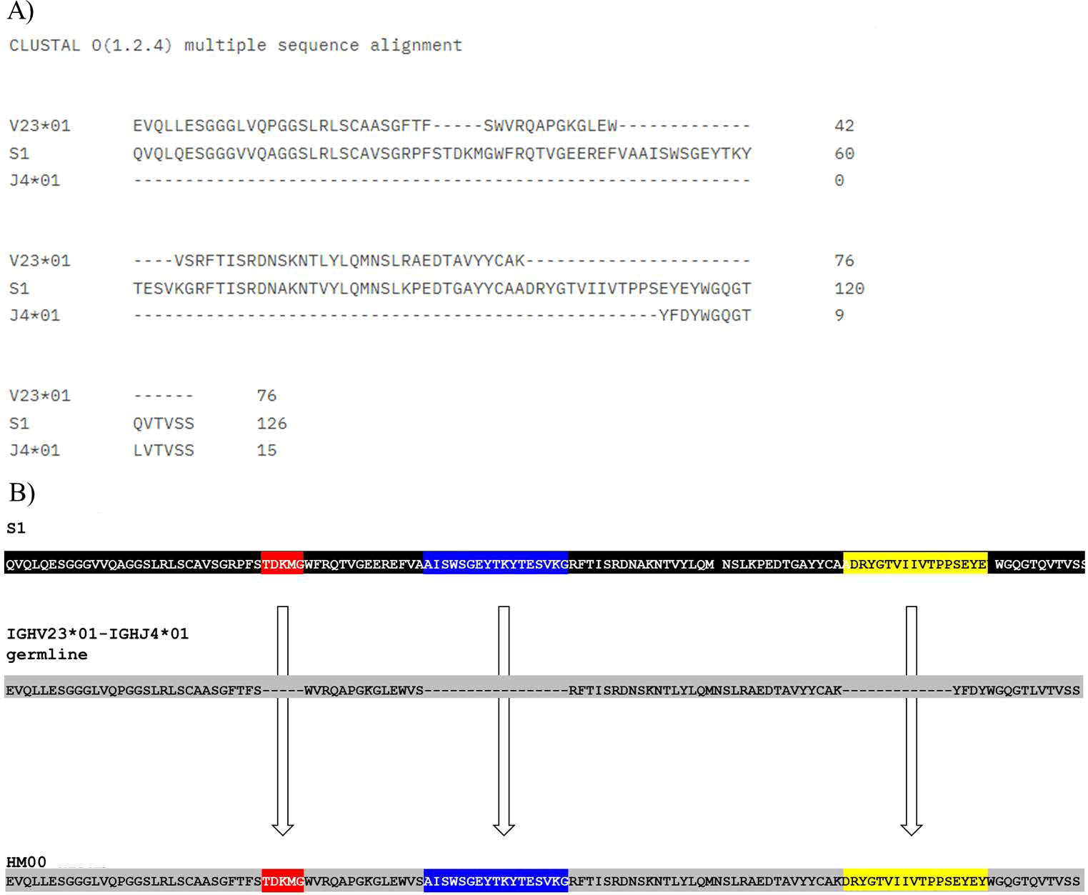
Figure 4: Humanization of S1 I-Dab by CDR grafting. Panel A; sequence alignment of amino acid sequences of S1 I-DAb with the most homologous human immunoglobulin consensus sequence and humanized HM00 sequence. Panel B; construction of the humanized, CDR grafted variant by swapping CDRs of S1 I-DAb with VH23-03* and J4-01* germlines. CDR residues are highlighted in red, blue and yellow color and defined according to the convention of Kabat and colleagues. FR residues are highlighted in blue colour.
Fabrication of back-mutant humanized S1 I-DAb variants for affinity restoration
As described in the methodology section, an empirical score for each attribute depends on their ability to influence the antigen binding ability of I-DAbs. Different attributes contributing to the antibody affinity along with the positions governing these attributes and the scores assigned to them are described in Table 1.
| Sr. no. | Attribute | Representation | Kabat position | Assigned BM score |
| 1 | Residues in FR3 directly affecting CDR 3 conformation | C | 94 | 5 |
| 2 | Residues in FR3 indirectly affecting CDR 3 conformation | c | 93 | 4 |
| 3 | Stability and conformation of CDR3 | S | 37, 47 | 4 |
| 4 | VHH solubility | s | 44, 45 | 3 |
| 5 | Residues belonging to CDR4 | A | 71-78 | 1 |
| 6 | Residue with the propensity of antigen contact | a | 1, 2, 47, 94 | 1 |
| 7 | Vernier zone | V | 2, 27-30, 47-50, 67, 69, 71, 73, 78, 93, 94, 103 | 1 |
| 8 | FR residue adjacent to CDR but not part of vernier zone | v | 2, 4, 24, 27, 28, 29, 30, 26, 37, 38, 46, 47, 49, 50, 66, 67, 68, 69, 70, 71, 73, 78, 83, 86, 91, 92, 93, 94, 103, 104 | 1 |
Table 1: Attributes governed by multiple positions across I-DAb sequence and their respective allotted scores.
These were mapped on the S1 I-DAb sequence and the nonconserved positions in the humanized variant (HM00) with respect to S1 I-DAb were considered for further scoring. Total BM scores and ranking of these positions as per their scores are described in Tables 2 and 3 respectively.
| Residue number | Kabat position | Residue | Attributes | Total BM score | |||||||
| C | c | S | s | A | a | V | v | ||||
| 1 | 1 | Q | - | - | - | - | - | 1 | - | - | 1 |
| 24 | 24 | V | - | - | - | - | - | - | - | 1 | 1 |
| 27 | 27 | R | - | - | - | - | - | - | 1 | 1 | 2 |
| 28 | 28 | P | - | - | - | - | - | - | 1 | 1 | 2 |
| 37 | 37 | F | - | - | 4 | - | - | - | - | 1 | 5 |
| 44 | 44 | R | - | - | - | 3 | - | - | - | - | 3 |
| 45 | 45 | R | - | - | - | 3 | - | - | - | - | 3 |
| 47 | 47 | F | - | - | 4 | - | - | 1 | 1 | 1 | 7 |
| 49 | 49 | A | - | - | - | - | - | - | 1 | - | 1 |
| 75 | 74 | A | - | - | - | - | - | - | 1 | - | 1 |
| 79 | 78 | V | - | - | - | - | 1 | - | 1 | 1 | 3 |
| 87 | 83 | K | - | - | - | - | - | - | - | 1 | 1 |
| 98 | 94 | A | 5 | - | - | - | - | 1 | 1 | 1 | 8 |
Table 2: Calculated BM scores for the hallmarked positions across S1 I-DAb.
| Residue number | Kabat position | Residue | Total BM score | Rank |
| 98 | 94 | A | 8 | 1 |
| 47 | 47 | F | 7 | 2 |
| 37 | 37 | F | 5 | 3 |
| 79 | 78 | V | 3 | 4 |
| 44 | 44 | R | 3 | 5 |
| 45 | 45 | R | 3 | 5 |
| 27 | 27 | R | 2 | 6 |
| 28 | 28 | P | 2 | 7 |
Table 3: Ranking of scored residues on the basis of their respective BM scores.
According to the ranking obtained using the empirical scoring system for BM scores, the BMs were inserted in an incremental order each at a time in such a way that HM01 consisted of a single mutation till HM07 which consisted of 8 BMs, whereas HM04-27 and HM04-28 (designed by excluding the residues affecting nanobody expression in E. coli) each contain about 5 BMs. The mutations present in each variant are described in Table 4 and Figure 5.
| Sr. no. | Variant name | BMs introduced | Total no. of BMs |
| 1 | HM01 | K98A | 1 |
| 2 | HM02 | K98A | 2 |
| W47F | |||
| 3 | HM03 | K98A | 3 |
| W47F | |||
| V37F | |||
| 4 | HM04 | K98A | 4 |
| W47F | |||
| V37F | |||
| L78V | |||
| 6 | HM05 | K98A | 6 |
| W47F | |||
| V37F | |||
| L78V | |||
| G44E | |||
| L45R | |||
| 7 | HM06 | K98A | 7 |
| W47F | |||
| V37F | |||
| L78V | |||
| G44E | |||
| L45R | |||
| F27R | |||
| 8 | HM07 | K98A | 7 |
| W47F | |||
| V37F | |||
| L78V | |||
| G44E | |||
| L45R | |||
| T28P | |||
| 9 | HM04-27 | K98A | 5 |
| W47F | |||
| V37F | |||
| L78V | |||
| F27R | |||
| 10 | HM04-28 | K98A | 5 |
| W47F | |||
| V37F | |||
| L78V | |||
| T28P |
Table 4: Designed variants of humanized S1 I-DAb containing BMs in incremental order.
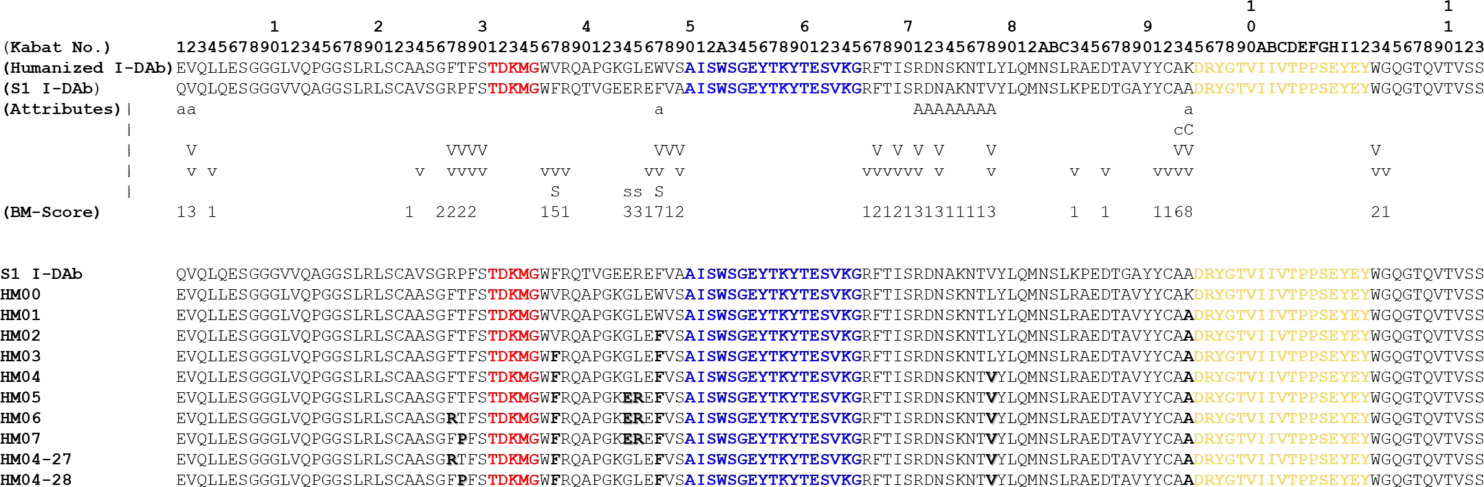
Figure 5: Tailoring of the back-mutated variants. Residues governing critical attributes are demarcated along the wild type sequence and their respective BM scores are denoted below them. BMs introduced in each variant are highlighted in each sequence. CDR residues are highlighted in red, blue and yellow color and defined according to the convention of Kabat and colleagues.
Humanness evaluation of I-DAbs
The T20 score was used to evaluate the immunogenicity of the IDAbs.
The T20 score for the wild-type antibody was 73.1 (S1 I-DAb). After CDR-grafting the T20 score increased to 93.04 (HM00). A T20 score above 85 indicates that an antibody is a humanized antibody, thus it was confirmed that HM00 is humanized. When the T20 score for HM01-07 was calculated, the incremental BMs resulted in a sequential decrease in the T20 score. Despite the lowering of the T20 scores, all the scores were above 85 and classified as humanized (nonimmunogenic) sequences. T20 scores for all the variants have been summarized in Table 5.
| Variant name | T20 score |
| S1 I-DAb | 73.1 |
| HM00 | 93.04 |
| HM01 | 91.89 |
| HM02 | 90.8 |
| HM03 | 89.54 |
| HM04 | 88.5 |
| HM05 | 86.26 |
| HM06 | 86.2 |
| HM07 | 86.26 |
| HM04-27 | 88.5 |
| HM04-28 | 88.5 |
Table 5: Humanness scores calculated for all the variants of I-DAb using a T20 score analyzer.
Assessment of I-DAb and hTNF-α interactions using molecular docking
HADDOCK v2.4 was used to study the binding affinities of the I-DAb variants with h TNF-α. PDBsum r evealed thein terfacial interactions between the epitopes and paratopes of the docked complexes. For the docking of the wild type S1 I-DAb, the docking pose with the most negative binding score (highest target affinity) and the lowest RMSD was selected. The interactions studied using PDBsum revealed the substantial involvement of CDR3 residues in the electrostatic interactions (salt bridges and hydrogen bonds) which stabilize the overall docking pose. All the energies of these interactions are summarized in Table 6.
| Variant name | HADDOCK score |
| S1 I-DAb | -138.8 |
| HM00 | -85.5 |
| HM01 | -113.2 |
| HM02 | -104.1 |
| HM03 | -113.8 |
| HM04 | -117.7 |
| HM05 | -94 |
| HM06 | -131.3 |
| HM07 | -110.1 |
| HM04-27 | -129.1 |
| HM04-28 | -115.4 |
Table 6: Binding scores predicted for all the I-DAb variants-hTNF-α complex using HADDOCK v2.4.
Overlap of back-mutated humanized structures with the wild type S1 I-DAb is portrayed in Figure 6. Superimposing the structures of HM00 and S1 I-DAb revealed the distorted CDR3 conformation in HM00, which may alter its binding affinity (Figure 7). This resulted in a 38% decrease in the binding score as compared to the wild type. A lesser electrostatic energy suggests a weaker interaction between antigen and I-DAb, as seen in Figure 8.
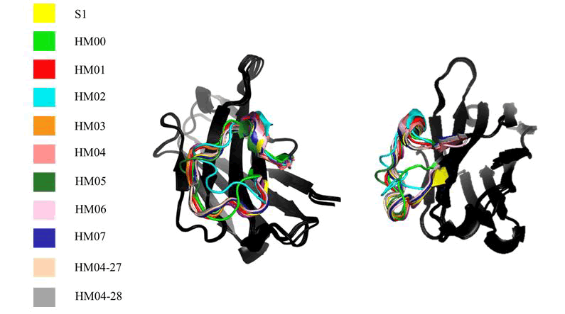
Figure 6: Structural differences observed in CDR 3 loop of I-DAb variants with different BMs at each position. Each CDR of a I-DAb is colored with a distinctive color as shown in the figure.
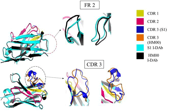
Figure 7: Structural dissimilarities observed within the wild type and humanized I-DAbs using the 3D constructed models. Both of these models were superimposed on each other using PyMol v2.5.5.
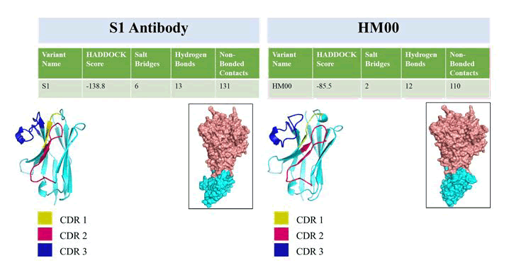
Figure 8: Comparative analysis of S1 I-DAb (wild type) and HM00 (humanized) intermolecular interactions. The humanized variant showed a drastic decrease in the number of inter atomic interactions. The HADDOCK generated complex of I-DAb (cyan) and hTNF-α (pink) was visualized using PyMol v2.5.5. All the interactions are determined using PDBsum. (In figure replace S1 antibody to S1 IDAb)
In the case of HM01, the K98A mutation led to some improvement in the docking score. Although the next mutation W47F in HM02 was thought to improve the binding affinity, it reduced the binding affinity by 24%. With the incremental mutations, the total binding energy as well as the electrostatic interactions from HM03 to HM06 increased significantly. The mutation F27R in HM06 dramatically restored the binding affinity. The binding energy of HM06 was found to be the nearest to the binding energy of the wild-type S1 I-DAb. The binding poses of HM06 and the wild type S1wild type I-DAb are also similar as depicted in Figures 9 and 10. However, in the case of HM07, replacing the mutation F27R with T28P resulted in a substantial loss of binding affinity. HM04-27 and HM04-28 do not have the mutations at position no. 44 and 45, which are responsible for the solubility of the I-DAb. The binding affinity for HM04-27 is the nearest to HM06 as well as S1 I-DAb (Figure 11). However, in this case, too, the T28P mutation in HM04-28 resulted in decreased binding affinity. Structures and the binding information of other variants are provided in the supplementary Figure S6 and Table S1.
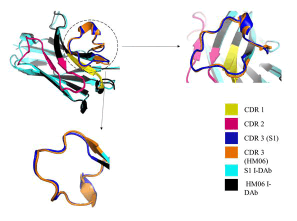
Figure 9: Restoration of the previously distorted structure of CDR 3 loop after introduction of BMs in HM06 as visualized with the aid of PyMol v2.5.5.
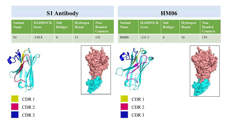
Figure 10: Resemblance in the docking poses of I-DAb (cyan) and hTNF-α (pink) as well as number of interactions as observed with HM06 and S1 I-DAb. This was visualized using PyMol v2.5.5. All the interactions are determined using PDBsum.
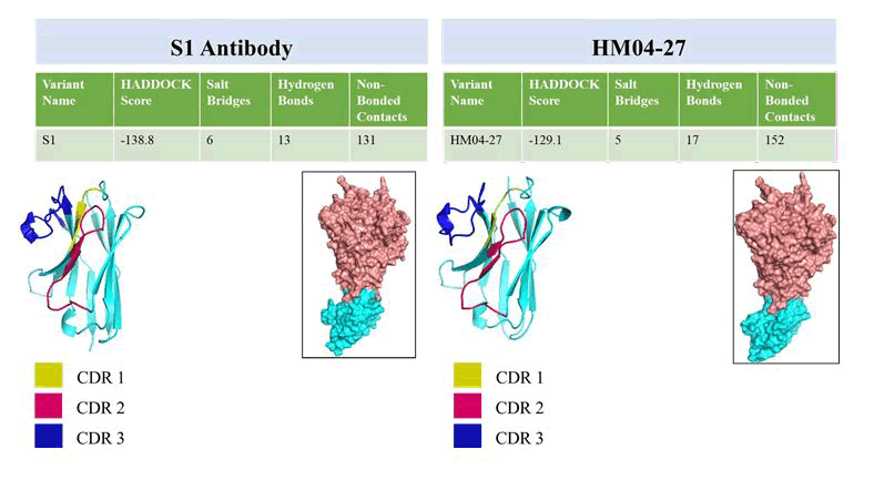
Figure 11: Similarities observed within the docking poses of I-DAb (cyan) and hTNF-α (pink) between HM04-27 and S1 I-DAb using PyMol v2.5.5. All the interactions are determined using PDBsum.
HM00 was the humanized variant which demonstrated poor binding potential towards the antigen. Analysis of protein-protein interactions indicated drastically reduced hydrogen bonds and salt bridges. When the structure of HM00 was compared with S1 I-DAb, a considerable difference was seen in the CDR 3 loop (Figure 7). This distorted structure of the CDR 3 loop is anticipated to affect the binding potential of the I-DAbs. Thus, the introduction of BMs within the FR is believed to facilitate the restoration of binding potentials. A BM scoring system as described and exemplified in this article, eases this crucial step by ranking them. When these BMs were introduced within the HM00 sequence in an incremental order, 9 different types of sequences: HM01-07, HM04-27 and HM04-28 were obtained. BMs within the humanized I-DAb sequence resulted in the improvement of the binding affinity of the back-mutated variants. Structural comparison of the back-mutated variants and wild type I-DAbs showcased an increasing resemblance of the CDR 3 loop of HM06 and HM04-27 with the wild type (Figure 9). Increased structural similarity of the CDR 3 loop also contributed in improving the binding potential of these back-mutants (Figure 6 and Table 6). Thus, these back-mutant variants were considered as successful. In our first attempt, seven variants were designed wherein HM01 contains a single mutation and HM07 contains the highest number of mutations. To further reduce the necessary BMs in HM06 while retaining its binding potential, different combinations of the mutations were tried this time. HM05 variant precedes the HM06 variant but their binding potential is poor as compared to HM04 and HM06. Thus, HM04 was chosen for this particular step. BMs at residue number 27 and 28 were introduced separately. Through docking interactions, it was confirmed that the HM04-27 variant has better binding potential as compared to HM04 and HM04-28. The predicted 3D structures, docking poses as well as the docking scores of the best binders; HM06 and HM04-27 variants are nearly equivalent. However, they differ in their immunogenic potential. T20 scores indicate that HM04-27 is less immunogenic than HM06, so one may opt for HM04-27.
Discussion
Humanization of I-DAbs is one of the crucial steps that is necessary to escalate their therapeutic potential in human subjects. Here, an example of humanization of I-DAb, followed by the selection of critical BMs for the affinity restoration of the humanized I-DAb and designing of the back-mutant humanized I-DAbs was presented. It has been long evident that CDR grafting of xenogeneic antibodies results in the considerable loss of binding potential of the antibodies. A similar pattern was observed in the studies carried out groups while working on I-DAb humanization. Extensive modification in the FR sequences of the I-DAb during the humanization process results in the altered folding of the I-DAb’s CDR loops. Without a protocol to begin with, it is a herculean task to decide which residues one may consider for BM thereby restoring the binding affinity of the humanized antibody. The protocol by Sulea 2022 provides a systematic way for BM insertion and affinity restoration of humanized I-DAbs. In our case study, we included an extra step in this protocol, i.e., molecular docking. Molecular docking aids in understanding the possible binding site, binding pose, binding energy as well as the possible intermolecular interactions. This aids in designing mutants of humanized I-DAbs which may have binding affinity equivalent to the parent I-DAb with least number of BMs required. If desired one may also opt for molecular dynamic simulations to gain a deeper understanding of the I-DAb dynamics pre humanization and post humanization.
Conclusion
Using a simple yet improved back-mutation scoring approach, this article exemplified an attempt to restore the binding affinity of a single domain antibody post humanization. A total of 8 attributes were selected that can majorly affect the CDR loop conformation and expression of I-DAbs. After calculating BM scores, a total of 8 types of back mutations were obtained, which were inserted one at a time in an incremental manner. About 9 back-mutated variants were obtained out of which, HM06 and HM04-27 variants have binding scores almost equivalent to the wild type S1 I-DAb. Additional to the preexisting protocol, an extra step of molecular docking, which aided in understanding the possible antigen-antibody interactions. Along with that, a tool for examining the humanness of I-DAbs, T20 analyzer, was also included throughout the experiment in this case study. With this approach, the selection of mutants with the least number of BMs and restored binding affinity of the humanized variant was possible. The combined approach of CDR grafting, molecular modeling, docking and humanness evaluation using computational methods helps to strike out an optimal balance with affinity restoration and lower BM insertion.
Acknowledgment
This work was partially supported by a research grant BT/ PR42965/PBD/26/821/2021 from, Department of Biotechnology, New Delhi, Government of India and the National Institute of Pharmaceutical Education and Research, S.A.S. Nagar (NPLC-AHP) for providing financial support.
References
- Abhinandan KR, Martin AC (2008) Analysis and improvements to Kabat and structurally correct numbering of antibody variable domains. Mol Immunol 45: 3832-3839.
[Crossref] [Google Scholar] [PubMed]
- Apgar JR, Mader M, Agostinelli R, Benard S, Bialek P, et al. (2016) Beyond CDR-grafting: Structure-guided humanization of framework and CDR regions of an anti-myostatin antibody. mAbs 8: 1302-1318.
[Crossref] [Google Scholar] [PubMed]
- Bond CJ, Wiesmann C, Marsters Jr JC, Sidhu SS (2005) A structure-based database of antibody variable domain diversity. J Mol Biol 348: 699-709.
[Crossref] [Google Scholar] [PubMed]
- Boulianne GL, Hozumi N, Shulman MJ (1984) Production of functional chimaeric mouse/human antibody. Nature 312: 643-646.
[Crossref] [Google Scholar] [PubMed]
- Conrath K, Vincke C, Stijlemans B, Schymkowitz J, Decanniere K, et al. (2005) Antigen binding and solubility effects upon the veneering of a camel VHH in framework-2 to mimic a VH. J Mol Biol 350: 112-125.
[Crossref] [Google Scholar] [PubMed]
- Chiu WC, Lai YP, Chou MY (2011) Humanization and characterization of an anti-human TNF-a murine monoclonal antibody. PLoS One 6: e16373.
[Crossref] [Google Scholar] [PubMed]
- Foote J, Winter G (1992) Antibody framework residues affecting the conformation of the hypervariable loops. J Mol Biol 224: 487-499.
[Crossref] [Google Scholar] [PubMed]
- Gao SH, Huang K, Tu H, Adler AS (2013) Monoclonal antibody humanness score and its applications. BMC Biotechnol 13: 55.
[Crossref] [Google Scholar] [PubMed]
- Honorato RV, Koukos PI, Jimenez-Garcia B, Tsaregorodtsev A, Verlato M, et al. (2021) Structural biology in the clouds: the WeNMR-EOSC ecosystem. Front Mol Biosci 8: 729513.
[Crossref] [Google Scholar] [PubMed]