Case Report, J Surg Clin Pract Vol: 2 Issue: 2
Laparoscopic Removal of Foreign Body from Colon
Chinmay S. Gandhi* and Ashok M. Dhonde
Bharati Vidyapeeth Deemed University, Medical College And Hospital, Sangli, Maharashtra, India
*Corresponding Author : Chinmay Gandhi
Bharati Vidyapeeth Deemed University, Medical College and Hospital, Sangli, Maharashtra, India
Tel: +910233 2305350
E-mail: chinmaygandhi00@yahoo.co.in
Received: March 15, 2018 Accepted: April 07, 2018 Published: April 14, 2018
Citation: Gandhi CS, Dhonde AM (2018) Laparoscopic Removal of Foreign Body from Colon . J Surg Clin Pract 2:2.
Abstract
Foreign bodies in gastrointestinal tract are common. Most don’t require treatment. Sharp foreign body in hepatic flexure causing intestinal perforation is very rare. We are presenting a chronic alcoholic patient under the effect of acute alcoholic intoxication unknowingly swallowed a ball pen. he developed acute abdominal symptoms such as pain in right hypochondrium, fever and nausea of three days. On examination he had a tender mass in right hypochondrium. On diagnostic laparoscopy he had chronic inflammatory mass and perforation at hepatic flexure of colon. We had done successful retrieval of ball pen by laparoscopy and done laparoscopic intra corporeal closure of colonic perforation. In view of minimal contamination and collection we had kept a wide bore drain adjacent to perforation. Patient had excellent post operative recovery and was discharged on 5th post operative day. Laparoscopy is safe and feasible for retrieval of foreign body from colon.
Keywords: Foreign body; Laparoscopy; Colonic perforation; Localized peritonitis
Introduction
Toddlers aged 2 to 3 years are most commonly affected by swallowed foreign body. Most of the ingested foreign bodies 90% pass through the gastrointestinal tract spontaneously and don’t require any treatment. Only 1% to 14% of the foreign bodies which cause intestinal obstruction or perforation require treatment in the form of endoscopy, laparoscopy or laparotomy [1,2]. Perforation is one of its rare complication found in less than 1% of the patients. According to Goh the most common site of intraabdominal perforation is the terminal ileum. Sharp foreign body may perforate and blunt foreign body may cause intestinal obstruction and patient may land up in emergency. Laparoscopy can be attempted if skilled expertise is available. Here we report a young adult swallowed ball pen accidently. It was found in hepatic flexure of colon causing inflammatory mass formation and perforation.
Case Report
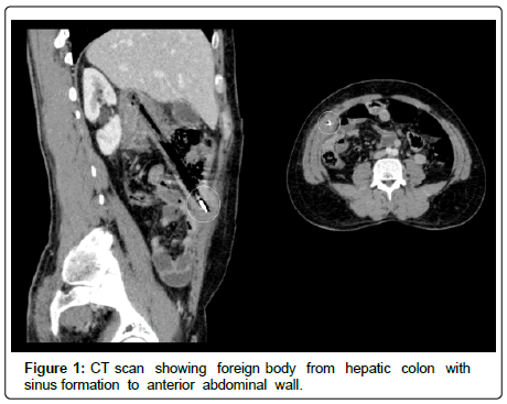
A 48 year old chronic alcoholic male patient presented with right upper quadrant pain since 3 days, it was associated with fever and nausea. On examination his vitals were normal, he had tender lump in right hypochondrium. He had total white blood cell count of 18,500/cmm. Ultrasound was suggestive of localized perforative peritonitis. CT scan of abdomen confirmed an elongated foreign body in hepatic flexure of colon with sinus tract to anterior abdominal wall. There was localized collection and mass formation around (Figure 1). After getting the CT scan finding we asked retrospectively about accidental ingestion of foreign body, but patient was unable to give history.
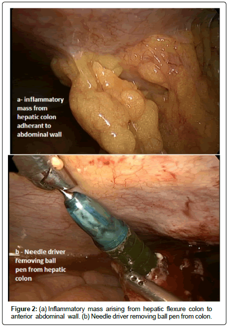
Diagnostic laparoscopy was done under general anesthesia with 10 mm umbilical camera port. There was a omental inflammatory mass in relation with hepatic flexure of colon adherent to anterior abdominal wall (Figure 2a). Afterward two 5 mm port one in right lower mid-clavicular and other in left upper midclavicular region were placed. Adhesiolysis was gently performed and to our surprise a glistening sharp tip was visualized. The tip was grasped with needle driver and pulled gently. The whole of ball pen was retrieved from hepatic flexure of colon (Figure 2b). The 5 mm right trocar was changed to 10mm at the time of extraction of the Ball pen from abdomen. Ball pen was extracted thorough that trocar. The colonic perforation was sutured intra -corporeally with 2-0 vicryl interrupted sutures. A wide bore drain was placed thorough 10 mm port adjacent to perforation. The post operative recovery was uneventful and patient was discharged on 5th post operative day.
Discussion
The management of foreign body ingestion is based on collective anecdotal experience of surgeons. Most of the ingested foreign bodies pass spontaneously from gastro intestinal tract. The highest incidence of foreign body ingestion occurs in children’s between one to three years age and coins are the most common ingested objects. In adults foreign body ingestion is common in edentulous , drug abusers, alcoholic and psychiatric individuals [3,4]. Our patient was under the effect of acute alcoholic intoxication at the time of ingestion of ball pen , hence unable to give history. Sharp objects like chicken, fish bones, tooth , needles, open safety pins and dental implant can cause perforation. Removal of all sharp objects is recommended even in asymptomatic patients. All sharp objects should be removed by upper gastrointestinal endoscopy before they pass the pylorus of stomach as 15 to 35% cause intestinal perforation [5]. Blunt objects like tooth brush, longer than 7cm from stomach need endoscopic or surgical removal [6,7]. Large blunt objects can cause intestinal obstruction. The perforation occurs usually at physiological sphincters, acute angulations and areas of previous surgery [8]. Foreign body with sharp edges can cause complications by penetrating hollow viscous leading to peritonitis, abscess, fistula or formation of inflammatory mass [4,5]. The pre operative diagnosis in OUR case was challenging as the patient had presented with acute abdomen and no history of foreign body ingestion. In the present case, ball pen was trapped in hepatic flexure of colon. It resulted in to penetration of colon and adherence to anterior abdominal wall. It had caused localized peritonitis. Considering a case of acute abdomen, we had done emergency laparoscopy. Foreign body was retrieved successfully. In view of perforation and minimal contamination we approximated the perforation with sutures and kept a wide bore drain. Use of laparoscopy has been described for removal of intra abdominal foreign bodies either intraperitoneal or intraluminal [6]. Laparoscopic removal of foreign bodies from the peritoneal cavity (IUCD) and needle from pelvis were reported earlier. Accurate localization of foreign body in small bowel is a challenge. Fluoroscopy can help intraoperatively to localize small bowel foreign body. Murshid and Khairy removed a metallic pin from small bowel laparoscopicaly under fluoroscopy guidance. Wichmann et al laparoscopicaly removed a tooth pick causing small bowel perforation followed by lavage of the abdominal cavity and laparoscopic closer of perforation. Similarly foreign bodies like safety pin were removed by enterotomy laparoscopicaly.
Conclusion
Laparoscopy is safe and feasible option for retrieving foreign bodies from colon. laparoscopy can be attempted if adequate expertise and good endo vision system is available. In view of acute abdomen with local tenderness and lumpiness we preferred laparoscopy over colonoscopy. We had removed local collection around perforation and prevented residual abscess by laparoscopy.
References
- Chia DKA, Ramesh W, Andrew W, Su-Ming T (2015) Laparoscopic Management of Complicated Foreign Body Ingestion: A Case Series. Int Surg 100: 849-853.
- Ginsberg GG (1995) Management of ingested foreign objects and food bolus impactions. Gastrointest Endosc 41: 33-38.
- Velitchkov NG, Grigorov GI, Losanoff JE, Kjossev KT (1996) Ingested foreign bodies of gastrointestinal tract: retrospective analysis 542 cases. World J Surg 20: 1001-1005.
- Selivanov V, Sheldon GF, Cello JP, Crass RA (1984) Management of foreign body ingestion. Ann Surg 199:187-191.
- Melissa H, Steven S, Michael G (2017) Pointing towards colonoscopy: sharp foreign body removal via colonoscopy. Annals of Gastroenterology 30: 254-256.
- Chin EH, Hazzan D, Herron DM, Salky B (2007) Laparoscopic retrieval of intraabdominal foreign bodies. Surg Endosc 21: 1457.
- Wishner JD, Roger AM (1997) Laparoscopic removal of swallowed toothbrush. Surg Endosc 11: 472-473.
- Golash P (2008) Laparoscopic Removal of Large and Sharp Foreign Bodies from the Stomach. Oman Med J 23: 42-45.
 Spanish
  Chinese
  Russian
  German
  French
  Japanese
  Portuguese
  Hindi
Hindi 


