Clinical Image, J Otol Rhinol Vol: 8 Issue: 2
Neuroendocrine Carcinoma Arising in a Wound after Endoscopic Sinus Surgery for Maxillary Sinusitis
Takeshi Kusunoki1*, Hirotomo Homma1, Yoshinobu Kidokoro1, Aya Yanai1, Satoshi Hara1, Ryo Wada2, Kazuya Saito3 and Katsuhisa Ikeda4
1Department of Otorhinolaryngology, Juntendo University of Medicine, Shizuoka Hospital, Izunokuni, Shizuoka 410-2211, Japan
2Department of Pathology, Juntendo University of Medicine, Shizuoka Hospital, Izunokuni, Shizuoka 410-2211, Japan
3Department of Otorhinolaryngology, Kinki University School of Medicine, Higashiosaka, Osaka, Japan
4Department of Otorhinolaryngology, Juntendo University of Medicine, Faculty of Medicine, Shizuoka Hospital, Izunokuni, Shizuoka 410-2211, Japan
*Corresponding Author : Kusunoki T
Department of Otorhinolaryngology, Juntendo University of Medicine, Shizuoka Hospital 1129 Nagaoka Izunokuni-shi, Shizuoka 410-2295, Japan
Fax: +81-55-948-5088
E-mail: ttkusunoki001@aol.com
Received: July 08, 2019 Accepted: July 29, 2019 Published: August 05, 2019
Citation: Kusunoki T, Homma H, Kidokoro Y, Yanai A, Hara S, et al (2019) Neuroendocrine Carcinoma Arising in a Wound after Endoscopic Sinus Surgery for Maxillary Sinusitis. J Otol Rhinol 8:2. doi: 10.4172/2324-8785.1000371
Abstract
Sinonasal tumors with neuroendocrine differentiation are a rare group of neoplasms that account for only 5% of all sinonasal malignancies. Paranasal neoplasms very rarely arise postoperatively from the maxillary sinus. We encountered a case of neuroendocrine carcinoma arising in a wound after endoscopic sinus surgery for maxillary sinusitis. At the first medical examination, we tried to discriminate between maxillary tumor and postoperative recurrence of maxillary sinusitis.
Keywords: Sinonasal tumors
About the Study
Sinonasal tumors with neuroendocrine differentiationare a rare group of neoplasms that account for only 5% of all sinonasal malignancies [1,2]. Paranasal neoplasms very rarely arise postoperatively from the maxillary sinus. We encountered a case of neuroendocrine carcinoma arising in a wound after endoscopic sinus surgery for maxillary sinusitis. At the first medical examination, we tried to discriminate between maxillary tumor and postoperative recurrence of maxillary sinusitis.
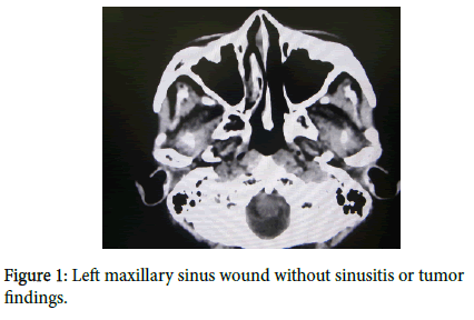
A 66-year-old Japanese woman visited our hospital with one month history of swelling of the left cheek. On her past history, 6 years previously, she received only drainage operation of the left maxillary sinusitis by endoscopic sinus surgery, but without a histopathology examination. One year before (5 years after endoscopic sinus surgery for maxillary sinusitis, when 65-years-old), postoperative CT scan demonstrated that the left maxillary sinus wound was clear without sinusitis or tumor findings (Figure 1).
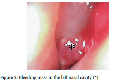
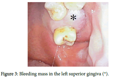
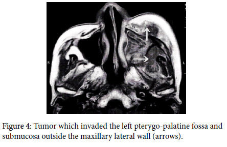
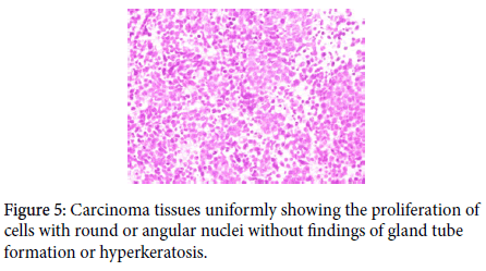
At this first medical examination, a bleeding mass was observed in the left nasal cavity (Figure 2) and left superior gingiva (Figure 3). Therefore, we suspected a neoplasm rising from the postoperative maxillary sinus in addition to the postoperative recurrence of maxillary sinusitis. T1 and T2 weight MRI, and enhanced MRI in left maxillary sinus showed tumors with uneven low and high intensities. This tumor invaded the left pterygo-palatine fossa and submucosa outside the maxillary lateral wall (Figure 4). We examined a biopsy of the maxillary tumor obtained by endoscopic surgery. On the histological examination (H&E staining), the carcinoma tissues uniformly showed the proliferation of cells with round or angular nuclei without findings of gland tube formation or hyperkeratosis (Figure 5). Moreover, immunohistochemical studies showed positive staining for keratin, CAM5.2, and CD56, but not LCA (leukocyte common antigen). From the above results, we finally diagnosed this case as neuroendocrine carcinoma.
References
- Mitchell EH, Diaz A, Yilmaz T, Roberts D, Levine N, et al. (2011) Multimodality treatment for sinonasal neuroendocrine carcinoma. Head Neck 34: 1372-1376.
- Van der Laan TP, Lepsma R, Witjes MJ, van der Laan BF, Plaat BE, et al. (2016) Meta-analysis of 701 published cases of sinonasal neuroendocrine carcinoma: The importance of differentiation grade in determining treatment strategy. Oral Oncol 63: 1-9.
 Spanish
  Chinese
  Russian
  German
  French
  Japanese
  Portuguese
  Hindi
Hindi 