 Spanish
  Chinese
  Russian
  German
  French
  Japanese
  Portuguese
  Hindi
Hindi Research Article, Acute Med Res Vol: 1 Issue: 1
Proniosomal Gel Based Delivery System of Aceclofenac for Ocular Delivery with Improved Safety and Efficacy
Supriya Verma*, Bhupinder Singh and Om Prakash Katare
University Institute of Pharmaceutical Sciences, Panjab University, Chandigarh, India
*Corresponding Author : Supriya Verma
University Institute of Pharmaceutical Sciences, Panjab University, Chandigarh, India
Tel: +919463453377
E-mail: supriya_punjab@yahoo.com
Received: September 20, 2018 Accepted: November 13, 2018 Published: November 19, 2018
Citation: Verma S, Singh B, Katare OP (2018) Proniosomal Gel Based Delivery System of Aceclofenac for Ocular Delivery with Improved Safety and Efficacy. Acute Med Res 1:1.
Abstract
Aim: The study was envisioned to develop a proniosomal gel based carrier system of drug aceclofenac (ACE) for its effective ocular delivery at the inflammatory sites.
Summary of the problem: In the treatment of inflammatory conditions prevailing in eye, multiple treatment strategies have been employed like steroids and non steroidal antiinflammatory agents. But their use is delimited due to their downsides like low drug permeation and retention, local irritation, low drug availability, and stinging effect at the affected site in the eye.
Methodology and theoretical orientation: The chosen drug (ACE) was entrapped in a proniosomal system, which consists of sorbitan stearate (Span 60), membrane stabilizer (cholesterol), Coating agent (maltodextrin) and non-aqueous (proniosomal) gel as the secondary vehicle.
Observations: The particle size, polydispersity index (PDI) & zeta potential of the prepared system were obtained as 679.2 nm, 0.354 and -25.3 mV, respectively. FTIR studies proved the useful interaction of the drug with maximum absorption peak at 3275.90, 2921.01, 1770.51, 1717.81, 1417.95, 1149.39, 749.01 cm-1 in the bilayers of the proniosomal system. The prepared gel (non-aqueous) system showed shear-thinning nature. In in vivo and ex vivo release studies, superiority of the prepared system over conventional system have been witnessed. Moreover, the formulation was able to maintain the drug assay for the period of six months.
Summary and conclusion: The current findings provide the lead for the development of an effective ocular formulation of ACE with substantial stability in the proniosomal system.
Keywords: Aceclofenac; Proniosomal system; Proniosomal gel; Ocular delivery
Introduction
Inflammation is identified as one of the core challenges in the current time, involving a vast majority of the diseases and disorders, including cancer and diabetes. In fact, pain and inflammation are involved in every other disease and often, an acute inflammation turns into chronic inflammatory disease [1], a prolonged and persistent inflammatory state, marked chiefly by new connective tissue formation [2]. Diseases like osteoporosis, rheumatoid arthritis (RA), chronic inflammatory bowel disease, and ocular afflictions and ailments like uveitis, conjunctivitis, retinitis, corneal ulcer, diabetic retinopathy and diabetic macular edema [3-6] are instances of “difficult-to-treat” inflammatory conditions.
Eye inflammation is a common condition, involving people of all ages. It occurs in response to an infection, allergy, autoimmune disorder, irritation, injury or trauma to the eye or eyelid, and, can last from a few minutes to years, depending on the type and severity of the underlying cause. The symptoms include, blurred vision, pain, redness, swelling and tearing. These can be treated with various antiinflammatory agents, which are broadly classified as a) steroidal antiinflammatory agents, e.g. betamethasone, prednisolone and triamcinolone, etc and b) non-steroidal anti-inflammatory agents (NSAIDs), e.g. diclofenac, ibuprofen and ketorolac, etc.
The unique chemical structure of ACE makes it, a potent antiinflammatory drug molecule as well as lipophilic molecule that can better penetrate the ocular tissue if formulated properly, ensuring relief in eye from pain, redness, swelling after certain eye surgeries and other eye inflammations like uveitis, conjunctivitis, retinitis and corneal abrasion [7]. Thus, for the treatment of eye inflammation, the possibility of improving the availability of ACE at the intraocular level by making use of Novel Drug Delivery System would be explored.
ACE is a derivative of diclofenac, hence drug of class Non-steroidal anti-inflammatory drugs or NSAIDS, has been approved by US FDA in the early 90’s (1992) [8]. It is a selective COX-2 inhibitor (an enzyme responsible for the inflammatory mediators generation), potent in action and used in OA, rheumatoid arthritis, ankylosing spondylitis and musculoskeletal pain [9]. Orally administered NSAIDs leads to serious consequences which includes gastrointestinal ulceration followed by bleeding, hypertension, congestive heart failure, hepatotoxicity, renal failure, idiosyncratic and various skin reactions [10-12]. Hence, there is always a standby need to support the oral therapy by topical (ocular) application. The instability of ACE not only decreases its effectiveness and safety but also makes it difficult to put in a formulation. Therefore, the present work aims to develop a stable formulation of ACE for ocular delivery, prepared in non-aqueous environment, as with marked potential. It would deliver our chosen drug (ACE) in protected and successful manner for the confined treatment of pain-related disorders of eye [13].
Materials
Aceclofenac (ACE; M/s Ipca Laboratories Pvt. Ltd, Mumbai, India), Glycerylphosphorylcholine Hydrate 85% (GPC; M/s VAV Life Sciences Pvt. Ltd, Mumbai, India) were obtained ex-gratis by the respective companies. Proniosomal gel was obtained as a generous gift sample from M/s Ipca Laboratories Pvt. Ltd., Mumbai, India. Cholesterol (M/s Avanti Polar Lipids, Alabaster, USA), span 60 (Central Drug House, Pvt. Ltd., Mumbai, India) and maltodextrin (Himedia Lab Pvt. Ltd., Mumbai, India) were procured from the respective sources. All other chemicals used were of analytical grade. Ultrapure water (Milli-Q® Integral system; M/s Merck Millipore, Billerica, USA) was employed throughout the study.
Methods
By taking the photosensitivity of drug ACE into consideration, preparation of formulation and its characterization were carried out in amber colored glass wares.
Proniosomes were prepared by the well described slurry method. Weighed amount of drug, non-ionic surfactant (240 mg of span 60) and membrane stabilizer (i.e. 160 mg of CHL), were added in chloroform and methanol (1:1) in a round bottom flask and weighed amount of carrier (i.e. 800 mg of MLDX) was added to form slurry (as shown in Figure 1). The flask was attached to a rotary flash evaporator (Laborota 4011, Heidolph, Germany) and the organic solvent was evaporated at 45 ± 2°C under reduced pressure (at 70-80 rpm), until a dry, white lipid film was obtained. The resultant powders were further dried overnight in a vacuum oven at room temperature so as to obtain dry, free-flowing product. The obtained proniosomal powders were stored in a tightly closed container at 4˚C for further evaluation.

Figure 1: Preparation of proniosomes by slurry method.
Characterization of proniosomes
Differential scanning calorimetry (DSC): The DSC thermograms of the drug, excipients (span 60, GPC) and proniosomes were recorded with a DSC Q20 TA (M/s PerkinElmer Inc. USA) instrument by using standard aluminium pans. Accurately weighed samples (2-5 mg) were tightly sealed in the pans. An empty pan was used as a reference pan. Samples were heated at the scanning rate of 10°C/min over a temperature range between 20°C and 350°C [14].
Fourier transform infra-red spectroscopy (FTIR): Fourier transform infrared spectroscopy of the drug, excipients and proniosomes were carried out by using FT-IR instrument (Spectrum Two™ M/s PerkinElmer Inc, USA) at 25°C and wave number range from 3500 cm-1 to 500 cm-1. The samples were mixed thoroughly with potassium bromide (KBr) in the mass ratio of 2:98 [14].
Preparation of ACE-loaded proniosomes
Micromeritics and Zeta potential measurement: The vesicles size distribution analysis of the developed proniosomes were carried out using Delsa™ Nano C (M/s Beckman Coulter, India Pvt. Ltd., Mumbai, India) installed at Institute of Microbial technology (IMTECH), Chandigarh. The principle underlying the above analysis is Dynamic Light Scattering (DLS) method. An aliquot of 1ml of the suspension (proniosomes after hydration with PBS of pH 7.4) was diluted 100 times with distilled water and the size of vesicles was obtained from the instrument software. Zeta potential was determined by measuring the electrophoretic movement of the charged particles under an applied electric field from the Doppler shift of scattered light at 25°C and the electric field strength was around 23.2 V/cm. The average zeta potential were directly obtained from the instrument software [16,19].
Transmission electron microscopy (TEM): The surface morphology of niosomal vesicular suspension was examined by Hitachi H-7500, Japan, installed at Central Instrumentation Lab (CIL), Panjab University, Chandigarh. 1% phosphotungstic acid (PTA) was used as a staining agent. Pictures were taken at an excitation voltage of 200 kV [16].
Confocal laser scanning microscopy (CLSM): The coumarin-6 (3- (2-Benzothiazolyl)-N, N-diethylumbelliferylamine, 3-(2- Benzothiazolyl)-7-(diethylamino) coumarin) loaded proniosomes were prepared by mixing coumarin-6 at concentration of 0.15 μmol/mL with 240 mg of Span 60, 800 mg of MLDX, and 160 mg of cholesterol. Dye-loaded proniosomes without drug were applied to the excised pig eye for 6 h followed by rinsing it with PBS pH 7.4 and the fixed slides were prepared. Localization of fluorescence in the ocular layers after experimentation was examined using a confocal laser scanning microscope (Olympus FV10i, M/s Olympus Singapore Pvt. Ltd, Singapore). All confocal images were acquired in the same setting parameters [17].
Incorporation of proniosomes in secondary vehicle
To incorporate the prepared proniosomes in topically applicable vehicle, these were further gelled with 1% w/w of Proniosomal gel.
Release studies: Ex vivo transcorneal permeation studies were carried out using freshly excised pig corneas. The cornea was mounted between clamped donor and receptor compartments of an all-glass modified Franz diffusion cell in such a way that its epithelial surface faced the donor compartment. The corneal area available for diffusion was 0.50 cm2. The receptor compartment was filled with 10 ml freshly prepared tear fluid (pH 7.2), and all air bubbles were expelled from the compartment. An aliquot (1 ml) of formulation [Aceclofenac (0.1% w/v) ophthalmic solution (pH 7.2), was placed on the cornea, and the opening of the donor cell was sealed with a glass cover slip. Receptor fluid was kept at 37°C with constant stirring using a Teflon-coated magnetic stir bead. Permeation study was continued for 360 min, and samples were withdrawn from receptor and analyzed for aceclofenac content by analysing samples with HPLC. Results were expressed as amount permeated or in vitro ocular availability [18].
Characterization of the developed proniosomes-based Proniosomal gel
Rheological studies: Rheological characterization of proniosomal based gel and conventional gel was performed using a rotational type rheometer (Rheolab QC, M/s Anton Paar GmbH, Vienna, Austria) attached with a water jacket (C-LTD80/QC) for maintaining constant temperature. Data analysis was carried out using in-built instrument software Rheoplus/32, version 3.40. A DG26 spindle geometry was used for measuring the torque as well as viscosity of the samp006Ce. The optimized formulation was subjected to different shear stress and shear rate conditions and the measurements were carried out at room temperature. Temperature was kept at 30˚C and shear rate was selected from 0 to 100 s-1. All the tests were performed in triplicate and mean values were used in analysis [19,20].
Power-law (eqn 2) and Herschel–Bulkey model (eqn 3) were employed to analyze the data of stress (s) and shear rate (g) as per the following equations.
 … (2)
… (2)
 … (3)
… (3)
Where, k=consistency Index (Pa sec-n); τ=yield stress (Pa); n=Power-law exponent [21].
Stability studies
The physical stability of the prepared was evaluated after storage for 6 months under different temperature conditions i.e. 5°C ± 2°C (refrigerator), 30°C ± 2°C (room temperature) and 40°C ± 2°C (accelerated temperature). The samples kept under different temperature conditions were withdrawn at initial day, after 6 months and analyzed with HPLC for drug assay and particle size [18].
Statistical analysis
The results were statistically analyzed using one-way ANOVA followed by post hoc analysis using Student's t test. Statistical significance was considered at p<0.05.
Results and Discussion
Characterization of the prepared proniosomes
Differential scanning calorimetry (DSC): Figure 2 depicts the DSC curves of ACE, excipients and proniosomes. The DSC curve of Aceclofenac showed an endothermal peak with an onset temperature at about 153°C, as the melting point of ACE lies in the range of 149°C-153°C [20]. In the DSC curve of proniosomes, the characteristics endothermal peak has been disappeared, which showed that ACE had been completely merged into the proniosomes and they had some interactions, due to the combination of hydrogen bonds and/ or vander waals force [22].

Figure 2: DSC thermograms of (A) ACE (B) GPC (C) span 60, and (D) proniosomes.
Fourier transform infrared spectroscopy (FTIR): The use of FT-IR technique allows the detection of proniosomes formation in solid phase and to point out the implication of the different functional groups of drug and excipients in proniosomes formation process by analyzing the significant changes in the shape and position of the absorbance bands of ACE, Span 60, GPC and proniosomes. Figure 3 illustrates the IR peak of the solid ACE, Span 60, GPC and proniosomes. The Span 60 and GPC exhibited significant FTIR peak at wave number 2919.67, 2850.67, 1737.67, 1467.82, 1174.98, 101.68, 1652.38, 1481.64, 1231.72, 1087.20, 1053.14 cm-1 respectively while, ACE showed FTIR peaks at wave number of 3318 cm-1, 1770, 1583, 1505, 1252, 1053 cm-1, that confirms the purity of drug sample as per the establish standards. While proniosomes had maximum absorption peak at 3275.90, 2921.01, 1770.51, 1717.81, 1417.95, 1149.39, 749.01 cm-1. In proniosomes the peaks of ACE were masked/missing or shifting mainly due to overlapping and intermolecular interactions. All the IR spectra of drug with excipients showed the above mentioned characteristic peaks with insignificant shift and reduced intensity. This ratifies the formation of proniosomes [23].

Figure 3: FTIR-spectra of ACE, Span 60, GPC and proniosomes.
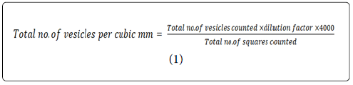
Vesicle count: The density of vesicle population of optimized unsonicated niosomal suspension after hydration of proniosomes with PBS pH 7.4 was found to be 18.4 ± 0.1 × 104 mm3. This confirmed the formation of vesicles is enough to entrap the drug molecules [16].
Micromeritics and Zeta potential measurement: As vividly apparent from the Figure 4, portraying the vesicles size distribution of presonicated and sonicated niosomes after hydration of prepared proniosomes with PBS 7.4. The average vesicles size in case of unsonicated niosomes was found to be 784 nm and PDI was found to be 0.395. Smaller vesicles, however, were obtained with optimal sonication (i.e. for 3 min) with average vesicle diameter of 679.2 nm. Further, it was found that sonication while reducing vesicle size also decreased the value of PDI from 0.395 to 0.354. This value of PDI for prepared optimized formulation confirmed the polydispersity of vesicles population. However, low value of PDI ratifies the narrow variation in size range of the vesicles within suspension.

Figure 4: PDI of proniosome based niosomes.
Zeta potential characterized the surface charge of particle and thus gives an information about repulsive forces between particles and droplets (Figure 5). To obtain stable vesicular suspension by preventing flocculation and coalescence, zeta potential should usually reach a value above 25-30 mV [13,24]. In the present case the Zeta Potential of the developed system was found to be -34.4 mV.

Figure 5: Zeta potential of proniosome based niosomes.
Transmission electron microscopy (TEM): The uniform shape and spherical nature of proniosomes after hydration was further evidenced by TEM, as depicted in Figure 6. TEM micrographs confirmed that prepared niosomes have a closed spherical shape, the aggregation of vesicles and phase separation of the formulation almost become negligible. The morphological evaluations corroborated the formation of circular SUVs and integrity of vesicle lamellae [15].

Figure 6: TEM photomicrographs of proniosome based niosomes.
Confocal laser scanning microscopy (CLSM): CLSM studies revealed that the prepared vesicles (proniosome based niosomes) were uniform in size, round in shape and unilamellar or multilamellar in nature [25]. The studies were carried out to determine the distribution and penetration pattern of the coumarin-6 fluorescent dye-entrapped vesicles into the excised pig eye after 6 h of ocular application. The spherical fluorescent structures in ocular layers as shown in Figure 7, clearly indicated the transport of entrapped-dye, plausibly along with the carriers [26]. The study consents that the prepared vesicles can penetrate the deep layers of eye and can lead to the deep-sited targets.

Figure 7: CLSM images of the proniosome based niosomes.
Release studies: Figure 8(a) and 8(b) depicted the superiority of Proniosomal gel based system over carbopol and conventional gel.

Figure 8: In vitro release studies of different gel systems.

Figure 9: (A) Rheological studies of the formulated proniosome based gel (B) Conventional gel.
Rheological studies
The developed proniosome based gel exhibited non-Newtonian behaviour as shown in Figure 7, with significant magnitude of the yield value. It enlists the various rheological attributes, as per the Herschel- Bulkley model of the developed gels. For the developed proniosomal based gel, value of ‘n’ was found to be less than 1, hence the system was found shear thinning in nature. Consistency index was found to be 6.97 Pa.sn [21]. The viscosity of the prepared proniosomal based gel was found to be higher than the conventional gel thereby, indicating greater contact time of the former with the eye, hence more the therapeutic effect.
Stability studies
Table 1 is depicting the stable nature of the prepared system at varied temperature conditions, i.e. 4 ˚C ± 2˚C, 30 ˚C ± 2˚C, and 40 ˚C ± 2˚C.
| Time (months) | ACE-Proniosome based gel | |
|---|---|---|
| Drug Assay | Mean Size (nm) | |
| 5 ± 2˚C | ||
| Initial | 101.27 ± 2.19 | 369.6 |
| 1 | 101.25 ± 1.07 | 369.5 |
| 3 | 101.19 ± 1.59 | 366.0 |
| 6 | 101.09 ± 1.76 | 367.6 |
| 30 ± 2˚C/65% ± 5% RH | ||
| Initial | 101.27 ± 1.09 | 369.6 |
| 1 | 101.07 ± 1.16 | 372.8 |
| 3 | 100 ± 1.01 | 372.6 |
| 6 | 99.9 ± 0.98 | 389.9 |
| 40 ± 2˚C/75% ± 5% RH | ||
| Initial | 101.27 ± 1.10 | 369.6 |
| 1 | 100.04 ± 1.12 | 372.8 |
| 3 | 99.69 ± 0.87 | 387.8 |
| 6 | 99.61 ± 0.23 | 401.7 |
Table 1: Effect of storage conditions and time on various formulation parameters of ACE-loaded proniosome based gel as per ICH guidelines.
Conclusion
The twin challenges of the drug Aceclofenac, relating to inappropriate physicochemical properties i.e. solubility, permeability and stability (hydrolytic cleavage of ester bond leading to the loss of drug content) were really been the tough proposition to face on. But the innovative ideas and creativity led to design a unique proniosomal system with the formation of closed bilayer assemblies (of vesicles). The later owned the responsibility of transportation across cornea into deep-seated inflammatory pain receptors of the prevailing diseased conditions. But the most gratifying outcome, besides the delivery advantage, has been of overcoming the degradation of the molecule which is otherwise very difficult to prevent. This stability attainment for the drug can be explained on the basis of protective effect of the carrier, which is constructed using appropriate components, while choosing the right experimental conditions and process. The uniqueness in the delivery system, as understood, relates to the creation of such self-assembling supramolecular host assemblies which resulted in achieving the goals, as set in the beginning. And the final outcome was in the form of Aceclofenac bearing proniosome based gel with improved efficacy, safety and stability.
References
- Krishnamoorthy S, Honn KV. Inflammation and disease progression. Cancer Metastasis Rev. 2006;25(3):481-491.
- Cid YP, Pedrazzi V, de Sousa VP, Pierre MB. In vitro characterization of chitosan gels for buccal delivery of celecoxib: influence of a penetration enhancer. AAPS Pharm Sci Tech. 2012;13(1):101-111.
- Adamis AP. Is diabetic retinopathy an inflammatory disease? Br J Ophthalmol. 2002;86(4):363-365
- Rangasamy S, McGuire PG, Das A. Diabetic retinopathy and inflammation: novel therapeutic targets. Middle Afr J Ophthalmol. 2012;19(1):52-59.
- Park UC, Park JH, Yu HG. Long-term outcome of intravitreal triamcinolone acetonide injection for the treatment of uveitis attacks in behcet disease. Ocul Inflamm. 2014;22(1):27-33.
- Cicchitti L, Martelli M, Cerritelli F. Chronic inflammatory disease and osteopathy: a systematic review. PLoS One. 2015;10(3):e0121327.
- Dave V, Paliwal S. A novel approach to formulation factor of aceclofenac eye drops efficiency evaluation based on physicochemical characteristics of in vitro and in vivo permeation. Saudi Pharm J. 2013;22(3):240-245.
- Legrand E, 2004. Aceclofenac in the management of inflammatory pain. Drug Evaluation. 5, 1347-1357.
- Yamazaki R, Kawai S, Matsuzaki T, Kaneda N, Hashimoto S, Yokokura T, Okamoto R, Koshino T, Mizushima Y, 1997. Aceclofenac blocks prostaglandin E2 production following its intracellular conversion into cyclooxygenase inhibitors. Eur J Pharmacol. 329, 181-187.
- Boelsterli UA, 2002. Mechanisms of NSAID-induced hepatotoxicity: focus on nimesulide. Drug Saf. 25, 633-648.
- Jones RH, Tait CL, 1995. Gastrointestinal side-effects of NSAIDs in the community. Br J Clin Pract. 49, 67-70.
- Manjanna KM, Shivakumar B, Pramod Kumar TM, 2010. Microencapsulation: an acclaimed novel drug-delivery system for NSAIDs in arthritis. Crit Rev Ther Drug Carrier Syst. 27, 509-545.
- Bhatia A, Singh B, Rani V, Katare OP, 2009. Phospholipid -based formulation with improved attributes of coal tar. J Cosmetic Dermatol 8, 282-288.
- Yap KL, Liu X, Thenmozhiyal JC, Ho PC, 2005. Characterization of the 13-cis-retinoic acid/cyclodextrin inclusion complexes by phase solubility, photostability, physicochemical and computational analysis. Eur J Pharm Sci. 25, 49-56.
- Jain S, Jain P, Umamaheshwari RB, Jain NK, 2003. Transfersomes--a novel vesicular carrier for enhanced transdermal delivery: development, characterization, and performance evaluation. Drug Dev Ind Pharm. 29, 1013-1026.
- Raza K, Singh B, Mahajan A, Negi P, Bhatia A, Katare OP, 2011. Design and evaluation of flexible membrane vesicles (FMVs) for enhanced topical delivery of capsaicin. J Drug Target. 19, 293-302.
- van Kuijk-Meuwissen ME, Junginger HE, Bouwstra JA, 1998. Interactions between liposomes and human skin in vitro, a confocal laser scanning microscopy study. Biochim Biophys Acta. 1371, 31-39.
- Katara R, Sachdeva S, Majumdar DK. 2017. Enhancement of ocular efficacy of aceclofenac using biodegradable PLGA nanoparticles: formulation and characterization. Drug Deliv. and Transl. Res. 7, 632–641
- Patel D, Dasgupta S, Dey S, Ramani YR, Ray S, Mazumdar B, 2012. Nanostructured Lipid Carriers (NLC)-Based Gel for the Topical Delivery of Aceclofenac: Preparation, Characterization, and In Vivo Evaluation. Scientia Pharmaceutica 80, 749–764.
- Ibrahim MM,El-Nabarawi M, El-Setouhy DA, Fadlalla MA, 2011. Polymeric surfactant based etodolac chewable tablets: formulation and in vivo evaluation. AAPS PharmSciTech. 11, 1730-7
- Basu S, Shivhare US, Raghavan GSV, 2007 Time Dependent Rheological Characteristics of Pineapple Jam. Int. J. Food Eng. 3.
- Elzainy AA, Gu X, Simons FE, Simons KJ, 2004. Cetirizine from topical phosphatidylcholine-hydrogenated liposomes: evaluation of peripheral antihistaminic activity and systemic absorption in a rabbit model. AAPS J. 6, e18.
- Rupal J, Kaushal J, Mallikarjuna SC, Dipti P, 2009. Preparation and Evaluation of Solid Dispersions of Aceclofenac. Int. J. Pharm. Sci. Drug Res. 1 32–35.
- Kostarelos K, Luckham PF, Tadros TF, 1998. Rational design and engineering of delivery systems for therapeutics : biomedical exercises in colloid and surface science. J. Chem. Soc. Faraday Trans. 94, 2159–2168.
- Ahmed K, Gribbon PN, Jones MN, 2002. The application of confocal microscopy to the study of liposome adsorption onto bacterial biofilms. J. Liposome Res. 12, 285–300.
- Raza K, Shareef MA, Singal P, Sharma G, Negi P, Katare OP, 2014. Lipid-based capsaicin-loaded nano-colloidal biocompatible topical carriers with enhanced analgesic potential and decreased dermal irritation. J Liposome Res. 24, 290-296.