Clinical Image, J Otol Rhinol Vol: 8 Issue: 2
Tracheal Stenosis Due to an Abscess from Thyroid Tumor
Takeshi Kusunoki1*, Hirotomo Homma1, Yoshinobu Kidokoro1, Aya Yanai1, Kenji Sonoda1, Yuichiro Saikawa1, Ryo Wada2 and Katsuhisa Ikeda3
1Department of Otorhinolaryngology, Juntendo University of Medicine, Shizuoka Hospital, Shizuoka, Japan
2Department of Pathology, Juntendo University of Medicine, Shizuoka Hospital, Shizuoka, Japan
3Department of Otorhinolaryngology, Juntendo University, Faculty of Medicine, Tokyo, Japan
*Corresponding Author : Takeshi Kusunoki
Department of Otorhinolaryngology, Juntendo University of Medicine, Shizuoka Hospital-1129, Nagaoka Izunokuni-shi, Shizuoka 410-2295, Japan
Fax: +81-55-948-5088
E-mail: ttkusunoki001@aol.com
Received: April 22, 2019 Accepted: April 29, 2019 Published: May 06, 2019
Citation: Kusunoki T, Homma H, Kidokoro Y, Yanai A, Sonoda K, et al. (2019) Tracheal Stenosis Due to an Abscess from Thyroid Tumor. J Otol Rhinol 8:2.
Abstract
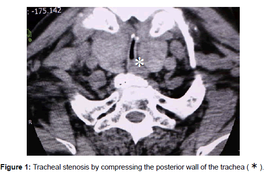
An 88-year-old Japanese man underwent cervical spondylotic surgery under general anesthesia at a referring hospital. He soon fell into dyspnea after extubation. He underwent tracheotomy to keep the airway patent and was transferred to our hospital. In the preoperative CT scan, tumors of the bilateral lobes of the thyroid were diffusely swollen and had grown into the space between the posterior wall of the trachea and esophagus, leading to the tracheal stenosis by compressing the posterior wall of the trachea .
Keywords: Tracheal Stenosis, Thyroid Tumor, Postoperative CT, Tracheomalacia
An 88-year-old Japanese man underwent cervical spondylotic surgery under general anesthesia at a referring hospital. He soon fell into dyspnea after extubation. He underwent tracheotomy to keep the airway patent and was transferred to our hospital.
In the preoperative CT scan, tumors of the bilateral lobes of the thyroid were diffusely swollen and had grown into the space between the posterior wall of the trachea and esophagus, leading to the tracheal stenosis by compressing the posterior wall of the trachea (Figure 1).
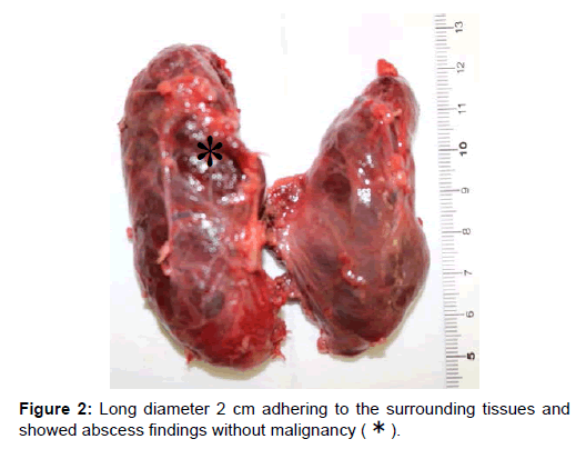
Moreover, an upper region of right thyroid lobe showed a low density area 2 cm in the long a diameter. We performed total thyroidectomy with preservation of the bilateral recurrent nerve. The postoperative histopathology was mostly adenomatous goiter in both lobes. By preoperative CT an upper region of the right thyroid lobe showed a low density area with the long diameter 2 cm adhering to the surrounding tissues and showed abscess findings without malignancy (Figure 2). Two regions (long diameter 4 mm and 12 mm) in the left lobe showed papillary carcinoma without capsular invasion.
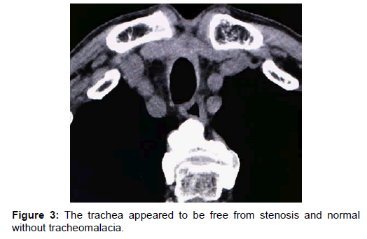
The progress after surgery was good, and there was no dyspnea or vocal cord paralysis. In the postoperative CT, the trachea appeared to be free from stenosis and normal without tracheomalacia (Figure 3).
Therefore, the stoma from the tracheotomy could be closed. Not all huge benign thyroid tumors with tracheal compression cause dyspnea. We reviewed the CT findings of tumors with dyspnea and found that tumors that occupied the space between the posterior wall of the trachea and esophagus lead to tracheal stenosis, as in our case [1,2].
References
- Sato K, Hanzawa H, Watanabe J, Takahashi S (2005) Differntial diagnosis and management of air way obstruction in Riedel’s thyroiditis: A case report. Auris Nasus Larynx 32: 439-443.
- Ayabe H, Kawahara K, Tagawa Y, Tomita M (1992) Upper airway obstruction from a benign goiter. Jpn J Surg 22: 88-90.
 Spanish
  Chinese
  Russian
  German
  French
  Japanese
  Portuguese
  Hindi
Hindi 


