Case Report, J Otol Rhinol Vol: 7 Issue: 1
Two Cases of Laryngeal Myxoma Misdiagnosed as a Vocal Fold Polyp
Mitsuyoshi Imaizumi*, Yasuhiro Tada, Akiko Tani, Masakazu Ikeda, Yuta Nakaegawa and Koichi Omori
Department of Otolaryngology, Fukushima Medical University, School of Medicine1 Hikarigaoka, Fukushima 960-1295, Japan
*Corresponding Author : Mitsuyoshi Imaizumi, MD
Department of Otolaryngology, Fukushima Medical University, School of Medicine1 Hikarigaoka, Fukushima 960-1295, Japan
Tel: +81-24-547-1321
Fax: +81-24-548-3011
E-mail: ima-mitu@fmu.ac.jp
Received: December 02, 2017 Accepted: January 08, 2018 Published: January 15, 2018
Citation: Imaizumi M, Tada Y, Tani A, Ikeda M, Nakaegawa Y, et al. (2018) Two Cases of Laryngeal Myxoma Misdiagnosed as a Vocal Fold Polyp. J Otol Rhinol 7:1. doi: 10.4172/2324-8785.1000332
Abstract
Laryngeal myxoma is a rare benign neoplasm of uncertain mesenchymal cell origin and often misdiagnosed as a vocal fold polyp [1]. The term ‘myxoma’ was introduced by Virchow in 1871. The histological findings of myxoma were described to resemble a mucinous substance of the umbilical cord [2]. To the best of our
knowledge, there are only eleven reported cases to date [1,3]. Here, we report two cases of laryngeal myxoma. Direct laryngomicrosurgery was performed under general anesthesia. The lesions have been stable without recurrence. Myxoma has a high incidence of local recurrence, because myxoma tends to infiltrate the surrounding tissue [4]. Further follow up might be necessary.
Keywords: Laryngeal myxoma, Myxoma, Laryngomicrosurgery
Introduction
Laryngeal myxoma is a rare benign neoplasm of uncertain mesenchymal cell origin and often misdiagnosed as a vocal fold polyp [1]. The term ‘myxoma’ was introduced by Virchow in 1871. The histological findings of myxoma were described to resemble a mucinous substance of the umbilical cord [2]. To the best of our knowledge, there are only eleven reported cases to date [1,3]. Here, we report two cases of laryngeal myxoma. Direct laryngomicrosurgery was performed under general anesthesia. The lesions have been stable without recurrence. Myxoma has a high incidence of local recurrence, because myxoma tends to infiltrate the surrounding tissue [4]. Further follow up might be necessary.
This study was approved by the Institutional Review Board of Fukushima Medical University on November 19th, 2013 (Confirmation Number: #1873), which is guided by local policy, national laws, and the World Medical Association Declaration of Helsinki.
Case Report 1
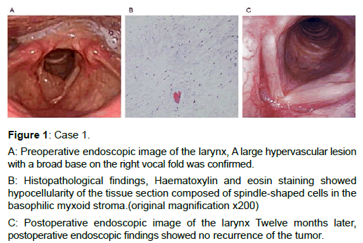
A 44-year-old man consulted a local ENT clinic due to five years of hoarseness and was diagnosed as having a vocal fold polyp. He was subsequently referred to our department. Examination by laryngeal flexible endoscopy (CLV-S40Pro, ENF-V2, OLYMPUS) revealed a large hypervascular lesion with a broad base on the right vocal fold (Figure 1A). No mucosal waves were observed on the mass on stroboscopic examination. Direct laryngomicrosurgery was performed under general anesthesia. An elastic hard mass was confirmed, which was suspected to be a tumor. The suspected laryngeal tumor was completely removed from the right vocal fold. The histopathological findings of the excised tumor were consistent with those of laryngeal myxoma (Figure 1B). After surgery, an improvement in voice quality was observed by vocal function analysis. Twelve months later, postoperative endoscopic findings showed no recurrence of the right vocal fold tumor (Figure 1C). Stroboscopic examination revealed an improvement in the vibratory amplitude and mucosal wave. Perceptual evaluation of the grade, roughness, breathiness, asthenia, strain (GRBAS) scales, mean airflow rate (MFR), AC/DC ratio using phonation analyzer PA-1000 (Minato Medical Science Co., Ltd, Osaka, Japan) and acoustic analysis using the Multi-Dimensional Voice Program (MDVP, Kay Elemetrics [KayPentax], Lincoln Park, NJ) to assess F0, Jitter (jitt), shimmer (shim), noise-to-harmonics ratio (NHR) were also improved (Table 1). No postoperative complications were observed, and no recurrence of the tumor has been observed for one and a half years.
Figure 1: Case 1.
A: Preoperative endoscopic image of the larynx, A large hypervascular lesion with a broad base on the right vocal fold was confirmed.
B: Histopathological findings, Haematoxylin and eosin staining showed hypocellularity of the tissue section composed of spindle-shaped cells in the basophilic myxoid stroma.(original magnification x200)
C: Postoperative endoscopic image of the larynx Twelve months later, postoperative endoscopic findings showed no recurrence of the tumor.
| Measure | pre-operation | post-operation |
|---|---|---|
| Perceptual | ||
| GRBAS | G2 R1 B2 A0 S0 | G0 R0 B0 A0 S0 |
| Aerodynamic | ||
| MPT (sec) | 24 | 23 |
| AC/DC ratio (%) | 32.2 | 68.5 |
| MFR(mL/s) | 227 | 178 |
| Acoustic | ||
| F0 (Hz) | 152.418 | 105.276 |
| Jitt (%) | 0.615 | 0.817 |
| Shim (%) | 4.32 | 1.784 |
| NHR | 0.137 | 0.131 |
MPT = maximum phonation time
AC/DC ratio = vocal efficiency index (alternating current/direct current [AC/DC])
MFR=mean airflow rate
F0= fundamental frequency
Jitt = jitter
Shim = shimmer
NHR = noise-to-harmonics ratio
Table 1: Examinations of voice before and after surgery.
Case Report 2
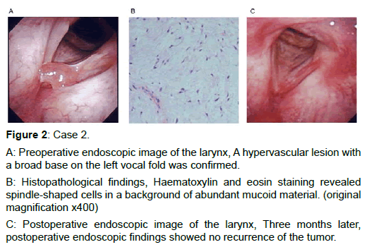
A 77-year-old man presented with one and a half years of hoarseness upon consultation at a local ENT clinic where he was diagnosed as having a vocal fold polyp. He was subsequently referred to our department. Examination by laryngeal flexible endoscopy revealed a hypervascular lesion with a broad base on the left vocal fold (Figure 2A). No mucosal waves were observed on the mass on stroboscopic examination. Direct laryngomicrosurgery was performed under general anesthesia. The laryngeal tumor was completely removed from the left vocal fold. The histopathological findings of the excised tumor were consistent with those of laryngeal myxoma (Figure 2B). After surgery, an improvement in voice quality was observed by vocal function analysis. Three months later, postoperative endoscopic findings showed no recurrence of the left vocal fold tumor. (Figure 2C). Stroboscopic examination revealed an improvement in the vibratory amplitude and mucosal wave. Maximum phonation time (MPT), perceptual evaluation of the GRBAS scale, MFR, AC/DC ratio using phonation analyzer PA-1000 and acoustic analysis using the Multi-Dimensional Voice Program to assess F0, Jitter (jitt), shimmer (shim), noise-to-harmonics ratio (NHR) were also improved (Table 2). No postoperative complications were observed, and no recurrence of the tumor has been observed for three months.
Figure 2: Case 2.
A: Preoperative endoscopic image of the larynx, A hypervascular lesion with a broad base on the left vocal fold was confirmed.
B: Histopathological findings, Haematoxylin and eosin staining revealed spindle-shaped cells in a background of abundant mucoid material. (original magnification x400)
C: Postoperative endoscopic image of the larynx, Three months later, postoperative endoscopic findings showed no recurrence of the tumor.
| Measure | pre-operation | post-operation |
|---|---|---|
| Perceptual | ||
| GRBAS | G2 R2 B1 A0 S0 | G1 R1 B0 A0 S0 |
| Aerodynamic | ||
| MPT (sec) | 6 | 15 |
| AC/DC ratio (%) | 22.3 | 57.6 |
| MFR(mL/s) | 583 | 302 |
| Acoustic | ||
| F0 (Hz) | 211.774 | 150.464 |
| Jitt (%) | 1.363 | 0.777 |
| Shim (%) | 14.803 | 3.745 |
| NHR | 0.182 | 0.149 |
MPT = maximum phonation time
AC/DC ratio = vocal efficiency index (alternating current/direct current [AC/DC])
MFR=mean airflow rate
F0= fundamental frequency
Jitt = jitter
Shim = shimmer
NHR = noise-to-harmonics ratio
Table 2: Examinations of voice before and after surgery.
Discussion
Laryngeal myxoma generally presents as a large polypoid mass of the vocal fold. It is for this reason that in both of our cases the patients were misdiagnosed as having a vocal fold polyp at a local ENT clinic. Myxoma is a rare benign tumor of uncertain mesenchymal cell origin, typically involving the heart. Myxomas of the head and neck most commonly occur in the mandible and maxilla, and are likely to be odontogenic in origin [1]. They arise innocuously as a painless, slow growing mass [5]. Furthermore, myxomas lack a fibrous capsule and tend to infiltrate the surrounding tissue. Therefore, they have a high incidence of local recurrence. Surgical excision together with a rim of surrounding tissue has been reported to reduce the risk of recurrence [6]. In the present case, the findings of preoperative stroboscopic examination revealed that the lesion could be different from a usual vocal polyp, and a laryngeal benign tumor was considered as one of the differential diagnoses. Therefore, we completely excised the mass, not as a laryngeal polyp, but as a laryngeal tumor. The vocal fold has since remained tumor free. In a previous paper reported by Sena T et al. [5], local recurrence may occur during the first three years postoperatively. Because we did not excise the mass together with a rim of surrounding tissue, further follow up might be necessary.
The histologic differential diagnosis includes myxoid liposarcoma, myxoid chondrosarcoma, and laryngeal polyp [4]. In lipoblasts and chondroblast, S-100 protein is positive. In our Case 1, immunohistochemical analysis showed the absence of S-100 protein (data not shown). More than half of myxomas have been reported to show immunoreactivity for CD34 [7]. In our Case 1, immunohistochemical analysis showed the presence of CD34 (data not shown). As Ritchie et al. [1] reported, a laryngeal polyp is the entity most difficult to distinguish from a laryngeal myxoma. When examination by laryngeal flexible endoscopy and stroboscopy suggests laryngeal tumors, biopsies should be considered in order to avoid misdiagnosis.
Conclusion
We report two cases of laryngeal myxoma misdiagnosed as a vocal fold polyp. It is important to keep in mind that laryngeal myxoma often shows findings and characteristics of vocal fold polyp.
References
- Ritchie A, Youngerman J, Fantasia JE, Kahn LB, Cocker RS (2013) Laryngeal myxoma: a case report and review of the literature. Head Neck Pathol 8: 204-208.
- Chen KT, Ballecer RA (1986) Laryngeal myxoma. Am J Otolaryngol 7: 58-59.
- Tang CG, Monin DL, Puligandla B, Cruz RM (2015) Glottic myxoma presenting as chronic dysphonia: a case report and review of the literature. Ear Nose Throat J 94: E30-33.
- Nakamura A, Iguchi H, Kusuki M, Yamane H, Matsuda M, et al. (2008) Laryngeal myxoma. Acta Otolaryngol 128:110-112.
- Sena T, Brady MS, Huvos AG, Spiro RH (1991) Laryngeal myxoma. Arch Otolaryngol Head Neck Surg 117: 430-432.
- Hadley J, Gardiner Q, Dilkes M, Boyle M (1994) Myxoma of the larynx: a case report and a review of the literature. J Laryngol Otol 108: 811-812.
- Idrees MT, Hessler R, Terris D, Mixson C, Wang BY (2005) Unusual polypoid laryngeal myxoma. Mt Sinai J Med 72: 282-284.
 Spanish
  Chinese
  Russian
  German
  French
  Japanese
  Portuguese
  Hindi
Hindi 


