Research Article, Biomater Med Appl Vol: 1 Issue: 2
Zinc-doped Calcium Phosphate Coating on Titanium Surface Using Ostrich Eggshell as a Ca2+ Ions Source
Ferreira JRM1,2, Louro LHL2, Costa AM2, Marçal RLSB2, Navarro da Rocha D2, Barbosa RM1,4, Campos JB3 and Prado da Silva MH2*
1R-CRIO Criogenia S.A., Rua Cumaru 204, CEP 13098-324, SP, Brazil
2Military Institute of Engineering-IME, Materials Science Post Grad Programme, Pça Gen. Tiburcio, 80, Praia Vermelha, Urca, Rio de Janeiro, R.J., Brazil
3Universidade do Estado do Rio de Janeiro - UERJ, Rua Fonseca Teles, 121, 2ºandar, São Cristovão, Rio de Janeiro, RJ, Brazil
4Universidade Estadual de Campinas - UNICAMP, Av. Albert Einstein, 500, Campinas, São Paulo, SP, Brazil
*Corresponding Author : Marcelo H Prado da Silva
Instituto Militar de Engenharia – IME, Pça. Gen. Tibúrcio, 80, P. Vermelha, Urca, Rio de Janeiro, R.J, Brazil
E-mail: marceloprado@ime.eb.br
Received: September 13, 2017 Accepted: October 13, 2017 Published: October 20, 2017
Citation: Ferreira JRM, Louro LHL, Costa AM, Marçal RLSB, Navarro da Rocha D, et al. (2017) Zinc-doped Calcium Phosphate Coating on Titanium Surface Using Ostrich Eggshell as a Ca2+ Ions Source. Biomater Med Appl 1:2.
Abstract
Natural calcium source materials can be thermochemically processed and directly transformed into hydroxyapatite. In this work, ostrich eggshell was used as Ca2+ ions source in the preparation of zinc-calcium phosphate coatings on Ti substrates promoted by a two-step deposition method. The obtained coatings were characterized by X-ray diffraction (XRD) analysis, scanning electron microscopy with field emission gun (FEG-SEM), bioactivity using McCoy medium and toxicity with chicken embryo chorioallantoic membrane (CAM). The results showed the presence of Znbrushite and Zn-hydroxyapatite as coating phases. Bioactivity was observed in all samples showing bone-like apatite on their surface. The toxicity test showed inflammatory response between mild to moderate confirming the material’s biocompatibility. The use of ostrich eggshells was shown to be effective as a Ca2+ ions source to obtain zinc-containing hydroxyapatite coatings on Ti substrates.
Keywords: Calcium phosphates; Zinc ion substitution; Ostrich eggshell; Bioactivity; Toxicity
Introduction
The rehabilitation of mutilated dentition has always represented a great challenge in implantology and restorative dentistry. The modification of implant surfaces (e.g. microroughness, nanostructures features and wettability) have demonstrated improvement of osseointegration phenomena and changed the dynamics of the surgical procedures [1]. Nowadays, a high degree of previsibility and patient satisfaction can be achieved to many patients. However, improvement of biological response from dental implants is still necessary for those patients with functionally debilitated condition (e.g. uncontrolled diabetes) or inadequate bone dimension [1,2]. The concept of a new implant design applied to health area involves the different material properties (thread size and length, surface energy and morphology, screw design) and surgical procedures (prognostic tool and socket preservation techniques) to prevent structural/functional failure, bacterial infections and immune responses [2,3].
On the 80´s, Branenmark defined the term osseointegration as the process whereby bone grows directly on implant surfaces, allowing implant anchorage [4]. The Osseointegration phenomena has gained better understanding of biological processes surrounding the implant surface. Recent approaches consider the presence of ions and proteins in the bone-metal interface, followed by biological signaling that promotes cell recruitment and the formation of a new extracellular matrix in the interface [5,6]. In fact, the implant must also be “osteoconductive” i.e., allow bone growth on its surface [6]. However, the concept of bioactivity establishes another level of bone-implant bonding: osteoconductivity with chemical bonding [7,8]. In the 90´s Kokubo et al. [7] reported synthetic body fluid solutions able to reproduce in vivo surface-structure changes in bioactive glass-ceramic. This process involves mainly ion exchange and dissolution-precipitation mechanisms, and promotes bonelike apatite formation [9]. Recent trends on bioactive coatings with bioactive glass-ceramic or calcium phosphate coatings may include the concept of osteoinduction [10,11]. Nowadays clinical trials for hip and ankle prosthesis demonstrated promising results using dicalcium phosphate dihydrate (CaHPO4.2H2O, brushite) coatings. This phase is more soluble than tricalcium phosphate (TCP) and, when immersed in physiological fluids, could promote also an appetite formation [10].
Zinc (Zn) is a biologically relevant ion. The effect of Zn presence on calcium phosphate coatings produced by a two-step hydrothermal treatment was investigated in a recent study [12]. Zn-containing bioactive coatings produced by various techniques have been reported and characterized recently [12-15]. Besides the biological relevance of Zn, this element influences nucleation and crystal growth affecting coating morphology, density and homogeneity [12-15]. According to Zn content, Zn-containing hydroxyapatite can show modified thermal stability when compared to pure HA. Miyaji et al. [16] reported the effect of Zn addition on HA lattice parameter: the lattice parameter “a” is reduced with the addition of up to 5 mol% Zn, and starts to increase over 5 mol% Zn. The more the lattice parameter is displaced, the lower thermal stability is [17].
Natural source materials can be thermochemically processed, being directly transformed into hydroxyapatite. In a recent study, Ferreira et al. [18] used eggshell as a calcium source for the synthesis of pure hydroxyapatite. Similarly, Zhao et al. [19] used cytidine 5′-triphosphate disodium salt as a phosphate source in a microwaveassisted hydrothermal rapid synthesis of amorphous calcium phosphate nanoparticles and hydroxyapatite microspheres.
Experimental
Zn-calcium phosphate deposition
In this work, ostrich eggshell was used as Ca2+ ions source in the preparation of zinc-calcium phosphate coatings on Ti substrates and the deposition method was a two-step route, based on previous studies [20-22]. The precursor solution was prepared by mixing three solutions: 0.5 mol/L Ca(OH)2 and 0.3 mol/L H3PO4 and 1 mol/L lactic acid. Briefly, ostrich eggshell powder obtained by maceration and sieving was dispersed in deionized water under stirring in order to produce a 0.5 mol/L CaO3 solution, according to the route previous described [18]. Then, a 1 mol/L lactic acid solution was dropped to the eggshell dispersion solution under stirring. To obtain a CaP precursor solution with 2% Zn (mol Zn/mol Ca), zinc nitrate Zn(NO3)2 with 98% purity was added to the solution considering a theoretical calcium site substitution of 2% Zn in the HA structure. Finally, a 0.3 mol/L H3PO4 solution was slowly dropped to the obtained solution, and a pH around 3 was reached. This precursor solution was left under stirring for 24 hours at room temperature.
The first step consists of a chemical deposition method, and the titanium substrates are immersed into the saturated precursor solution and heated at 80 ºC, during 4 hours, to precipitate a zinccalcium phosphate coating. In the second step, so called conversion, the samples are left a chemical bath of 0.1 mol/L potassium hydroxide (KOH) at 60 ºC for a period of 24 hours, to convert the deposited Znacidic calcium phosphate to Zn-doped hydroxyapatite.
Characterization
The obtained coatings were characterized by X-ray diffraction (XRD) analysis, scanning electron microscopy with field emission gun (FEG-SEM), bioactivity and toxicity.
XRD analysis was performed in a PANalytical X’Pert X-ray diffraction system, operating with CuKα (λ = 0.1542 nm) with 45 kV and 40 mA, step 0.05° at 100 seconds per step from 10° to 80°.
FEG-SEM analysis was performed in a FEI Quanta 250 FEG, operating on high and low vacuum modes, between 1 and 30kV.
The bioactivity assessment was performed into McCoy solution analyzing the apatite formation and modification on the coatings surface. The samples were autoclaved and immersed in 20 ml of McCoy solution. The tests were performed at an incubator (Panasonic, COM- 19AIC-PA) at 37ºC, at 5% CO2 atmosphere, for 14 days.
The toxicity test was performed in the chicken embryo chorioallantoic membrane (CAM) that is a preclinical model widely used for short-term toxicity bioassays, recognized as an intermediate model that can bridge the gap between cell and animal assessment. The experiment was carried out in triplicate, in a total of 4 fertile Specific-Pathogen-Free chicken eggs.
Fresh fertilized chicken eggs were disinfected with ethanol. The eggs were then incubated with the narrow apex down in a 90° swinging incubator at 37°C and 65% relative humidity for embryogenesis (day 1). On day 3, a “window” on the apex of the eggs was performed and then sealed with transparent adhesive tape to avoid contamination. The eggs were then further incubated in a stagnant position with the apex upright until day 11. The viability of the embryos and the vasculature of the CAM were then visually inspected and were used for Zn-hydroxyapatite powder deposition. 70 mg of Zn-HA powder, previously sterilized by autoclaving at 121ºC for 20 minutes, was deposited on the chorioallantoic membrane with the aid of a small sterile device. The eggs were closed with transparent adhesive tape (polypropylene) and incubated again. After 07 days of inoculation, the eggs were removed from the incubator. All chorioallantoic membranes were removed, identified, immersed in 10% formalin, embedded in paraffin, sectioned, and finally stained with H&E for histopathological analysis.
Results and Discussion
XRD analyses
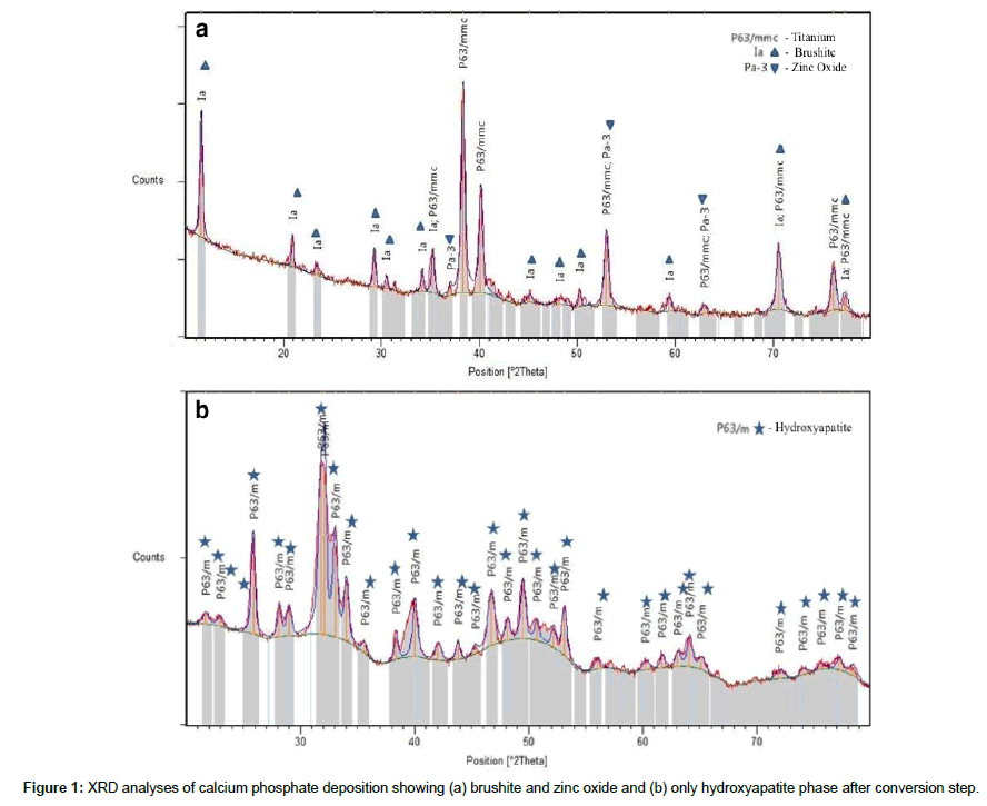
After the deposition first step, the XRD patterns showed brushite as the only calcium phosphate phase on the surface of Ti substrates (Figure 1a). However, a minor phase, zinc oxide was observed, probably as an excess of unreacted zinc. As expected, after the conversion the XRD showed only hydroxyapatite as the only calcium phosphate phase (Figure 1b). The absence of zinc oxide is explained by the process of conversion, performed into a chemical bath with a strong alkali solution that dissolves the oxide, contributing to the dissolution-precipitation conversion mechanism.
FEG-SEM analysis and bioactivity assessment
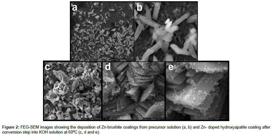
FEG-SEM analysis, Figure 2 (a, b), showed platelet crystals on all samples, in accordance to the literature, characteristic of brushite crystals morphology [21,22]. These results indicated a Zn influence on the precipitation of acidic calcium phosphate in this deposition method, in opposition to the results found by Prado da Silva et al. [20], that indicated the formation of monetite coatings, instead of brushite.
After the second step, in an alkali chemical bath, the coatings showed a nanometric needle-like morphology, characteristic of HA, on the former brushite crystal surface, as can be seen in the Figure 2 (c, d, and e). HA is the thermodynamically stable calcium phosphate phase under highly alkali pH level, since, PO43- is the ion with high relative concentrations in these conditions, among the four polymorphs of phosphoric acid used in the present study [23].
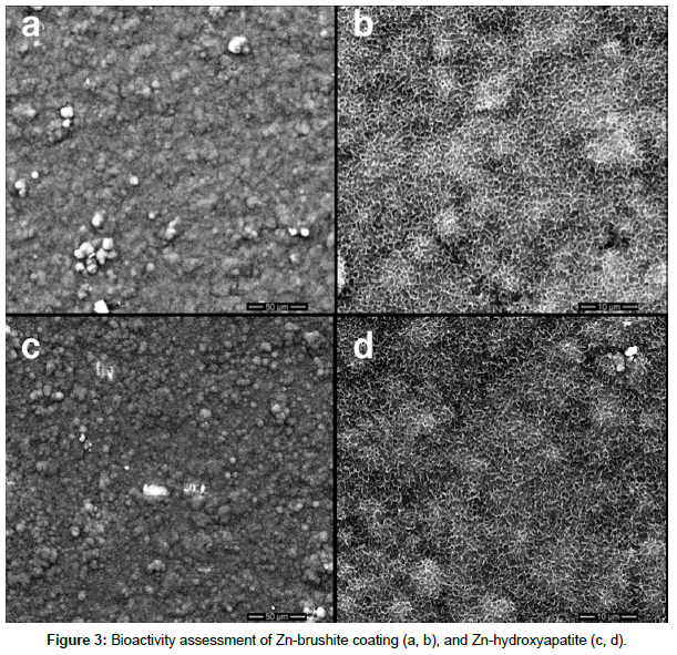
Figure 3 shows the results of bioactivity assessment. After 14 days, the precipitation of bone-like apatite on the surface of Zn-brushite and Zn-hydroxyapatite coatings was observed. The globule-like morphology is characteristic of bioactive surfaces when immersed into SBF solution. The precipitation of this thin layer is an essential prerequisite for biomaterials to make a direct and chemical bond with living bone [24].
Zn-brushite phase is a metastable calcium phosphate and hence transforms into crystalline bone-like apatite. The incorporation of Zn ions into hydroxyapatite structure is known to induce distortions in its crystal lattice and lead to the Ca-deficient hydroxyapatite, increasing its solubility and bioactivity. In a previous work, Marçal et al. [22] studied the deposition of calcium phosphates on titanium plates, it was found that brushite/monetite coatings did show bioactivity, whereas, pure hydroxyapatite did not.
Toxicity assessment
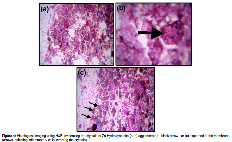
Figure 4 shows the presence of a mild to moderate inflammatory response and absence of fibroplasia, which is desired in regeneration processes. Histological images using H&E demonstrated inflammatory cells involving the crystals of Zn-Hydroxyapatite, dispersed in the membrane (arrows in the Figure 4(c)). Note that the HA crystals can also be agglomerated Figure 4(b).
According to VALDES et al. [25] and ZIJLSTRA et al. [26], the inflammatory processes, hyperemia (increased blood volume in the region) and angiogenesis are expected. These conditions are related to the absence of toxicity. The presence of inflammatory cells in a mild response is fundamental for the activation of endothelial factors that allow angiogenesis process, which is essential in clinical events of wound repair or regeneration.
Conclusion
The use of ostrich eggshells was shown to be effective as a Ca2+ ions source to obtain zinc-containing hydroxyapatite coating on Ti substrates. The two-step method showed to be efficient to produce Zn-brushite coatings and after conversion Zn-hydroxyapatite.
Titanium plates coated with Zn-brushite and Zn-hydroxyapatite showed satisfactory bioactivity with the precipitation of bone-like apatite.
The obtained powder showed no toxicity on CAM test, exhibiting an inflammatory response between mild to moderate, a characteristic response for absence of toxicity.
Acknowledgements
The authors would like to acknowledge CNPq, CAPES, IME and R-Crio for the support. The authors declare no conflict of interest.
References
- Gittens RA, Olivares-Navarrete R, Schwartz Z, Boyan BD (2014) Implant Osseointegration and the Role of Microroughness and Nanostructures: Lessons for Spine Implants. Acta Biomater 10: 3363-3371.
- Dubey RK, Gupta DK, Singh AK (2013) Dental implant survival in diabetic patients; review and recommendations. Natl J Maxillofac Surg 4: 142-150.
- Chappuis V, Araújo MG, Buser D (2017) Clinical relevance of dimensional bone and soft tissue alterations post-extraction in esthetic sites. Periodontol 73: 73-83.
- Branemark PI (1983) Osseointegration and its experimental background. J Prosthet Dent 50: 399-410.
- Albrektsson T, Wennerberg A (2004) Oral implant surfaces: Part 1-review focusing on topographic and chemical properties of different surfaces and in vivo responses to them. Int J Prosthodont 17: 536-543.
- Albrektsson T, Wennerberg A (2004) Oral implant surfaces: Part 2-review focusing on clinical knowledge of different surfaces. Int J Prosthodont 17: 544-564.
- Kokubo T (1998) Apatite formation on surfaces of ceramics, metals and polymers in body environment. Acta Mater 46: 2519-2527.
- Kokubo T, Yamaguchi S (2010) Novel Bioactive Titanate Layers Formed on Ti Metal and Its Alloys by Chemical Treatments. Materials 3: 48-63.
- El-Ghannam A, Ning CQ (2006) Effect of bioactive ceramic dissolution on the mechanism of bone mineralization and guided tissue growth in vitro. J Biomed Mater Res A 76: 386-397.
- Zhang BGX, Myers DE, Wallace GG, Brandt M, Choong PFM (2014) Bioactive Coatings for Orthopaedic Implants-Recent Trends in Development of Implant Coatings. Int J Mol Sci 15: 11878-11921.
- Ripamonti U, Roden LC, Renton LF (2012) Osteoinductive hydroxyapatite-coated titanium implants. Biomaterials 33: 3813-3823.
- Prado da Silva MH, Moura FN, Navarro da Rocha D, Gobbo LA, Costa AM, et al. (2014) Zinc-modified hydroxyapatite coatings obtained from parascholzite alkali conversion. Surf Coat Technol 249: 109-117.
- Furko M, Jiang Y, Wilkins T, Balázsi C (2016) Development and characterization of silver and zinc doped bioceramic layer on metallic implant materials for orthopedic application. Ceram Int 42: 4924-4931.
- Huang Y, Zhang X, Mao H, Li T, Zhao R, et al. (2015) Osteoblastic cell responses and antibacterial efficacy of Cu/Zn co-substituted hydroxyapatite coatings on pure titanium using electrodeposition method. RSC Adv 5: 17076-17086.
- Guangfei S, Jun M, Shengmin Z (2014) Electrophoretic deposition of zinc-substituted hydroxyapatite coatings. Mater Sci Eng C. 39: 67-72.
- Miyaji F, KonoY, Suyama Y (2005) Formation and structure of zinc-substituted calcium hydroxyapatite. Mater Res Bull 40: 209-220.
- Moreira MP, Soares GDA, Dentzer J, Anselme K, Sena LA, et al. (2016) Synthesis of magnesium-and manganese-doped hydroxyapatite structures assisted by the simultaneous incorporation of strontium. Mater Sci Eng C61: 736-743.
- Ferreira JRM, Navarro da Rocha D, Louro LHL, Prado da Silva MH (2014) Phosphating of Calcium Carbonate for Obtaining Hydroxyapatite From the Ostrich Egg Shell. Key Eng Mater 587: 69-73.
- Zhao J, Zhu YJ, Cheng GF, Ruan YJ, Sun TW, et al. (2014) Microwave-assisted hydrothermal rapid synthesis of amorphous calcium phosphate nanoparticles and hydroxyapatite microspheres using cytidine 5′-triphosphate disodium salt as a phosphate source. Mater Lett 124: 208-211.
- Prado da Silva MH, Lima JHC, Soares GA, Elias CN, de Andrade MC, et al. (2001) Transformation of monetite to hydroxyapatite in bioactive coatings on titanium. Surf Coat Technol 137: 270-276.
- Navarro da Rocha D ,Cruz LRdO, de Campos JB, Marçal RLSB, Mijares DQ, et al. (2017) Mg substituted apatite coating from alkali conversion of acidic calcium phosphate. Mater Sci Eng 70:408-417.
- Marçal RLSB, Ferreira JRM, Louro LHL, Costa AM, Navarro da Rocha D, et al. (2017) Apatite Coatings from Ostrich Eggshell and its Bioactivity Assessment. Key Engineering Materials 720: 185-188.
- Lynn AK, Bonfield W (2005) A novel method for the simultaneous, titrant-free control of pH and calcium phosphate mass yield. Acc Chem Res 38: 202-207.
- Davies JE (2003) Understanding peri-implant endosseous healing. J Dent Educ 67: 932-949
- Valdes TI, Kreutzer D, Moussy F (2002) The chick chorioallantoic membrane as a novel in vivo model for the testing of biomaterials. J Biomed Mater Res. 62: 273-282.
- Zijlstra A, Seandel M, Kupriyanova TA, Partridge JJ, Madsen MA, et al. (2006) Proangiogenic role of neutrophil-like inflammatory heterophils during neovascularization induced by growth factors and human tumor cells. Blood.107: 317-327.
 Spanish
  Chinese
  Russian
  German
  French
  Japanese
  Portuguese
  Hindi
Hindi 