Review Article, Clin Dermatol Res J Vol: 3 Issue: 1
A Study on Stability of Acrofacial Vitiligo
Megha Chandrashekar* and Umashankar Nagaraju
Rajarajeshwari medical college, RGUHS, Karnataka, India
*Corresponding Author : Megha Chandrashekar
Rajarajeshwari medical college, RGUHS, Karnataka, India
Tel: +918095196167
E-mail: mghzoots@gmail.com
Received: December 19, 2017 Accepted: January 11, 2018 Published: January 16, 2018
Citation: Chandrashekar M, Nagaraju U (2018) A Study on Stability of Acrofacial Vitiligo. Clin Dermatol Res J 3:1. doi: 10.4172/2576-1439.1000121
Abstract
Introduction: Acrofacial vitiligo being cosmetically disfiguring, can affect patients self-esteem, employability and interaction with society. The resistant nature if this form of vitiligo adds to anger
and disillusionment in the affected individual. The defiant form in addition to its unstable nature necessitates the further study on acrofacial vitiligo.
Objectives: To determine the clinical morphology with respect to stability of Vitiligo. Materials and Methods: A cross sectional study was done over a period of one year comprising of patients with acro-facial vitiligo. Relevant investigations including histopathology and patch test were done wherever necessary.
Results: A total of 191 cases of acrofacial vitiligo were included in the study. Among stable acral vitiligo (59.48%), ill-defined margins were seen in 84.61% and dorsum was the commonest
site (23.57%). Among unstable Vitiligo, joint was predominantly involved (27.03%).
Conclusion: Being prone for injuries, acrofacial areas are unstable. Well defined margins do not strike to the stability of acral vitiligo and signs of activity can be seen even in stable vitiligo. Hence unlike vitiligo of other areas, where well defined margins can be a marker of stable Vitiligo, margins may not be a scale for defining the stability of acral Vitiligo. Involvement of bony prominences could indicate unstable course and least involvement of proximal area of the phalanges may indicate its unstable nature.
Keywords: Acrofacial vitiligo; Stability; Morphology
Introduction
Pigmentary changes of skin particularly vitiligo can be psychologically devastating, affecting individual’s self-esteem, employability and interaction with people which are due to the disgrace associated with it and the fear of being an outcaste. This stigma is more commonly encountered with depigmentary disorders particularly when it affects exposed parts of the body. It is more frequent in darker skin type like that of Indians as there is a striking color contrast between the affected skin and the normal skin.
Acro-facial areas include the dorsum of hands and feet, fingers, toes, palms, soles and periorificial areas of face [1]. Vitiligo is the most common depigmenting disorder, which affects 0.5-1% of the worldwide population [2]. Recently, the prevalence of acrofacial depigmentation is on rise. This may be attributed to increased social and cosmetic awareness; increased immigrants, occupational and household exposure to products which are irritant to skin.
The depigmentation occurring secondary to Vitiligo typically affects the exposed areas like the fingers, knuckles, around the eyes and mouth [3]. Resistance to treat acral vitiligo is basically due to unstable nature of its stability leading for further challenges which need to be answered. Also the stability of Vitiligo may vary with different areas in same individual hence, detailed study on stability and its association with morphological aspects of acrofacial vitiligo is important which is lacking in English literature. We attempted to address all these issues in our study.
Objectives of the Study
To determine the clinical morphology with respect to the stability of acral vitiligo.
Methodology
The present study was a cross-sectional study conducted over a period of one year from January 2013 to December 2013. The study population comprised of all patients presenting with acro-facial vitiligo. The sample size was 191 cases.
Inclusion criteria
Patients of all age groups and both the sexes presenting with acrofacial Vitiligo were included.
Exclusion criteria
Vitiligo universalis and segmental vitiligo other than acro-facial areas were excluded.
Ethical clearance for the present study was obtained from the Institutional Ethical Committee Rajarajeshwari Medical College and Hospital Bangalore.
In the present study, detailed history regarding occupational exposure, onset of lesions, infection, insect bites, trauma and reaction following topical medications, surgeries and family history of similar lesions was elicited, this was followed by cutaneous examination in all cases presenting with acrofacial vitiligo. This was followed by complete cutaneous and systemic examination. Written consent from the patient or from the parent/guardian in case of minor was obtained. Blood investigations including complete haemogram, Erythrocyte Sedimentation Rate, liver function test, blood Urea, serum creatinine, and urine examination was done in all patients. Anti-Herpes simplex 1 and 2 IgG /IgM antibodies and Tzanck smear was done in presence of depigmentation associated with vesicles. Patch test was performed if depigmentation is suspected to contact dermatitis. Digital photography of the depigmentary lesions was taken in all patients. In doubtful cases, skin biopsy for histopathological evaluation, was carried out to establish the diagnosis.
Data analysis
The data collected in this study was analyzed statistically by entering the data in MS Excel. The quantitative variables were described by computing Mean, SD, percentages. The qualitative variables were described by computing proportion and percentages. The association between two categorical variables was found by computing Chisquare test and where ever the expected cell frequencies in a 2 × 2 contingency tables less than 5. The Fishers exact probabilities were computed. The differences between two proportions were tested using Standard Normal Test (Z-test). The results were considered statistically significant whenever the p-value is ≤ 0.05 and statistically highly significant whenever it is ≤ 0.001.
Results and Observations
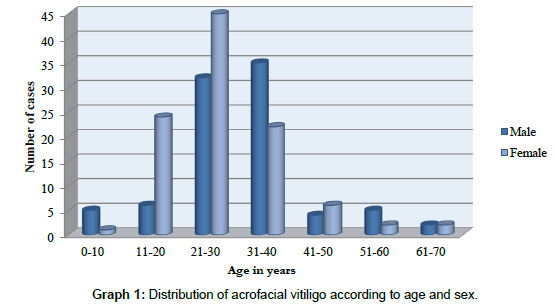
Maximum number of patients 127 (36.28%) belonged to third decade, followed by 96 (27.43%) in the fourth decade. Least was seen in the first decade 13(3.71%).
Male to female ratio was 1.1:1.= (Graph 1).
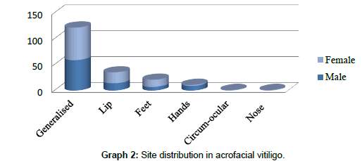
Out of the 191 cases of acrofacial vitiligo, Generalized vitiligo involving more than one site was seen in 122(63.87%) and localized vitiligo was seen in 69 (38.13%). Among localized vitiligo, face- 37 (53.62%), lip formed the major group 35 (50.72%); followed by feet 21(30.43%); hand 11 (15.94%), nose 1 (1.45%) and none were seen involving the ears (Table 1) (Graph 2).
| Vitiligo site | Male | Female | Total (%) | ||
|---|---|---|---|---|---|
| Generalized | 59 | 63 | 122(63.87) | ||
| Localized | Lip | 14 | 21 | 35 (50.72) | 69 (38.13) |
| Feet | 07 | 14 | 21(30.43) | ||
| Hands | 09 | 02 | 11 (15.94) | ||
| Circum-ocular | 00 | 01 | 01 (1.45) | ||
| Nose | 00 | 01 | 01 (1.45) | ||
| Total | 89 | 102 | 191(100) | ||
Table 1: Site distribution in acrofacial vitiligo.
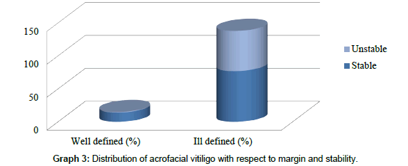
To assess the stability emphasis was laid on the morphology of acral vitiligo, Out of 153 cases of acral vitiligo, 91cases (59.48%) had stable vitiligo; 62(40.52%) had unstable vitiligo. Out of 91 cases of stable vitiligo, 77(84.61%) had ill-defined margins and 14 (15.38%) had well defined margins.
Out of 62 cases of unstable vitiligo, 61 (98.39%) cases had ill-defined margins and 1 (1.61%) had well defined margins. Chi –square test- 7.909, p-value ≤ 0.001 and is highly statistically significant (Table 2).
| Clinical stability | Well defined (%) | Ill defined (%) | Total (%) |
|---|---|---|---|
| Stable | 14 (15.38) | 77 (84.61) | 91 (59.48) |
| Unstable | 1(1.61) | 61 (98.39) | 62 (40.52) |
| Total | 15 | 138 | 153 (100) |
Table 2: Distribution of acrofacial vitiligo with respect to margin and stability.
In stable acral vitiligo involving the hand, combined distribution was predominantly noted in 40(32.52%), followed by involvement of the dorsum in29 (23.57%). In unstable acral vitiligo, dorsum was predominantly involved in 22(17.88%), followed by combined distribution in 21(17.07%) cases.
In stable acral vitiligo involving the feet, dorsum was predominantly involved and was seen in 47(36.15%) cases, followed by combined involvement in 31 (23.84%)
In unstable acral vitiligo too, dorsum was predominantly involved in 39(30%), followed by combined distribution in 7(5.38%) cases (Table 3) (Graph 3).
| Stability | Hand (%) | Feet (%) | ||||||
|---|---|---|---|---|---|---|---|---|
| Dorsum | Palm | Combined | Total | Dorsum | Palm | Combined | Total | |
| Stable | 29(23.57) | 8(6.50 ) | 40 (32.52 ) | 77(66.60) | 47(36.15) | 5(3.84) | 31(23.84) | 83(63.84) |
| Unstable | 22(17.88) | 3( 2.43) | 21(17.07 ) | 46(37.39) | 39(30) | 1(0.77) | 7 (5.38) | 47(36.15) |
| Total | 51(41.46) | 11(8.94) | 61( 49.59) | 123(100) | 86(66.15) | 6(4.61) | 38(29.23) | 130(100) |
Table 3: Site distribution of acral vitiligo involving both hands and feet.
Total number of acral vitiligo involving the hands was 123 (64.39%) cases out of 191 cases. Involvement of dorsum of the hand was seen in 111 (58.12%) cases.
In stable vitiligo involving dorsum of hand, distal phalanx was predominantly involved in 43 (47.25%) In unstable vitiligo involving the dorsum, distal phalanx involvement was noted in 27 (43.55%). (Z-value=0.452; p value=0.327-not significant.) In unstable vitiligo involving the dorsum, joint was predominantly involved in 30 (48.39%); in stable vitiligo involving dorsum of hand, joint involvement was noted in 32 (35.16%) (Z-value=1.636, p value=0.050- significant.) Involvement of dorsum was noted in 25(27.47%) in stable vitiligo and in unstable vitiligo 10 (16.13%). (Z-value=1.640, p value=0.050-significant). Involvement of proximal phalanx was seen in 31 (34.07%) cases in stable Vitiligo whereas it was only 13 (20.97%) in unstable vitiligo. (Z-value=1.757, p value=0.050- significant).
Total number of acral vitiligo involving the feet was 130 (68.06%) cases out of 191 cases. Involvement of dorsum of the feet was seen in 125 (65.45%) cases. In stable vitiligo involving dorsum of feet, distal phalanx was most commonly involved in 52 (57.14%) cases, in unstable vitiligo, distal phalanx was involved in 24 (38.71%) cases (Z-value=2.239; p value=0.013- significant). This was followed by dorsum involvement in stable vitiligo 46 (50.55%) and in unstable vitiligo, dorsum was involved in 21 (33.87%) cases. (Z-value=2.041, p value=0.023-significant). In unstable vitiligo, joint was predominantly involved in 42 (67.74%), in stable vitiligo joints in 18 (19.78%) cases. (Z-value=5.965, p value=0.001- significant) (Table 4).
| Sites | Stability(Hand) | Stability(Feet) | ||
|---|---|---|---|---|
| Stable(n=91) | Unstable(n=62) | Stable(n=91) | Unstable(n=62) | |
| Pp (%) | 31 (34.07) | 13(20.97) | 15(16.48) | 6(9.68) |
| Mp (%) | 30(32.97) | 13(20.97) | 17(18.68) | 6(9.68) |
| Dp (%) | 43(47.25) | 27(43.55) | 52(57.14) | 24(38.71) |
| Dor (%) | 25(27.47) | 10 (16.13) | 46(50.55) | 21(33.87) |
| Joint (%) | 32(35.16) | 30(48.39) | 18(19.78) | 42(67.74) |
Table 4: Site distribution of acral vitiligo involving the dorsum of hand and dorsum of feet.
Involvement of palmar aspect of the hand was seen in 70 (36.65%) cases. In stable vitiligo involving palmar aspect of hand, distal phalanx was predominantly involved in 29(31.87%), in unstable vitiligo also distal phalanx was predominantly involved in 22 (35.48%) cases (Z value=0.466, p value=0.311- not significant), followed by thenar eminence in 26 (28.57%); and 12 (19.35%) in stable and unstable cases respectively (Z-value=1.295, p value =0.097- not significant).
Involvement of soles of the feet was seen in 49 (25.65%) cases. In stable Vitiligo involving the soles, heels was predominantly involved in 25 (27.47%) cases In unstable vitiligo, all areas were equally involved with heels being marginally high in 7 (11.29%) cases (Z-value-2.416; p value-0.008-highly significant). In stable vitiligo, soles involvement was followed by middle phalanx and distal phalanx in 19(20.88%) cases each. In contrast, in unstable vitiligo almost all areas are equally affected on soles. (Z-value=1.840; p value=0.033 and Z-value=1.687; p value- 0.050 respectively both being significant) (Table 5).
| Sites | Stability(Hand) | Stability(Feet) | ||
|---|---|---|---|---|
| Stable(n=91) | Unstable(n=62) | Stable(n=91) | Unstable(n=62) | |
| Pp (%) | 18 (19.78) | 11(17.74) | 18(19.78) | 6 (9.68) |
| Mp (%) | 22(24.18) | 10(16.13) | 19(20.88) | 6 (9.68) |
| Dp (%) | 29(31.87) | 22(35.48) | 19(20.88) | 6 (9.68) |
| Medial (%) (Hypothenar) | 26(28.57) | 12(19.35) | 15(16.48) | 6 (9.68) |
| Lateral (%) (Thenar) | 23(25.27) | 10(16.13) | 18(19.78) | 6 (9.68) |
| Middle (%) | 17(18.68) | 11(17.74) | 25(27.47) | 7(11.29) |
Table 5: Site distribution of acral vitiligo involving palms and soles.
Discussion
Disorders of depigmentation are of great concern to patients of darker skin shade like that of Indians because of the marked contrast between the affected skin and normal skin. Stability of vitiligo may vary with different areas in an individual [4]. We tried to correlate morphological variations with stability of each area on both hands and feet in acral vitiligo. This helps us to decide on choosing treatment options either medical or surgical mode.
This study helps in localizing the site distribution and helps to correlate its association with stability in vitiligo cases. Such elaborate characterization of the profile of acral vitiligo with respect to site distribution has not been attempted so far.
Incidence of vitiligo was found to be 1095 patients in the year of 2013. The total number of acrofacial vitiligo cases was 191 (17.44%). Incidence of acrofacial vitiligo in our study was 17.4%, however this incidence varies in different studies; in a study by Krupashankar et al. [5] 8.8%, Hita Shah et al. [6] 0.8%, Jacintha Martis et al. [7] 18%, in a study by Agarwal et al., [8] acrofacial vitiligo was the most common clinical type of vitiligo affecting 44.5% of the patients. This higher incidence of acrofacial vitiligo can be explained by the fact that exposed areas of the body are most frequently affected due to repeated physical insults resulting in Koebner’s phenomenon.
Maximum number of acrofacial vitiligo was seen in third decade in 77(40.31%) cases. These findings were similar to the studies conducted on vitiligo by Gopal et al. [9] and Shajil et al. [10] where mean age was 23 years and 25.59years respectively. Conversely in studies conducted on vitiligo by Kar et al. [11] and Rajpal et al. [12] the peak incidence of vitiligo was noted in age group of 0-20years and 11-20 years respectively. According to a study by R. Speeckaert et al [13] it was noted that the depigmentation involving the acral areas occurs commonly at a relatively long disease time and hence seen in older age group. Though Vitiligo is a disorder that can affect all age groups, the peak incidence is in the younger age group. This could be due to cosmetic concern and due to early reporting of cases. In our study, the females outnumbered the males (1:0.8). The following studies conducted on vitiligo, show female preponderance which was found to be consistent with our study, Reghu et al. [14], (M:F -:1:1.1), Shajil et al. [10] (M: F 1: 1.6) and Shah et al. [6] (M: F 1: 2.1). However, there are studies showing males marginally outnumbering females which include Krupashankar et al. [11] (1.05: 1), Gopal et al. [9] (1.17: 1).Conversely vitiligo can affect both the sexes equally as seen in studies conducted by Sarin et al [15], Kar et al. [11]. The female preponderance in our study can be explained by the growing concerns with respect to cosmetic awareness and apprehension of matrimonial alignment.
Similar to our results (table 1) study on distribution patterns of generalized vitiligo by Speeckaert et al. [13], revealed face to be the most commonly involved site in 87.0%.This was followed by hands (68.4%) and feet (45.7%); In contrast, study carried out by Kar et al. [11] at Military Hospital on 120 cases of vitiligo revealed the commonest site to be the leg (50%) followed by hands (36.6%), face (36%), and bony prominences (28%).
Face being the most exposed part of the body and hence its involvement would initiate the need for medical consultation. It might also be due to young patients with allergic rhinitis or atopic eczema who often have tendency to rub their eyes [13]. However; in our study we found only one case involving the eyelids.
We classified our patients as stable and unstable vitiligo based on IADVL task force which defines stability as a patient reporting no new lesions, no progression of the existing lesions and absence of koebner phenomenon during the past one year. In a study by Jha et al. [16] 1 year was taken a period of stability.
Unlike vitiligo of other areas, where well defined margins can be a marker of stable vitiligo, margins may not be a scale for defining the stability of acral vitiligo. In stable acral vitiligo involving the hand, localized involvement of dorsum was seen in 23.57% whereas only 6.50% cases showed localized involvement of palms. Similarly in unstable acral vitiligo too, dorsum was predominantly involved in 17.88% compared to palms 2.43%.In stable acral vitiligo involving the feet, predominant distribution was seen on the dorsum in 36.15% cases, compared to sole 3.84%
In unstable acral vitiligo, dorsum was predominantly involved in 30%, followed by sole 0.77%. We attribute that the predominant involvement of the dorsum of hands and feet in vitiligo to be the same as the depigmentation involving these sites as described earlier. The least number of cases involving the palms and soles could be explained by the fact that the involvement of this site could be overlooked as it is normally lighter shaded compared to dorsum and it may not cosmetically alert the patients with palm and soles involvement to visit dermatologist when compared to involvement of dorsum. In addition, involvement of the dorsum has strikingly distinct margins due to the color contrast between diseased skin and the normal skin and also the skin on the dorsum of hand and feet being thinner can be more prone to develop depigmentation.
In unstable vitiligo involving the dorsum, joint was predominantly involved in 48.39%; in stable vitiligo involving dorsum of hand, joint involvement was noted in 35.16%. Hence, predominant joint involvement in acral vitiligo of the dorsum of hands indicates unstable vitiligo compared to stable which is statistically significant.
Involvement of dorsum was noted in 27.47% in stable vitiligo and in unstable vitiligo 16.13%. Involvement of proximal phalanx was seen in 31 (34.07%) cases in stable Vitiligo whereas it was only 13 (20.97%) in unstable vitiligo. The dorsum of hand included part of dorsum of hand excluding fingers. In both stable and unstable vitiligo, distal phalanx and joint was commonly involved. Predominant joint distribution can be explained due to Koebner’s phenomenon involving the acral areas.
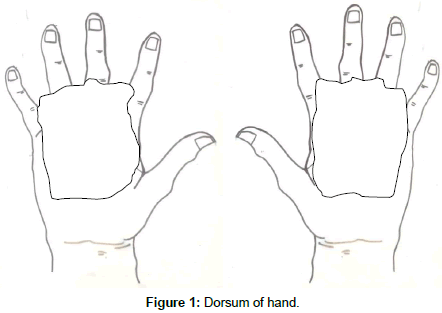
Involvement of proximal phalanx may play significant role in pointing at stable vitiligo. Least involvement of proximal may indicate its unstable nature. Acral vitiligo involving palmar aspect of the hand was seen in 70 (36.65%) cases. In both stable and unstable vitiligo involving the palms, distal phalanx was predominantly involved and the differentiation was insignificant statistically. Next common site involved was thenar eminence 28.57% and 19.35% in stable and unstable cases respectively which was also not significant. All these factors points towards the role of koebnerization in acrofacial vitiligo where distal phalanx is more prone for trauma. Hence, site distribution play minimal role as a deciding factor on stability if palmar vitiligo (Figure 1).
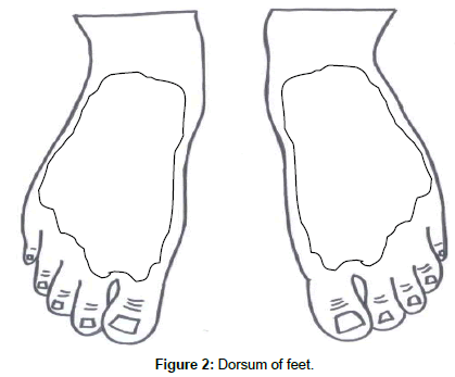
In stable vitiligo involving dorsum of feet, distal phalanx was most commonly involved (Figure 2). In unstable vitiligo, joint was predominantly involved in 67.74%, in stable vitiligo joints in 19.78% cases. Hence, similar to dorsum of the hand, predominant joint involvement of dorsum of feet too, indicates unstable vitiligo compared to stable which is very highly statistically significant. Hence, predominant joint involvement in acral vitiligo of dorsum of the feet indicates unstable vitiligo.
Involvement of vitiligo involving soles was seen in 49 (25.65%).In stable vitiligo involving the soles, heels was predominantly involved in 27.47% cases. In unstable vitiligo, all areas were equally involved with heels being marginally high in 11.29% cases. Hence, heel involvement in stable vitiligo is highly statistically significant when compared to unstable vitiligo. We propose that predominant involvement of heels with minimal involvement of other parts of soles indicates stability.
We don’t have similar studies in literature to compare our findings.
Conclusion
Acral vitiligo more commonly affected dorsum of the hands and feet compared to palms and soles. Higher incidence of acral vitiligo in our study is attributed to acral areas being prone for injuries and subsequent development of koebnerization.
Well defined margins do not strike to the stability of acral vitiligo and signs of activity can be seen even in stable vitiligo. Hence, unlike Vitiligo of other areas, where well defined margins can be a marker of stable vitiligo, margins may not be a scale for defining the stability of acral vitiligo.
In both stable and unstable acral vitiligo involving the hand and feet too, dorsum was predominantly involved than palms and soles. Predominant joint involvement in acral vitiligo of dorsum of hands indicates unstable course. Involvement of proximal phalanx may play significant role in pointing at stable vitiligo. Least involvement of proximal region may indicate its unstable nature.
Site distribution play minimal role as a deciding factor on stability in palmar vitiligo. Predominant joint involvement in acral vitiligo of dorsum of feet indicates unstable vitiligo. Predominant involvement of heels with minimal involvement of other parts of soles indicates stability.
Study on larger population is essential for further affirmation of these clinical findings.
Conflict of Interests
The authors declared no potential conflict of interests with respect to the research, authorship, and/or publication of this paper.
References
- Hee-Young P, Marinya P, Jin L, Mina Y (2008) Disorders of Melanocytes (7th edtn.) Mc Graw Hill, New York, USA: 591-640.
- Taieb A, Picardo M VETF Members (2007) The definition and assessment of vitiligo: a consensus report of the Vitiligo European Task Force. Pigment cell Res 20: 27-35.
- Nordlund JJ (2011) Vitiligo: A review of some facts lesser known about depigmentation. Indian J Dermatol 56: 180-189.
- Falabella R (2003) Surgical treatment of vitiligo: Why, when and how. J Eur Acad Dermatol Venereol 17: 518-520.
- Krupashankar DS, Shashikala K, Madala R (2012) Clinical patterns of vitiligo and its associated co-morbidities: A prospective controlled cross-sectional study in south India. Ind Dermatol J 3: 114-118
- Shah H, Mehta A, Astik B (2008) Clinical and sociodemographic study of vitiligo. Indian J Dermatol Venereol Leprol 74: 701.
- Martis J, Bhat R, Nandakishore B, Shetty JN (2002) A clinical study of vitiligo. Indian J Dermatol Venereol Leprol 68: 92-93.
- Agarwal S, Ojha A, Gupta S (2014) Profile of vitiligo in Kumaun region of Uttarakhand, India. Indian J Dermatol 59: 209.
- Gopal K, Rama Rao GR, Kumar YH, Appa Rao MV, Vasudev PS (2007) Vitiligo: A part of systemic autoimmune process. Indian J Dermatol Venereol Leprol 73:625.
- Shajil EM, Agrawal D, Vagadia K, Mafatia YS, Begum R (2006) Vitiligo: Clinical profiles in Vadodara, Gujarat. Indian J Dermatol 51: 100-104.
- Kar PK (2001) vitiligo: A study of 120 cases. Indian J Dermatol Venereol Leprol 67: 302-304
- Rajpal S, Atal R, Palaian S, Prabhu S (2008) Clinical profile and management pattern of vitiligo patients in a teaching hospital in Western Nepal. J Clin Diagnos Res 2: 1065-1068.
- Speeckaert R, Van Geel N (2014) Distribution patterns in generalized vitiligo. J Eur Acad Dermatol Venereol 28: 755-762.
- Reghu R, James E (2011) Epidemiological profile and treatment pattern of vitiligo in a tertiary care teaching hospital. Int J Pharm Pharm Sci 3: 137-141.
- Sarin RC, Kumar AS (1997) A clinical study of vitiligo. Indian J Dermatol Venereol Leprol 43: 300-314.
- Jha AK, Pandey SS, Shukla VK (1992) Punch grafting in vitiligo. Ind J Dermatol Venereol Leprol 58: 328-330.
 Spanish
  Chinese
  Russian
  German
  French
  Japanese
  Portuguese
  Hindi
Hindi 