Case Report, Clin Oncol Case Rep Vol: 3 Issue: 4
Acrometastases in Renal Cell Carcinoma: A Case Report and Review of Literature
Vassilis Milionis1, Dimitrios Vlachodimitropoulos2, Dimitrios Goutas3* and Nikolaos Goutas2
1Istomedica Private Anatomic Pathology Lab, Athens, Greece
2Laboratory of Forensic Medicine and Toxicology, The National and Kapodistrian University of Athens, Greece
3First Department of Pathology, School of Medicine, The National and Kapodistrian University of Athens, Greece
*Corresponding Author :Dimitrios Goutas
First Department of Pathology,
School of Medicine, The National and Kapodistrian University of Athens Laikon,
General Hospital of Athens, Greece
E-mail: goutas.dimitris@hotmail.com
Received: June 03, 2020 Accepted: August 25, 2020 Published: September 10, 2020
Citation: Citation: Milionis V, Vlachodimitropoulos D, Goutas D, Goutas N (2020) Acrometastases in Renal Cell Carcinoma: A Case Report and Review of Literature. Clin Oncol Case Rep 3:4.
Abstract
Abstract
Skin metastases represent a rare entity, with an overall incidence of <2 % among all metastatic lesions and 3% of all Renal Cell Carcinomas (RCC). They are more common in males. They usually get misdiagnosed, initially, due to their close resemblance with other inflammatory conditions of the skin. Most common sites of skin metastases include the head, neck and trunk in that order. We report the case of a Renal Clear Cell Carcinoma (RCCC) with a right index finger metastasis. Our case should raise clinicians’ awareness for digital lesions in patients with a known history of renal cell carcinoma.
Keywords: Carcinoma; Inflammatory; Renal cell; Metastatic; Lesions
Introduction
Acrometastasis represent a rare entity, constituting 0.1% of metastases. Most commonly, the primary cancer site that can lead to acrometastasis is the lung, followed by colorectal, breast and genitourinary tract. Occasionally, it can imitate a benign condition and be the primary presentation of an occult cancer. However, they are usually seen in patients at a critical state, with advanced disseminated disease [1,2]. Treatment remains debatable.
Case Report
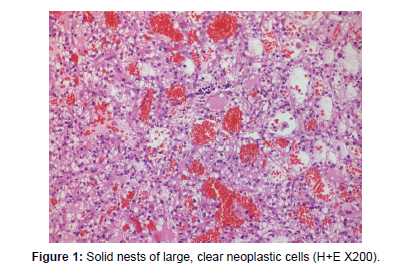
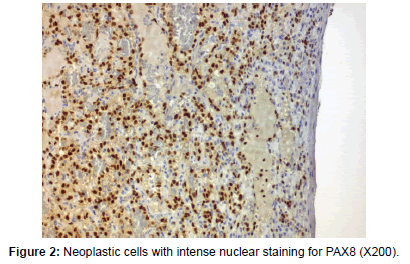
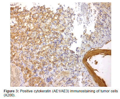
A 76 year old male patient, with a history of RCCC, for which he had undergone a radical nephrectomy 5 years ago, was examined by his dermatologist for a rapidly growing ulcerated lesion in his right index finger. A skin lesion biopsy was performed and the sample was sent to our laboratory for histopathologic evaluation. The lesion was composed of clear cells and a capillary rich network invading the stroma and the subcutaneous adipose tissue, with focal areas of abscess formation and necrosis. The findings were suggestive of a RCCC, which was confirmed by immunohistochemistry for CK-PAN(+) and PAX-8(+). Two months later, the patient developed a new tan brown lesion in his left temporal lobe, with pathologic examination being positive for a new metastasis from the known RCCC. The histological features were similar to those described earlier. Within a few days after the lesion was excised, the patient experienced a stroke episode as a result of metastasis of the RCCC to the brain. Brain metastases cause significant peritumoral oedema and as a result of these tumors’ vascularity, they have the ability to progress to intracranial hemorrhage [3] (Figures 1-3).
Discussion
The rare event of acrometastasis to the hand was first described by Handley [4], a British surgeon, who reported the case of a woman with breast cancer who later developed multiple metastases at the metacarpals. Clinical presentation includes an erythematous, swollen, warm and painful lesion, which, in case of advanced tumors, it can become severely irritated and ulcerated with functional impairment. When appreciating the radiologic appearance of such irregular osteolytic lesions, with absence of periosteal involvement and a known history of malignancy, it should always raise suspicion for acrometastasis. Nevertheless, other inflammatory conditions, such as gout, osteomyelitis, rheumatoid arthritis, whitlow, fistula and osteoarthropathy should be excluded first [5].
Renal cell carcinomas show a great geographic variability in their prevalence, with the highest incidence being found in Europe and North America. Smoking, hypertension, obesity and polycystic kidneys are among the most common risk factors for renal carcinomas [6].
High vascularization of malignant renal tumors renders them capable for metastasis, mostly along the hematogenous route. Among the most prevalent sites of metastases are the lungs (50%), lymph nodes (35%), liver (30%), bones (30%), adrenal glands (5.5%) and brain (2%) [7].
RCC represents 2%-3% of all malignant tumors [8], from which only 10% of cases present with the classic triad of urological symptoms; flank pain, an abdominal mass and hematuria. It is estimated that 40% of patients with localized advanced disease develop metastasis, while 20% of patients with RCC present with metastatic disease at the time of diagnosis. Although novel therapies have improved the outcome of patients with renal cancer, long term survival for those with advanced disease can only be achieved by aggressive surgical resection [9].
Flynn et al. [10] studied hand metastases among 257 cases of patients, with 31 (12%) of them being related to a primary renal carcinoma. The study revealed a two-fold higher incidence in males, most frequently involving the distal phalanx and fifth finger. The authors came to the conclusion that, although further investigation on the mechanism of metastasis of RCC to the hand needs to be made, there is increasing evidence that a physical trauma could precede the metastasis.
For purposes of pain palliation and survival prolongation, amputation is the suggestive way of treatment [11], in most cases with a poor, however, outcome. It is generally estimated that most patients, with hand metastasis, survive less than 6 months [12].
Conclusion
High vascularization of malignant renal tumors renders them capable for metastasis, mostly along the hematogenous route. Among the most prevalent sites of metastases are the lungs, lymph nodes, liver, bones, adrenal glands and brain.
For the purposes of the pain palliation and prolongation, amputation is the suggestive way of treatment, in most of the cases with a poor, however, outcome, it is generally estimated that most patients, with hand metastasis, survive less than six months.
References
- Reddy MP, Rao DMR, Verma RS, Srinath B, Katiyar RS (1998) Response of S13 mulberry variety to VAM inoculation under semi-arid condition. Ind J Plant Physiol 3: 194-196.
- Bloemberg GV, Wijfjes AHM, Lamers GEM, Stuurmam N, Lugtenberg BJJ (2000) Simultaneous imaging of pseudomonas fluoresens WCS 3655 populations expressing three different autofluorescent proteins in the rhizosphere: new perspective for studying microbial communities. Mol Plant Mic Int 13:1170-1176.
- Goel AK, Laura RD, Pathak DV, Anuradha G, Goel A (1999) Use of biofertilizers: potential, constraints and future strategies review. Int J Trop Agric 17: 1-18.
 Spanish
  Chinese
  Russian
  German
  French
  Japanese
  Portuguese
  Hindi
Hindi 


