Spanish
  Chinese
  Russian
  German
  French
  Japanese
  Portuguese
  Hindi
Hindi Research Article, Clin Dermatol Res J Vol: 11 Issue: 1
Biomimetic Marine-Derived Human-like Ceramides as a Natural Alternative to Synthetic Ceramides for Skin Barrier Restoration
Juan Pablo De la Roche1, 2*, ÃÂÂguila-Carricondo1, 2
1Microalgae Solutions S.L., Calle Dehesa Vieja 8. Nave 5. 28052. Madrid. Spain
2MC Actives GmbH, Calle Dehesa Vieja 8. Nave 5. 28052. Madrid. Spain
- *Corresponding Author:
- Juan Pablo De la Roche
Microalgae Solutions S.L., Calle Dehesa Vieja 8. Nave 5. 28052. Madrid. Spain
E-mail: jproche@ microalgaesolutions.com
Received: 08-June-2026, Manuscript No. CDRJ-26-190150, Editor assigned: 10-June-2026, PreQC No. CDRJ-26-190150(PQ), Reviewed: 24-June-2026, QC No. CDRJ-26-190150, Revised: 26-June-2026, Manuscript No. CDRJ-26- 190150 (R), Published: 30-June-2026, DOI: https://doi.org/10.5281/zenodo.21352306
Citation: Roche JPD, ÃÂÂguila-Carricondo (2026) Biomimetic Marine-Derived Human-like Ceramides as a Natural Alternative to Synthetic Ceramides for Skin Barrier Restoration. Clin Dermatol Res J 11:248
Copyright: © 2026 Roche JPDL. This is an open-access article distributed under the terms of the Creative Commons Attribution License, which permits unrestricted use, distribution and reproduction in any medium, provided the original author and source are credited.
Abstract
Skin barrier function relies on the integrity of the stratum corneum lipid matrix, composed of ceramides, cholesterol, and free fatty acids (FFA). Disruption of this system, due to aging or environmental stressors, leads to increased trans epidermal water loss (TEWL), reduced skin moisture, and impaired skin barrier function. Despite its widespread use, synthetic ceramide NP does not fully replicate the complexity of native epidermal lipids. This study aimed to evaluate the efficacy of Oceamides® (hereafter OCM or the active ingredient), an ingredient based on a marinederived natural biomimetic lipid complex obtained from the microalga Nannochloropsis gaditana, containing human-like ceramides (NP, NS, NG), FFA and cholesterol, combined with phosphatidylcholine (PC) and cyclodextrin (CD). Its efficacy was assessed through one in vitro study using a reconstructed human epidermis model and three clinical studies. OCM resulted in a significant improvement in skin barrier integrity (+37% at 48 h), enhanced protection (+12% at 6 h), and promoted regeneration (+18% at 6 h) in the in vitro study. In clinical studies, OCM (0.5%) reduced TEWL after 3 days of topical application (−15% forehead, −9% chin, −24% cheeks), increased skin moisture more rapidly than synthetic ceramide NP (+13% vs. +9% at 2 h), and improved skin firmness (+14% vs. +9.2%), with comparable reductions in skin fatigue (-22%). These findings suggest that OCM is an effective natural and biomimetic alternative to synthetic ceramide NP, providing faster and multiparametric effects of skin barrier function through a multi-component lipid approach.
Keywords: Skin barrier; ceramides; Marine bioactives; Nannochloropsis; TEWL; Hydration
Introduction
Marine microalgae have emerged as a sustainable source of bioactive lipids for cosmeceutical applications, representing an alternative to synthetic ingredients. Lipid extracts from marine species such as Nannochloropsis species are of interest due to their high content of glycolipids, phospholipids, sphingolipids and ceramides [1,2,3]. Marine-derived lipids can interact with the intercellular matrix of the stratum corneum, contributing to the maintenance and restoration of epidermal barrier function; however, their biomimetic relevance depends on their compositional similarity to native epidermal lipids [3]. Recent lipidomic studies have demonstrated that the application of Nannochloropsis extracts and other microalgae extracts not only supplies the skin with essential ceramides but also actively modulates the intracellular sphingomyelin-ceramide (SM-CER) pathway, leading to an increased endogenous synthesis of ceramides in human keratinocytes [3,4]. Furthermore, these marine extracts contain antioxidants and exhibit anti-inflammatory properties, protecting the skin against environmental stressors such as UV radiation by preventing lipid peroxidation and mitigating pro-inflammatory signaling [4,5,6]. The importance of such advanced, naturally sourced marine lipid complexes is further supported by examining the fundamental biology and structural characteristics of the skin barrier that they are designed to restore.
The epidermis is divided into four sublayers, which, from deepest to most superficial, are:
stratum basale, stratum spinosum, stratum granulosum, and stratum corneum [7].
The stratum corneum is the outermost layer of the epidermis and is therefore the most exposed to external agents. Two major lipid classes can be found in this layer: i) sebaceous lipids, found in approximately 25%, and mainly produced from skin microbiota metabolism [8], and ii) stratum corneum structural lipids which are essential for the skin barrier function and reduce TEWL. The predominant lipids are ceramides (50%), cholesterol (10%), and FFA (25%) [7], [Figure 1]. Ceramides found in the stratum corneum, in order of abundance are AH (22%), NS (21%), AS (18%), NP (13%), and AP (4%) [9].
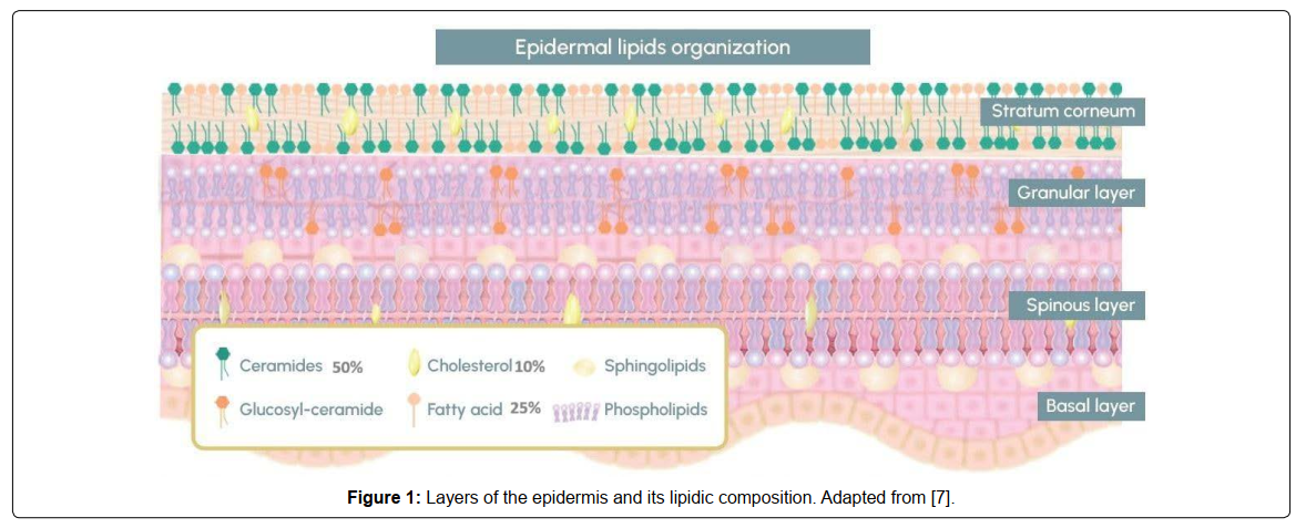
Figure 1: Layers of the epidermis and its lipidic composition. Adapted from [7]
The stratum corneum functions as the main barrier against external environmental factors. Its ability to retain moisture (measured as TEWL) and keep out irritants depends largely on an extracellular lipid matrix. This matrix consists of a densely packed lipid ‘mortar ‘of ceramides, cholesterol, and FFA [10,11].
Ceramides are key contributors to this process. They constitute most of these lipids and contribute to the formation of lamellar bilayers that keep the skin barrier cohesive. When ceramide levels drop or their composition shifts, the barrier dysfunction occurs. This is associated with characteristic features of atopic dermatitis and other inflammatory conditions [12,13]. Aging further alters this process; with aging, the human skin ceramide profile changes in both quality and quantity, resulting in the chronic dryness (xerosis) and sensitivity associated with aged skin [14].
Repairing this barrier typically involves topical ceramide-based formulations. Most formulations rely on synthetic versions, like Ceramide NP. However, these lab-grown versions struggle to match the sheer complexity of human skin, which is characterized by a diverse range of ceramide subclasses with varying chain lengths [15].
Natural biomimetic ceramides aim to close this gap by mimicking the structural diversity of native lipids. This structural diversity is functionally relevant. Emerging data suggests that these complex, biomimetic systems are an alternative to synthetic single molecules for restoring barrier function and deep hydration [16].
A healthy skin barrier depends not only on ceramides. It requires a specific balance of ceramides, cholesterol, and FFA acting synergistically. They form the highly ordered structures that support skin barrier resilience [17]. Disruption of this lipid balance compromises barrier integrity.
Current dermatological approaches increasingly focus on these multi-component systems. Research shows that the application of a complete lipid mix (matching the skin’s natural chemistry), accelerates barrier recovery and prolongs hydration [17,18].
This study evaluates an active ingredient (OCM) based on a marine-derived biomimetic lipid complex from de microalga Nannochloropsis gaditana. The active ingredient contains three human-like ceramides (NP, NS, NG) with lipids like FFA and cholesterol combined with PC and CD.
This study aims to evaluate whether this multi-component complex can serve as a natural alternative to synthetic ceramide NP to improve skin barrier function and associated clinical parameters, based on both in vitro and clinical data.
Methods and Materials
Lipid identification
The lipid profile of the active ingredient was characterized by High Performance Thin Layer Chromatography (HPTLC) at the Instituto de Acuicultura de Torre de la Sal (IATS-CSIC, Spain) with the [19].
High Performance Liquid Chromatography/ Mass Spectroscopy (HPLC/MS) was carried out by Ramon y Cajal University Hospital (Spain) to identify structurally anomalous lipids to those found in human skin.
In vitro studies
The active ingredient OCM was formulated by combining the microalgae lipid extract with PC and CD. A 3D reconstructed human epidermis (RHE) model (MatTek, Epiderm™, USA) was employed. The in vitro test was performed by GAIKER Technology Centre, Basque Research and Technology Alliance (BRTA, Spain), to evaluate the effects of topical application of OCM (0.5% w/w in cream) on parameters associated with skin barrier function, including barrier integrity, protection, and regeneration. Placebo and PC-based creams were used as controls.
The aim of this study was to evaluate the effects of the test formulations on skin barrier function in the damaged RHE. The repairing efficacy was assessed using three endpoints: transepithelial electrical resistance (TEER), resazurin reduction assay (RES), and lactate dehydrogenase (LDH) release.
TEER is widely used as an indicator of barrier integrity [20]. RES is commonly used as an indicator of metabolic activity and tissue regeneration [21], whereas LDH release reflects membrane damage and is used as an indicator of barrier disruption [22].
RHE models consist of normal human epidermal keratinocytes (NHEK) cultured at the air–liquid interface on specialized inserts in a chemically defined medium, forming a differentiated epidermal structure comprising basal, spinous, granular, and cornified layers.
Barrier disruption was induced by standardized mechanical injury, and each formulation was applied topically once daily for 3 consecutive days. Experimental groups included: untreated healthy control (healthy RHE), damaged RHE, damaged RHE + placebo cream, damaged RHE + PC cream, and damaged RHE+ OCM (0.5%). Six replicates per group were used. TEER, LDH, and RES were measured at 0, 24, 48, and 126 h.
Clinical studies
3 clinical studies were carried out. The first clinical study was conducted to evaluate the TEWL, comparing placebo with OCM at 0.5%, both formulated in cream. The study was performed with 19 volunteers. TEWL were measured was made day 0, before the application of cream and after 3 days. with probe Tewameter® TM Hex of Courage+Khazaka (Cologne, Germany) in forehead, chin and cheek, 4 replicates per area. The second clinical study was conducted to evaluate hydration effect, comparing synthetic ceramide NP at 0.5% with OCM at 0.5% in cream. The study was conducted with 22 volunteers (women and men) between 24 and 60 years. All the volunteers applied OCM cream at the left forearm, ceramide NP at the right forearm, and Placebo cream at the left biceps area with comparison purposes. Creams were applied twice per day (morning at night) for 3 days and hydration values were measured by triplicate in all the areas at 3 times: before the application, at 2 hours after the first application, and after 3 days of creams application. Skin hydration was measured using bt-analyze® device (Seattle, WA, USA).
Third clinical study was hemifacial, conducted to evaluate skin firming and skin fatigue. Two treatments were applied to a total of 20 volunteers: Ceramide NP at 0.5% and OCM at 0.5%, both in cream. Two biomechanical properties were determined at day 0 and day 28: skin fatigue and skin firmness using Cutometer MPA Dual 580 (Courage + Khazaka Electronic GmbH). Two-way ANOVA + Sídak’s were used for multiple comparisons. Study was performed by Dermaclaim Lab (Spain).
Results
Lipid composition
HPTLC analysis of the active ingredient revealed the presence of both neutral and polar lipid classes. Neutral lipids included sterol esters, triglycerides, FFA, sterols and pigments whereas polar lipids comprised sphingolipids, digalactosyldiacylglycerols (DGDG), phosphatidylglycerol and PC.
Ceramides (NP, NS, NG), PC, and cholesterol were identified by HPLC/MS and the chromatograms are shown in [Figure 2].
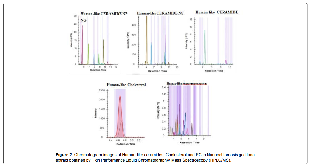
Figure 2: Chromatogram images of Human-like ceramides, Cholesterol and PC in Nannochloropsis gaditana extract obtained by High Performance Liquid Chromatography/ Mass Spectroscopy (HPLC/MS).
In vitro studies
Treatment with OCM significantly boosted skin barrier function in the RHE model. This effect was evidenced by an increase in TEER of more than 22% after 6 hours of application compared to damaged epidermis (with or without any treatment applied). In contrast, treatment with PC alone did not induce a measurable increase in TEER until 48 hours [Figure 3], indicating that the main effect was due to the effect of the biomimetic lipidic complex from Nannochloropsis gaditana.
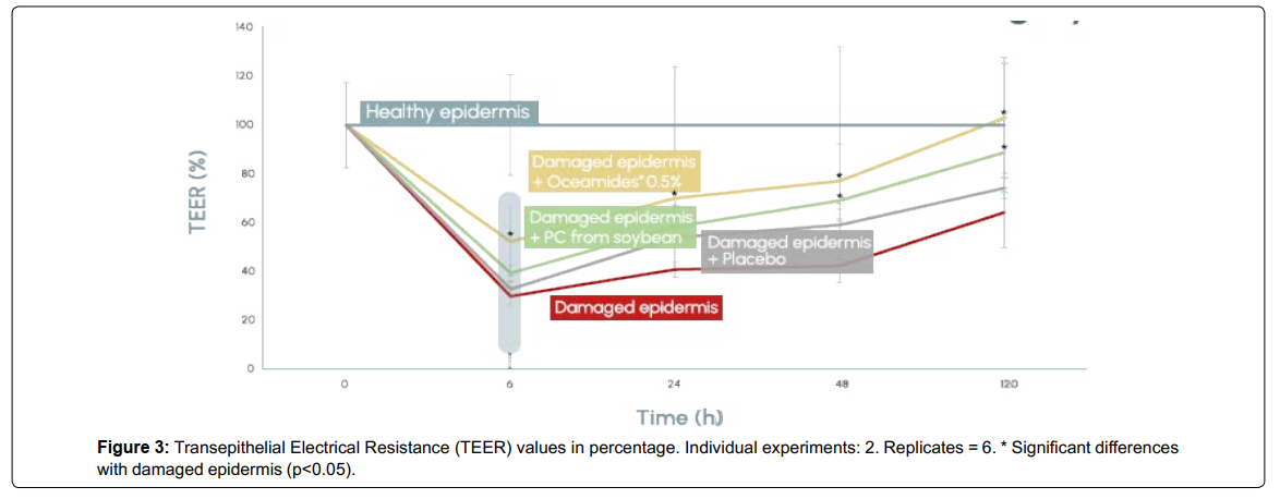
Figure 3: Transepithelial Electrical Resistance (TEER) values in percentage. Individual experiments: 2. Replicates = 6. * Significant differences with damaged epidermis (p<0.05).
Treatment with OCM (0.5%) reduced cellular damage, as indicated by decreased LDH release. LDH levels were reduced by 12% in 6 hours and by 28% in 24 hours [Figure 4].
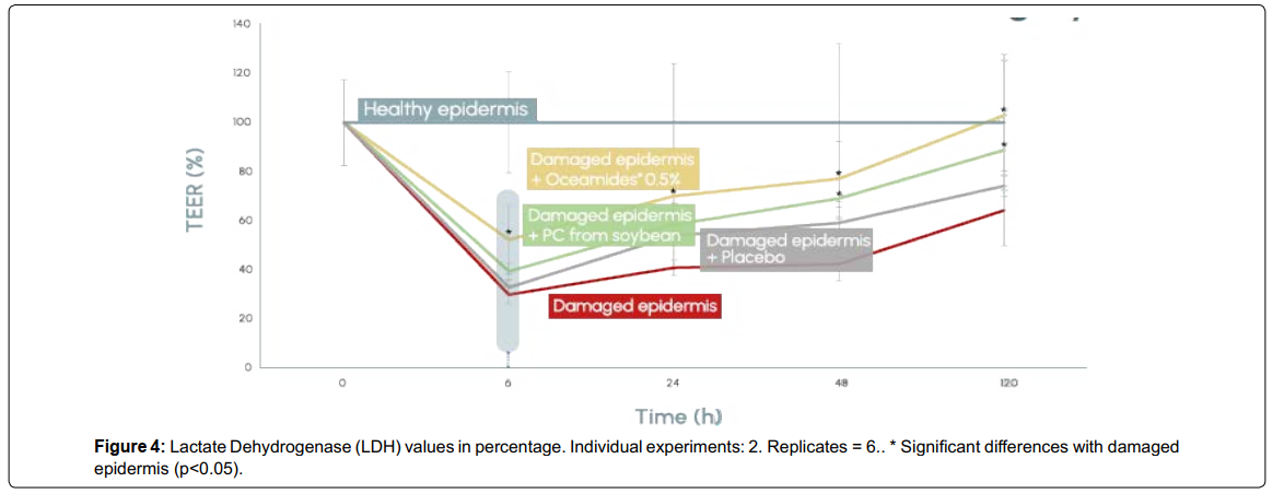
Figure 4: Lactate Dehydrogenase (LDH) values in percentage. Individual experiments: 2. Replicates = 6.. * Significant differences with damaged epidermis (p<0.05).
OCM also significantly enhanced epidermal regeneration, as demonstrated by a 35% increase in resazurin reduction (RES) after 24 hours. No significant effect was observed with PC alone at this point [Figure 5].
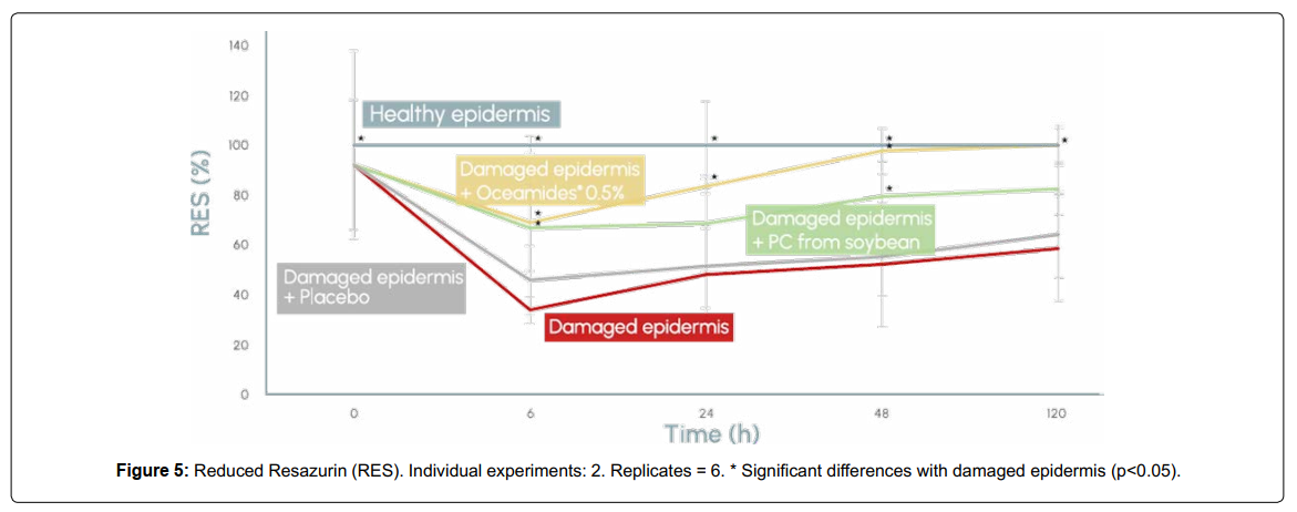
Figure 5: Reduced Resazurin (RES). Individual experiments: 2. Replicates = 6. * Significant differences with damaged epidermis (p<0.05).
Clinical studies
Clinical Study I demonstrated that topical application of OCM at 0.5% significantly reduced TEWL after 3 days across all evaluated facial areas. Specifically, TEWL decreased by 15% on the forehead, 9% on the chin, and 24% on the cheeks [Figure 6], indicating an overall improvement in skin barrier function.
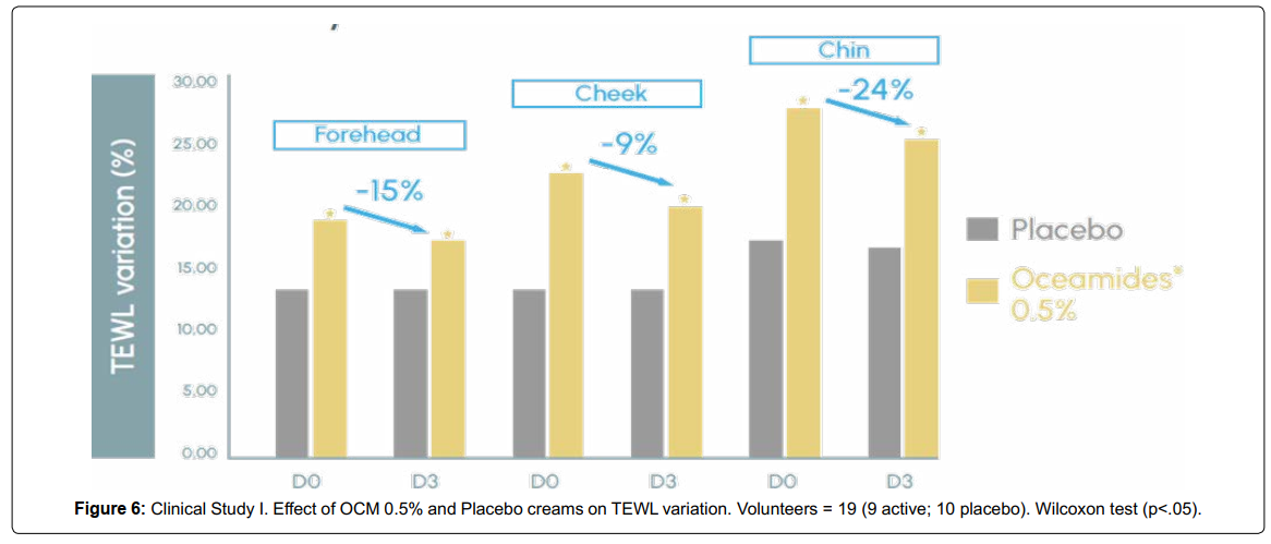
Figure 6: Clinical Study I. Effect of OCM 0.5% and Placebo creams on TEWL variation. Volunteers = 19 (9 active; 10 placebo). Wilcoxon test (p<.05).
Comparative Clinical Study II showed that OCM (0.5%) enhanced skin hydration more rapidly than synthetic ceramide NP. A 13% increase in moisture was observed after 2 hours with OCM, compared to a 9% increase with synthetic ceramide NP [Figure 7], suggesting a faster onset of moisturizing efficacy
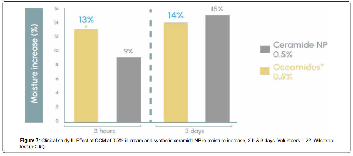
Figure 7: Clinical study II. Effect of OCM at 0.5% in cream and synthetic ceramide NP in moisture increase; 2 h & 3 days. Volunteers = 22. Wilcoxon test (p<.05).
Comparative Clinical Study III revealed that OCM (0.5%) led to a greater improvement in skin firmness (+14%) compared to synthetic ceramide NP (+9.2%). In terms of skin fatigue reduction, both treatments showed comparable effects, with reductions of 22% and 21% for OCM and synthetic ceramide NP, respectively, with no statistically significant differences between them [Figure 8, shows the visual comparative antiaging effect of OCM and synthetic Ceramide NP.
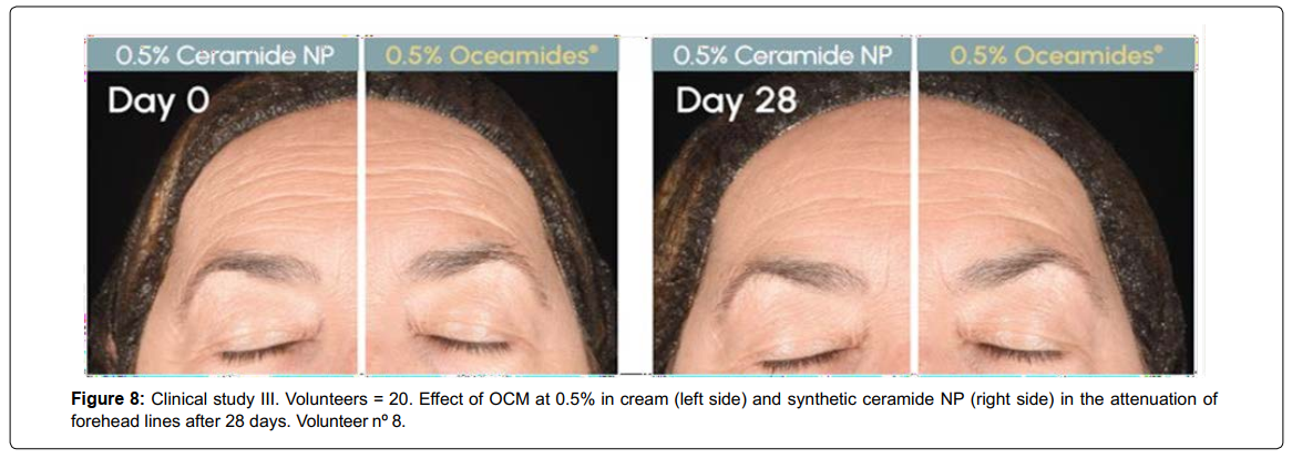
Figure 8: Clinical study III. Volunteers = 20. Effect of OCM at 0.5% in cream (left side) and synthetic ceramide NP (right side) in the attenuation of forehead lines after 28 days. Volunteer nº 8.
Discussion and Analysis
HPTLC of OCM revealed the presence of neutral lipids, including FFA, sterol esters, triglycerides, pigments and sterols. Polar lipids and included sphingolipids, PG, DGDG, and PC. Human-like ceramides (NP, NS, NG), cholesterol, and PC were identified by HPLC/MS, consistent with the presence of natural biomimetic epidermal lipid components. Lipidomic analyses of Nannochloropsis oceanica ethanol extracts [5] have shown a relative enrichment of polar lipids, including phospholipids and glycolipids such as PC, phosphatidylglycerol (PG), phosphatidylethanolamine (PE), and galactolipids (MGDG and DGDG), while neutral lipids (mainly triacylglycerols) were less represented in the extract. These extracts contained high levels of polyunsaturated fatty acids, particularly eicosapentaenoic acid (EPA), indicating preservation of bioactive lipid fractions.
These differences highlight the impact of extraction methodology and species selection on lipid composition and functional profile. The presence of sphingolipids and human-like ceramides in N. gaditana (OCM) supports its potential application in dermocosmetic formulations targeting skin barrier function. Overall, these findings suggest that combining the intrinsic lipid diversity of Nannochloropsis species with optimized extraction strategies may enable the development of ingredients with the potential to mimic the lipid architecture of the stratum corneum.
In vitro studies
The present study shows that OCM induces a rapid and functionally relevant restoration of epidermal barrier integrity, as evidenced by the early increase in TEER (>22% at 6 h), whereas PC alone showed a delayed and limited effect. This temporal advantage is consistent with previous in vitro studies reporting that marine-derived extracts improve epithelial cohesion and reduce permeability under stress conditions [23]. However, while such studies mainly describe barrier preservation, the current findings indicate an active and accelerated skin barrier reconstruction, suggesting that OCM contributes to lipid matrix reorganization through its biomimetic composition.
The reduction in LDH release observed with OCM (−12% at 6 h; −28% at 24 h) further supports its cytoprotective effect. In comparison, photodamage models have shown that UV exposure induces a substantial increase in LDH release (up to ~2.4-fold), which is only partially reversed by treatment [24]. The early and sustained reduction observed here indicates a more efficient stabilization of cellular membranes. These findings support the hypothesis that complex lipid systems outperform isolated compounds due to synergistic interactions among bioactive components.
In addition, the significant increase in metabolic activity (RES +35% at 24 h) suggests enhanced epidermal regeneration. This is consistent with studies reporting modulation of sphingolipid metabolism, including changes in ceramide and sphingomyelin levels, which are directly associated with barrier repair and epidermal homeostasis [1, 2, 25]. The absence of a comparable effect with PC alone further highlights the importance of multi-component lipid systems in promoting effective regeneration.
In vivo studies
The results obtained with OCM are consistent with the growing body of evidence on marine-derived bioactives(particularly macroalgae),in improving key parameters of skin barrier function and moisture. The significant reduction in TEWL observed after short-term application aligns with previous studies showing that marine lipid extracts can enhance epidermal barrier integrity and reduce water loss through reinforcement of the intercellular lipid matrix. These effects have been associated with the presence of bioactive lipids, including phospholipids, glycolipids, and fatty acids, which contribute to improved barrier cohesion and reduced permeability (26). This suggests that OCM may act through similar lipid-driven mechanisms, enhancing water retention and restoring barrier integrity.
The rapid increase in skin moisture observed within 2 hours further supports this mechanism and is consistent with evidence showing that marine-derived ingredients can provide both immediate and sustained moisturization, in some cases greater than established humectants such as hyaluronic acid (26). While hydration effects have often been attributed to polysaccharide-rich fractions, increasing evidence highlights the contribution of lipid fractions to barrier reinforcement and moisture retention. In addition, marine bioactives contain a diverse range of compounds, including lipids, amino acids, and antioxidant molecules, which support cellular protection and skin homeostasis. These bioactive components have been shown to improve skin resilience to environmental stress and contribute to overall skin condition, including moisture retention (27).
Regarding anti-aging effects, the superior improvement in skin firmness compared to synthetic ceramide NP is coherent with literature describing marine-derived compounds as modulators of dermal structure. Clinical and preclinical studies have shown that certain algae extracts, and marine carotenoids can stimulate collagen synthesis, improve elasticity, and reduce matrix degradation through antioxidant and anti-inflammatory pathways [28]. These mechanisms may underpin the enhanced biomechanical properties observed with OCM, suggesting a broader biological activity beyond simple barrier repair.
Finally, the comparable reduction in skin fatigue between OCM and synthetic ceramide NP indicates that both actives may converge on common pathways related to hydration status and oxidative stress mitigation. Indeed, microalgal compounds are known to exert antioxidant, anti-inflammatory, and cytoprotective effects, which contribute to improved skin appearance and resilience [28]. Collectively, these findings suggest that OCM is a multifunctional marine-derived ingredient with clinically relevant benefits in skin barrier function, hydration, and anti-aging performance.
Conclusion
The present study shows that OCM is an active ingredient based on marine-derived biomimetic lipid complex capable of boosting skin barrier function and improving parameters related to skin aging. The combination of human-like ceramides, FFA, and cholesterol may contribute to a multi-parametric effect of the stratum corneum lipid matrix compared with single-component synthetic ceramide NP.
The active ingredient improves skin barrier function increasing skin integrity, protection and regeneration and offers greater or comparable effect to pure ceramide NPenhancing hydration, increasing skin firmness and reducing skin fatigue. These findings support the use of multi-component lipid systems that mimic the natural composition of the skin, offering a physiologically relevant approach to skin barrier repair.
Overall, OCM may represent a natural and marine-derived alternative to synthetic ceramide for dermocosmetic formulations targeting skin barrier dysfunction and age-related skin changes.
Conflict of Interest
The authors are affiliated with organizations involved in the development of OCM. These affiliations may be considered a potential conflict of interest. All efforts were made to ensure scientific rigor and objectivity in the design, execution, and reporting of the study. No external writing assistance was used. All authors have approved the manuscript and declare that any potential competing interests have been disclosed.
References
- Biernacki M, Conde T, Stasiewicz A, SurażyÅ?ski A, Domingues MR, et al. (2023) Restorative effect of microalgae Nannochloropsis oceanica lipid extract on phospholipid metabolism in keratinocytes exposed to UVB radiation. International Journal of Molecular Sciences. 24(18): 14323
- GÄ?gotek A, Domingues MR, Domingues P, Skrzydlewska E (2025) The effect of lipid extract of Nannochloropsis oceanica marine microalgae on glutathione and thioredoxin-dependent antioxidant systems in UVB-irradiated keratinocytes. Marine Drugs. 23(12): 454.
- Grether-Beck S, Mühlberg K, Brenden H, Felsner I, Brynjólfsdóttir A, et al (2008) Bioactive molecules from the Blue Lagoon: In vitro and in vivo assessment of silica mud and microalgae extracts for their effects on skin barrier function and prevention of skin ageing. Experimental Dermatology. 17(9): 771–779.
- Lim Y, Park S-H, Kim E, Hwang I-H, Kim S, et al. (2023) Polar microalgae extracts protect human HaCaT keratinocytes from damaging stimuli and ameliorate psoriatic skin inflammation in mice. Biological Research. 56(1): 40.
- Melo T, Figueiredo ARP, da Costa E, Couto D, Silva J, et al. (2021) Ethanol extraction of polar lipids from Nannochloropsis oceanica for food, feed, and biotechnology applications evaluated using lipidomic approaches. Marine Drugs. 19(11): 593.
- Navalho S, Ferrer-Ledo N, Barbosa MJ, Varela J (2025) Nannochloropsis lipids and polyunsaturated fatty acids: Potential applications and strain improvement. Marine Drugs. 23(3): 128.
- Vietri Rudan M, Watt FM (2021) Mammalian epidermis: A compendium of lipid functionality. Frontiers in Physiology. 12: 804824.
- Pappas A (2009) Epidermal surface lipids. Dermato-Endocrinology. 1(2): 72–
- Knox S, O’Boyle NM (2021) Skin lipids in health and disease: A review. Chemistry and Physics of Lipids. 236: 105055.
- Pullmannová P, Vávrová L, Vávra K (2019) Recent advances in topical application of ceramides to restore skin barrier function. Cosmetics. 6(3): 52.
- Berdyshev E (2024) Skin lipid barrier: Structure, function, and metabolism. Allergy, Asthma & Immunology Research. 16(5): 445–461.
- Li Q, Fang H, Dang E, Wang G (2020) The role of ceramides in skin homeostasis and inflammatory skin diseases. Journal of Dermatological 97(1): 2–8.
- Cork MJ, Danby SG, Vasilopoulos Y, Hadgraft J, Lane ME, et al. (2009) Epidermal barrier dysfunction in atopic dermatitis. Journal of Investigative Dermatology. 129(8): 1892–1908.
- Schild J, Kalvodová A, Zbytovská, J, Farwick M, Pyko C (2024) The role of ceramides in skin barrier function and the importance of their correct formulation for skincare applications. International Journal of Cosmetic 46(4): 526–543.
- Uchida Y, Park K (2021) Ceramides in skin health and disease: An update. American Journal of Clinical Dermatology. 22: 853–866.
- Yong TL, Zaman R, Rehman N, Tan CK (2025) Ceramides and skin health: New insights. Experimental Dermatology. 34(2): e70042.
- Wertz PW (2018) Lipids and the permeability and antimicrobial barriers of the skin. Journal of Lipids. 2018: 5954034.
- Van SJ, Bouwstra, JA (2016) Stratum corneum lipids: Their role for the skin barrier function in healthy subjects and atopic dermatitis patients. Current Problems in Dermatology. 49: 8–26.
- Olsen RE, Henderson RJ (1989) The rapid analysis of neutral and polar marine lipids using double-development HPTLC and scanning densitometry. Journal of Experimental Marine Biology and Ecology. 129(2): 189–197.
- Srinivasan B, Kolli AR, Esch MB, Abaci HE, Shuler ML, et al. (2015) TEER measurement techniques for in vitro barrier model systems. Journal of Laboratory Automation. 20(2): 107–126.
- Czekanska EM, (2011) Assessment of cell proliferation with resazurin-based fluorescent dye. Methods in Molecular Biology. 740: 27–32.
- Kumar P, Nagarajan A, Uchil PD (2018) Analysis of cell viability by the lactate dehydrogenase (LDH) assay. Cold Spring Harbor Protocols. 2018: db.prot095497.
- Ko SH, Lim Y, Kim EJ, Ko YW, Hong IS, et al. (2022) Antarctic marine algae extracts as a potential natural resource to protect epithelial barrier integrity. Marine 20(9): 562.
- Smeriglio A, Lionti J, Ingegneri M, Burlando B, Cornara L, et al. (2023) Xanthophyll-rich extract of Phaeodactylum tricornutum Bohlin as new photoprotective cosmeceutical agent: Safety and efficacy assessment on in vitro reconstructed human epidermis model. Molecules. 28(10): 4190.
- Mourelle ML, Gómez CP, Legido JL (2025) Revealing the potential use of macro and microalgae compounds in skin barrier repair. Applied Sciences. 15(22): 11899.
- Bogdan C, Molnar M, Dima EI, Olteanu AA, Safta DA et al. (2025) Marine macroalgae in topical formulations: Bioactive compounds, variability, analytical challenges and skin benefits.Pharmaceutics, 17(9): 1143.
- Pallela R, Na-Young Y, Kim SK (2010) Anti-photoaging and photoprotective compounds derived from marine organisms. Marine Drugs 8(4): 1189–1202.
- Abu Bakar MF, Mohd Setapar SH (2025) Fucoxanthin: An emerging ingredient in cosmeceutical applications. Journal of Dermatologic Science and Cosmetic Technology 2: 100085.