Case Report, Clin Oncol Case Rep Vol: 6 Issue: 1
Carotid Canal: An Extremely Rare Site of Metastases from Carcinoma Breast Depicted by 18-F/ FDG PET/CT Imaging
1Department of Nuclear Medicine and PET/CT, Amrita Institute of Medical Sciences and Research Centre, Abudhabi, United Arab Emirates
2Department of Oncology, Lifeline hospital,Abudhabi, United Arab Emirates
*Corresponding Author: Anshu Tewari
Department of Nuclear Medicine and PET/CT,
Amrita Institute of Medical Sciences and Research Centre,
Burjeel Medical city, Abudhabi, United Arab Emirates
E-mail: anshutewari21@gmail.com
Received: December 27, 2022; Manuscript No: COCR-22-84810;
Editor Assigned: December 29, 2022; PreQC Id: COCR-22-84810 (PQ);
Reviewed: January 09, 2023; QC No: COCR-22-84810 (Q);
Revised: January 11, 2023; Manuscript No: COCR-22-84810 (R);
Published: January 15, 2023; DOI: 10.4172/cocr.6(1).269
Citation: Tewari A, Misra R, (2023)Carotid Canal: An Extremely Rare Site of Metastases from Carcinoma Breast Depicted by 18-F/ FDG PET/CT Imaging Clin Oncol Case Rep 6:1
Abstract
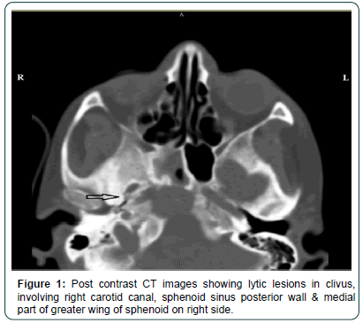
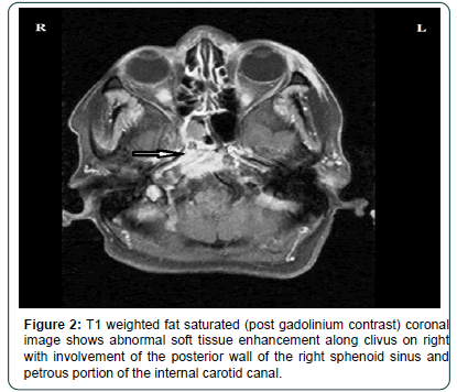
Diagnosing the disease earlier is of great importance in maximising the quality of life of the patient. We report a 60-year-old-female who presented with focal pain on right side of her face also involving eye and diplopia. Visual acuity, fundus and other ocular examination were within normal limits. Computerized Tomography (CT) detected lytic lesions in clivus, involving right carotid canal & medial part of greater wing of sphenoid on right side. Magnetic Resonance Imaging (MRI) reported tissue enhancement along clivus on right with involvement of petrous portion of the right internal carotid canal. Since patient was reluctant for any navigation-assisted surgery, whole body FDG PET/CT was offered. Whole body FDG PET/CT reported FDG avid lesion in right breast along with extensive distant deposits. Further mammogram and tissue biopsy was performed and FDG avid breast lesion was consistent with primary breast malignancy. FDG PET/CT played wonderful role not only in detecting primary site of malignancy but in upstaging and optimization of further treatment also. The ophthalmologists should be aware that breast metastases may be easily overlooked leading to a catastrophic late diagnosis when curative treatment may not be possible.
Keywords: Breast cancer; Internal carotid canal metastasis; Whole body FDG PET/CT.
Introduction
The most common lesion of Internal Carotid Canal (ICC) is vestibular schwannoma, which represents 91% of all cases. Other lesions at this site include meningioma, epidermoid cyst, secondary cholesteatoma, lipoma, angioma and cysts. Malignant tumour account for less than one per cent of all lesions, metastasis reaching the ICC are extremely rare in literatureWe present a rare case of a metastasis of a breast cancer within the ICC along with extensive skeletal metastases. We discuss the diagnostic role of whole body 18 FDG PET/CT (18- Flourodeoxyglucose Positron Emission Tomography/ Computed Tomography) to assess the extent of disease and its implications in staging breast cancer and its management.
Case Report
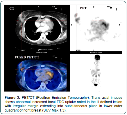
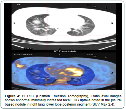
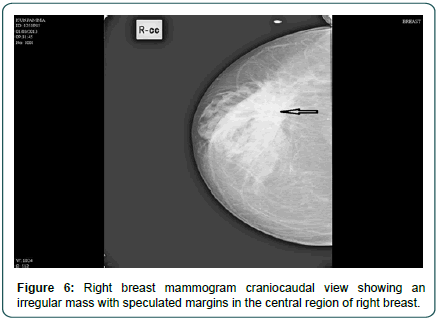
60-year-old-female presented to ophthalmology department with complaint of focal pain on right side of her face involving eye and diplopia. Visual acuity, fundus examination, vitreous and other ocular examination were normal. She was referred to neurology for further investigation. CT was done which reported lytic lesions in right clivus, involving right carotid canal, sphenoid sinus posterior wall & medial part of greater wing of sphenoid on right side (Figure 1).To assess any soft tissue impingement, MRI was suggested.MRI added tissue enhancement along clivus on right with involvement of the petrous portion of the internal carotid canal (Figure 2). Patient was in good health with no co-morbidities. In view of patient’s refusal for invasive investigation, whole body PET/CT was offered to detect culprit. Whole body PETCT reported abnormal increased focal FDG uptake in the right breast as an ill-defined lesion with irregular margin extending into subcutaneous plane in lower outer quadrant (Figure 3). FDG avid lesions found in clivus involving right carotid canal, posterior wall of sphenoid sinus, pleural based nodule in right lung (Figure 4) and extensive skeletal metastases (Figure 5). Mammogram of right breast concluded an irregular mass with speculated margins in the central region of right breast BIRADS5 (Figure 6). Patient was diagnosed with infiltrating ductal carcinoma after tissue biopsy. The tumour was ER/PR positive and HER2-neu negative.
Figure 5: 18 FDG PET/CT MIP (maximum intensity projection) images showing multiple metabolically active extensive skeletal metastases in Medial end of left clavicle, D5, D8, D10 & D11 dorsal vertebrae, L1, L2 & L5 lumbar vertebrae border of lower part of sacrum on the left, right iliac bone and bilateral acetabulum.
Once the diagnosis was established, treatment was mainly with palliative focusing on symptom relief and improvement of function as it is a high-grade tumour with extensive spread. The initial treatment consisted of neoadjuvant chemotherapy with epirubicin/ cyclophosphamide (EC-protocol) followed by surgical tumour ablation and dissection of the right axilla. Postoperative external beam radiation of the thoracic wall was performed. After completion of the initial treatment with complete remission the patients further clinical course was uneventful. Due to the positive receptor status antihormonal therapy with anastrozole was initiated. Local radiotherapy of face was reserved in case of progression of ocular symptoms.
Discussion
The carotid canal is the passage way in the temporal bone through which the internal carotid artery enters the middle cranial fossa from the neck. The canal's internal opening is near the foramen lacerum above which the internal carotid artery passes on its way to the cavernous sinus. It transmits into the cranium, the internal carotid artery, and the carotid plexus of nerves.
The lesion of Internal Carotid Canal (ICC) includes vestibular schwannoma, which represents 91% of all cases [1]. Other lesions at this site include meningioma, epidermoid cyst, secondary cholesteatoma, lipoma, angioma and cysts1. Malignant tumour account for less than one per cent of all lesions. Nasopharyngeal carcinomas and squamous cell carcinomas of external acoustic canal are known for ICC metastases [2,3]. Metastasis to the skull-base particularly affects patients with carcinoma of the breast and prostate [4]. This is first case of metastases of breast cancer to ICC in literature to our knowledge.
Typical manifestations of ICC metastases as such are not mentioned but internal carotid artery along with carotid plexus transmits through ICC and enters middle cranial fossa. Sympathetic to the head from the superior cervical ganglion also pass through the carotid canal. They have several motor functions: raise the eyelid (superior tarsal muscle), dilate pupil, innervate sweat glands of face and scalp and constricts blood vessels in head.Therefore, any pathology affecting ICC will affect the structures travelling through it. Symptoms may include ptosis, miosis, anhidrosis, hypoesthesia of face and transient loss of consciousness. Patients with ICC metastases usually have locoregional bone erosion also, like in present study tumour had involved medial part of greater wing of sphenoid on right side.
Treatment of ICC metastasis aims at improving patient's quality of life and restore or preserve function. The overall prognosis to skull base metastases is poor, with an overall median survival of about 2.5 years, probably because skull-base metastases appear late in the course of the disease [4]. The mean survival after diagnosis of specifically ICC metastases is unknown. Radiotherapy is the mainstay for skull metastases and appears to be safe and effective with objective response rates up to 79% [5]. Radiotherapy improves symptoms in 80% of cases [6]. Systemic chemotherapy is another mainstay in the palliative setting of bone metastases [7]; in addition, the initiation of bisphosphonate treatment is important if osseous metastasis have been diagnosed [8].
Conclusion
In summary, although rare, breast cancer patients can develop metastases to the ICC or other skull base regions. Patients with a history of breast cancer presenting with symptoms such as ptosis, meiosis, anhidrosis, diplopia or pain should be evaluated for skull base metastases. Once diagnosis is confirmed treatment for patients with ICC metastases is multidisciplinary.
References
- Font RL, Ferry AP (1976) Carcinoma metastatic to the eye and orbit. III. A clinicopathologic study of 28 cases metastatic to the orbit. Cancer 38: 1326-1335. [Google Scholar] [Cross Ref]
- Boamah H, Knight G, Taylor J, Palka K, Ballard B (2011) Squamous cell carcinoma of the external auditory canal: A case report. Case Reports Otolaryngol 2011. [Google Scholar] [Cross Ref]
- Han J, Zhang Q, Kong F, Gao Y (2012) The incidence of invasion and metastasis of nasopharyngeal carcinoma at different anatomic sites in the skull base. Anatomical Record: Advan Integrat Anat Evolution Biol 295: 1252-1259. [Google Scholar] [Cross Ref]
- Laigle-Donadey F, Taillibert S, Martin-Duverneuil N, Hildebrand J, Delattre JY (2005). Skull-base metastases. J Neuro-Oncol 75: 63-69. [Google Scholar] [Cross Ref]
- Ratanatharathorn V, Powers WE, Grimm J, Steverson N, Han I, et al. (1991) Eye metastasis from carcinoma of the breast: Diagnosis, radiation treatment and results. Cancer Treat Rev 18: 261-276. [Google Scholar] [Cross Ref]
- Char DH, Miller T, Kroll S (1997) Orbital metastases: diagnosis and course. British J Ophthalmol 81: 386-390. [Google Scholar] [Cross Ref]
- Eckardt AM, Rana M, Essig H, Gellrich NC (2011) Orbital metastases as first sign of metastatic spread in breast cancer: Case report and review of the literature. Head Neck Oncol 3: 1-4. [Google Scholar] [Cross Ref]
- Wong ZW, Phillips SJ, Ellis MJ (2004) Dramatic response of choroidal metastases from breast cancer to a combination of trastuzumab and vinorelbine. The Breast J 10: 54-56. [Google Scholar] [Cross Ref]
 Spanish
  Chinese
  Russian
  German
  French
  Japanese
  Portuguese
  Hindi
Hindi 