Short Communication, J Sleep Disor Treat Care Vol: 6 Issue: 2
Head of the Bed Elevation: An Alternative Treatment for Obstructive Sleep Apnea
Souza FJFB1,2*, Souza Filho AJ1 and Borges AN2
1Pulmonary Division, Hospital Sao Jose, Pulmonar Sleep Laboratory, Criciuma, Santa Catarina, Brazil
2Universidade do Extremo Sul Catarinense, Criciuma, Santa Catarina, Brazil
*Corresponding Author : Fabio Jose Fabricio de Barros Souza
Pulmonary Division - Hospital Sao Jose, Pulmonar Sleep Laboratory and Universidade do Extremo Sul Catarinense, Criciuma, Santa Catarina, Brazil
Tel: 55 48 34374088
E-mail: fsouzapneumo@hotmail.com
Received: March 06, 2017 Accepted: March 23, 2017 Published: March 30, 2017
Citation: Souza FJFB, Souza Filho A, Borges AN (2017) NHead of the Bed Elevation: An Alternative Treatment for Obstructive Sleep Apnea. J Sleep Disor: Treat Care 6:2. doi: 10.4172/2325-9639.1000194
Abstract
Obstructive sleep apnea (OSA) leads, in the long term, to important cardiovascular and neuropsychological changes. The prevalence of the disease is high and many therapies are being studied. In this context, this article aims to present an alternative treatment of OSA wich is well-tolerated, cheap and has few contraindications.
Keywords: Obstructive Sleep Apnea, OSA, Epworth Sleepiness Scale
Introduction
Obstructive sleep apnea (OSA) leads, in the long term, to important cardiovascular and neuropsychological changes [1]. The prevalence of the disease is high and many therapies are being studied [1-3]. In this context, this article aims to present an alternative treatment of OSA wich is well-tolerated, cheap and has few contraindications [2,3].
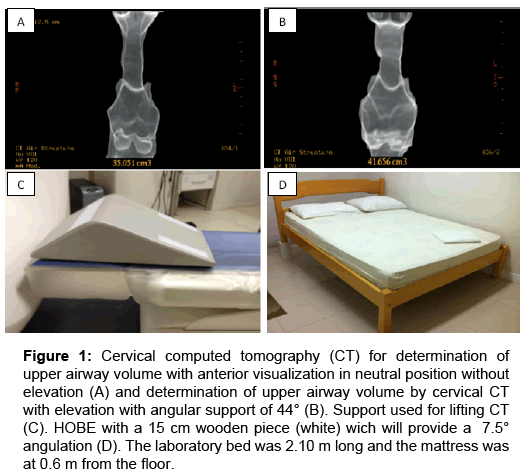
Patient A.A.B.S, 52 years-old, male, body mass index 35kg/ m2, neck circumference 43 cm, Epworth Sleepiness Scale 15 points and with previous history of OSA. In this study, a standard polysomnography (PSG) was performed, followed by another PSG with head of the bed elevation (HOBE), using a 15 cm wooden support with 7.5 degree angulation. The laboratory bed was 2.10 m long and the mattress was at 0.6 m from the floor. The results were positive: The increase in total was sleep time 340 to 363.5 minutes and the minimum saturation increased from 86 to 90%. There was also a reduction in the apnea and hypopnea index (10.8 to 4.1 events/hour) and the saturation time below 95% (from 148.8 to 38.1 minutes). Subsequently, in order to structure the results obtained from polysomnographies; the same patient was submitted to two tomographies (Figure 1), the first was a standard cervical computed tomography (CT) and the second was with an angulation of 44º (dimensions: 45.5 cm in its longest side×43.3 cm×18.5 cm). This resulted in an increase in the volume of the airway (measured from the hard palate to the base of the epiglottis) from 35.05 to 41.65 cm3.
Figure 1: Cervical computed tomography (CT) for determination of upper airway volume with anterior visualization in neutral position without elevation (A) and determination of upper airway volume by cervical CT with elevation with angular support of 44° (B). Support used for lifting CT (C). HOBE with a 15 cm wooden piece (white) wich will provide a 7.5° angulation (D). The laboratory bed was 2.10 m long and the mattress was at 0.6 m from the floor.
In a study comparing standard polysomnography (PSGs) with HOBE polysomnography (PSGHOBE) there were significant improvements with significant reduction in apnea-hypopnea index (AHI) total (PSGs 20 ± 14 vs PSGHOBE 15 ± 14, p=0.0003), duration of snoring percentage (PSGs 32 ± 21 vs PSGHOBE 21 ± 16, p=0.023) [2]. The minimum oxygen saturation showed a trend toward improvement (baseline PSGs83 ± 8 vs 85 ± 8 PSGHOBE, p=0.079) [2]. Souza et al. [3] analysed the upper airway by cervical CT with or without elevation. The mean upper airway volume was 7.9 cm3 greater when the head was with elevation than when it was in the neutral position, and that difference (17.5 ± 11.0%) was statistically significant (p=0.002) [3]. Thus, based on this case report and on previous studies [2,3], we suggest that raising the head of the bed leads to a greater area of air flow through the upper airway, reducing the rate of apnea and hypopnea and may be a possible management in mild to moderate degree of OSA patients.
References
- Young T, Peppard PE, Gottlieb DJ (2002) Epidemiology of obstructive sleep apnea: a population health perspective. Am J Respir Crit Care Med 165: 1217-1239.
- Souza FJFB, Souza Filho A, Lorenzi-Filho G (2011) The influence of bedhead elevation on patients with obstructive sleep apnea. Am J Respir Crit Care Med 183: A2732.
- Souza FJ, Evangelista AR, Silva JV, Périco GV, Madeira K (2016) Cervical computed tomography in patients with obstructive sleep apnea: influence of head elevation on the assessment of upper airway volume. J Bras Pneumol 42: 55-60.


